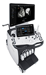
AngioPLUS is a significant advancement in colour doppler imaging. Conventional doppler is limited in its ability to show microvascular slow flow. Angio- PLUS significantly improves colour sensitivity and spatial resolution, resulting in the next level of microvascular imaging to visualize flows that couldn’t be seen before. Soon available on Aixplorer, this new imaging tool will provide highly detailed real-time information to physicians, which is key in diagnosing cancerous lesions. Lesion microvascularization and vessel flow are important indicators of a potential malignancy in areas such as breast, lymph nodes, thyroid and liver. This technique is also valuable for musculoskeletal assessments to help identify low-grade tendon inflammation. Read more
Azienda Socio Sanitaria Territoriale (ASST) Vimercate Hospital in Italy has implemented Fujifilm’s REiLI artificial intelligence system to assist healthcare practitioners on the frontline in the fight against the COVID-19 pandemic.
Due to the pandemic, operations and workflows in Italian hospitals were disrupted, which significantly increased workloads. As a consolidated partner of Vimercate, Fujifilm wanted to ensure its cutting-edge tools were available to reduce the impact caused by the rapid spread of COVID-19 by assisting in speeding up workflows.
REiLI’s processing of CT scans and chest x-rays provides important support for radiologists, offering them an extremely rapid, quantitative and objective assessment of the various zones of the lungs. The chest x-ray, in particular, is the best example of applying the new AI technology, in that it produces a quick and simple examination which provides a large amount of useful information for evaluating the presence of the pulmonary parenchymal consolidation caused by the virus.
The data obtained from artificial intelligence does not replace the molecular diagnoses performed using the nasopharyngeal swab (RT-PCR), nor does it replace the analyses and diagnoses performed by the radiologist. The data, however, does provide support in reporting on daily examinations conducted to monitor and study the development of the disease, and constitute a second opinion for the operators.
Commenting on the REiLI artificial intelligence system, Marcello Intotero, Head of Radiology Structure and Diagnostic Services Department at ASST Vimercate Hospital, said: “REiLI’s support has allowed our radiologists to report examinations with greater speed and efficiency. This new technology has given great support to the workflow; all images that the artificial intelligence system identified as suspected COVID-19 were subjected to processing by the radiologist in a faster and more urgent manner.”
REiLI is integrated with the Lunit Insight CXR module for the analysis and detection of the main types of pulmonary disease. The Lunit Insight CXR3 module was updated to provide support in specifically identifying the pulmonary parenchymal consolidation caused by the virus.
Giovanni Delgrossi, Head of the IT Department at ASST Vimercate noted that during the health emergency, more than 80 chest x-rays of patients were analysed each day at the hospital. “If no priority is set for the examinations to be reported, it may take hours to identify a particular x-ray which may require prompt, immediate action from doctors. In an emergency like the current one, we need to react and act even more quickly,” he said.
Discussing Fujifilm’s REiLI AI system, Nicola Bilibio, Clinical Specialist Medical Informatics (CSE) Fujifilm Europe GmbH explained that AI is a new operating concept projected towards collaborative intelligence between machine and man. “This is a fundamental tool for optimizing workflow and for screening patients for COVID-19. During the emergency, we promptly made our most advanced technologies available to make a concrete contribution to the crisis.”
For more information, visit: http://reili.fujifilm.com
About REiLI on YouTube: https://youtu.be/M6ISuRmvQbY
AMULET Innovality is Fujifilm’s flagship in digital mammography solutions. Images resulting from its innovative approach to tomosynthesis clinical application – two different acquisition modes, to better fulfil breast screening and diagnostic mammography needs – with unsurpassed 50 micron pixel size, will be shown together at the booth.
https://interhospi.com/wp-content/uploads/sites/3/2020/08/IH189_Fujifilm_Amulet-Innovality_crop.jpg8003823wmediahttps://interhospi.com/wp-content/uploads/sites/3/2020/06/Component-6-–-1.png3wmedia2020-08-26 14:31:322021-01-08 12:10:27Digital mammography system
Canon Medical Systems Corporation enters the premium 1.5T MRI market with the launch of its new Vantage Orian system. Vantage Orian is one of Canon Medical Systems’ first major product releases since officially changing their company name from Toshiba Medical on January 4, 2018, and represents a major foray into the premium wide bore 1.5T market along with new technology designed to boost productivity, enhance patient comfort and deliver diagnostic clinical confidence to users and patients alike. With a new slim gradient that delivers a gradient performance with a maximum amplitude of 45 mT/m, combined with a slew rate of 200 T/m/s that enhances high resolution and diffusion imaging, Vantage Orian offers high performance imaging capability previously not available on 1.5T systems from Canon Medical Systems and utilizes migrated high-end 3T technology. Canon Medical System’s PURERF and Saturn Gradient technology means that stable and consistent imaging performance is ensured, increasing signal to noise ratio (SNR) by up to 38%. The Vantage Orian offers a range of new Rapid Scan and Easy Tech technology that reduces scan time and helps improve workflow. A redesigned gantry interface and dockable table ensure seamless patient handling. New applications like Multiband SPEEDER, that shortens acquisitions such as high angular resolution diffusion imaging (HARDI), and up to x8 accelerated k-t SPEEDER allows high frame rate cardiac cine and perfusion imaging with free breathing. Vantage Orian also offers one of the smallest footprints in the 1.5T wide-bore class and low power consumption eco features that reduce energy use by up to 21%. The reliable system is equipped with excellent maintenance programs to satisfy the needs of hospital administrators. A relaxed patient is key in MRI, and Vantage Orian features quiet scan sequences that reduce ambient noise by Pianissimo Zen technology. The 71cm wide bore and immersive virtual MR Theater are designed to put patients at ease. Vantage Orian also addresses challenging patients with Pediatric SPEEDER coils, contrast- free applications and ForeSee View for simplified scan planning. Read more
https://interhospi.com/wp-content/uploads/sites/3/2020/08/IH262_Canon.jpg4486183wmediahttps://interhospi.com/wp-content/uploads/sites/3/2020/06/Component-6-–-1.png3wmedia2020-08-26 14:31:322021-01-08 12:10:35Premium 1.5T MRI system
Generally, endoscopic retrograde cholangiopancreatography (ERCP) procedures remain very safe for patients and allow the least-invasive manner for diagnosis and treatment for many biliopancreatic disorders and conditions. Yet, post-ERCP infections typically range between 2% and 4%. In August of this year, the FDA recommended healthcare facilities and manufacturers to begin transitioning to duodenoscopes with disposable components to reduce the risk of patient infection. A solution to the risk of infection, is to modify current endoscopes so that they have disposable parts. Especially the parts that are most receptable to contamination. Another solution would be to have disposable endoscopes. This of course introduces questions about pricing and the environmental impact which need to be solved by manufacturers and medical facilities before this solution can be used in daily practice. PENTAX Medical recently introduced two innovative product solutions that reduce the risk for infection transmission. The DEC Duodenoscope (ED34-i10T2) aims to achieve this by eliminating the riskiest components of the endoscope, the distal end and elevator, and replacing it by a single-patient use sterile, disposable distal cap with integrated elevator. Additionally, using the PlasmaTYPHOON to ensure the device is fully dry reduces the risk for microbial growth and subsequent potential con- tamination that can result from moisture. Drying endoscopes after reprocessing is recognized to be crucial for reducing the risk of infection. Read more
https://interhospi.com/wp-content/uploads/sites/3/2020/08/Pentax_DSCF0383.jpg5338003wmediahttps://interhospi.com/wp-content/uploads/sites/3/2020/06/Component-6-–-1.png3wmedia2020-08-26 14:31:322021-01-08 12:10:22Duodenoscope using disposable distal cap with integrated elevator
A study published in the Journal of Diabetes Science and Technology found that “Quo-Lab met all criteria” and “has no bias compared to the mean of four secondary reference measurement procedures.”* Quo-Lab is a semi-automated analyser designed to meet the needs of diabetes clinics and labs that demand low cost of operation and ease of use. It provides HbA1c results in four minutes using a small drop of blood. EKF has developed Quo-Lab in conjunction with industry experts to deliver a product that meets the tightening requirements of NGSP certification and CAP EQA criteria. Find out what Quo-Lab can do for your practice today.https://www.ekfdiagnostics.com/quo-lab.html *Lenters-Westra E, English E. Evaluation of Four HbA1c Point-of-Care Devices Using International Quality Targets: Are They Fit for the Purpose? Journal of Diabetes Science and Technology. 2018; 12: 762-770. Read more
https://interhospi.com/wp-content/uploads/sites/3/2020/08/EKF_Quo-Lab_In-use-scaled.jpg191725603wmediahttps://interhospi.com/wp-content/uploads/sites/3/2020/06/Component-6-–-1.png3wmedia2020-08-26 14:31:322021-01-08 12:10:30EKF Diagnostics’ Quo-Lab analyser is setting new standards in HbA1c measurement
Representing an evolution of the original detector-based IQon Spectral CT scanner, the IQon Elite advances the capabilities of its predecessor through a series of key benefits that support emergency/trauma department care and oncology care. This new configuration provides a number of enhanced features, including faster reconstruction speeds and better visualization of bone marrow pathology which increases diagnostic confidence within emergency and trauma departments, providing the opportunity to reduce the need for subsequent scans. The system’s ability to estimate electron density provides enhanced tissue characterization. IQon Elite’s faster reconstruction speeds have been shown to enable up to 200 CT patients per day. This is an important benchmark for healthcare facilities experiencing a high volume of patients, especially in the emergency department where CT can be a commonly-used imaging modality. The system’s new radiation therapy planning couch and bariatric table top allow larger patients to be scanned with increased positioning controls, providing greater clinical breadth.
Xenios AG, a company of the Fresenius Medical Care Group, reports that it has seen a significant growth in demand for its extracorporeal membrane oxygenation (ECMO) devices, which can be used for the treatment of patients who develop severe pneumonia and acute respiratory distress syndrome (ARDS) due to COVID-19 infection.
Jürgen Böhm, CMO of Xenios, explained that for critically ill COVID-19 patients with acute lung failure and refractory hypoxemia – despite use of standard therapy – “our treatment often remains the last therapeutic option and has been a lifesaver for many patients”.
Xenios’s ECMO therapy bypasses the function of the lungs. The patient’s blood is freed from carbon dioxide outside the body and enriched with oxygen. The lungs are thus given time to heal. Because of the increase of critically ill COVID-19 patients, more physicians are opting for ECMO therapy, and thus the increase in demand for Xenios’s ECMO devices.
To meet the demand, the company has increased production of its ECMO devices. “We have put many measures in place to maximize the utilization of our capacity to manufacture ECMO devices as well as patient kits. Our biggest challenge right now is the availability of specific components for our products,” said Andreas Terpin, CEO of Xenios.
For more information, visit: www.xenios-ag.com Read more
https://interhospi.com/wp-content/uploads/sites/3/2020/08/PRODUCT_XENIOS.jpg74910003wmediahttps://interhospi.com/wp-content/uploads/sites/3/2020/06/Component-6-–-1.png3wmedia2020-08-26 14:31:322021-01-08 12:10:17Xenios sees growing demand for ECMO devices
Radiation measurement often requires different devices for varying applications adding to the cost and complexity of data collection. The new RaySafe 452 Survey Meter is a versatile, powerful tool that can be used for multiple situations, reducing the number of devices technicians need to carry, learn, and calibrate. The 452 Survey Meter is the ideal tool to measure radiation in a wide variety of applications, including wall leakage, scatted radiation in a room, tube leakage, nuclear medicine, and control of treated patients. The meter helps technicians make data collection accurately and easily with fast response times and detect variations from background radiation levels due to its high sensitivity. There is no need for post processing of the data due to a flat, wide energy response range. With the automatic saving of dose rate values every second data is easily saved to be analysed later. The RaySafe 452 features an optimized modern design that provides a big, clear, easy-to-read display with all the parameters in a single backlit display. Its ergonomic and lightweight design makes it easy to hold, carry, and use even over extended periods of time. And it has an IP 64 rating so it can easily be cleaned with water without damaging the device. The survey meter comes with RaySafe View software that makes it easy to transfer data to a PC and log data quickly, easily, and efficiently. All data is time stamped and dose rate waveforms can be further analysed later. Read more
https://interhospi.com/wp-content/uploads/sites/3/2020/08/IH274_Fluke_Biomed_RaySafe_452.jpg5338003wmediahttps://interhospi.com/wp-content/uploads/sites/3/2020/06/Component-6-–-1.png3wmedia2020-08-26 14:31:322021-01-08 12:10:25Radiation meter for variety of applications
The Magnetom Sola Cardiovascular Edition is a 1.5 Tesla magnetic resonance imaging (MRI) scanner designed specifically for cardiovascular examinations. Magnetom Sola Cardiovascular Edition incorporates the latest technologies with the specific aim of providing the maximum diagnostic information in cardiovascular examinations. This leads to faster, more reliable, and definitive diagnoses for a larger number of patients with underlying ischemic, structural, and arrhythmogenic conditions. The MRI scanner’s innovative applications can cut examination times down to 20 minutes and clarify a broad range of clinical questions in cardiology. Guidelines issued by the European Society of Cardiology (ESC) have long described magnetic resonance imaging as the gold standard for assessing the morphology and function of the heart. Over 25 ESC Guidelines now contain specific recommendations describing instances where MRI should be used in cardiac diagnosis. This is primarily due to the accuracy of MRI when quantifying cardiac volume, mass, and wall motion, as well as its ability to diagnose ischemia and myocardial viability. There are also diseases such as myocarditis (inflammation of the heart muscle), for which cardiovascular MRI is the only non-invasive diagnostic option with the pre-requisite sensitivity. By receiving the right diagnosis at the right time, this group of patients can avoid unnecessary invasive procedures. The Compressed Sensing Cardiac Cine application accelerates MRI scanning sequences so that cardiac function can be measured while the patient is breathing freely. Previous scanners required patients to hold their breath for up to 20 seconds several times during the process. This means that the benefits of the gold standard are now available to new groups of patients, including individuals with arrhythmia or dyspnea who were previously not eligible for CMR. The MyoMaps application provides quantitative information on the tissue composition of the cardiac muscle. This can be used to diagnose diseases of the heart muscle, scar tissue and edema at a very early stage of the condition, allowing the right treatment option to be chosen for the patient as soon as possible. Clinical conditions such as myocarditis can even be detected without using contrast agents. Cardiac Dot Engine is a unique software package which provides step-by-step guidance for standardized diagnostic cardiac MRI exams. It is based on artificial intelligence algorithms, allows users to navigate confidently through the cardiovascular MRI scanning process. Also, BioMatrix Technology automatically adjusts to the biovariability of different patients to prevent undesirable variations in the scanning process. As a result, BioMatrix Technology reduces examination times, cuts down on the number of repeat scans, and leads to consistent, high quality scanning results. Read more
We may ask you to place cookies on your device. We use cookies to let us know when you visit our websites, how you interact with us, to enrich your user experience and to customise your relationship with our website.
Click on the different sections for more information. You can also change some of your preferences. Please note that blocking some types of cookies may affect your experience on our websites and the services we can provide.
Essential Website Cookies
These cookies are strictly necessary to provide you with services available through our website and to use some of its features.
Because these cookies are strictly necessary to provide the website, refusing them will affect the functioning of our site. You can always block or delete cookies by changing your browser settings and block all cookies on this website forcibly. But this will always ask you to accept/refuse cookies when you visit our site again.
We fully respect if you want to refuse cookies, but to avoid asking you each time again to kindly allow us to store a cookie for that purpose. You are always free to unsubscribe or other cookies to get a better experience. If you refuse cookies, we will delete all cookies set in our domain.
We provide you with a list of cookies stored on your computer in our domain, so that you can check what we have stored. For security reasons, we cannot display or modify cookies from other domains. You can check these in your browser's security settings.
.
Google Analytics Cookies
These cookies collect information that is used in aggregate form to help us understand how our website is used or how effective our marketing campaigns are, or to help us customise our website and application for you to improve your experience.
If you do not want us to track your visit to our site, you can disable this in your browser here:
.
Other external services
We also use various external services such as Google Webfonts, Google Maps and external video providers. Since these providers may collect personal data such as your IP address, you can block them here. Please note that this may significantly reduce the functionality and appearance of our site. Changes will only be effective once you reload the page
Google Webfont Settings:
Google Maps Settings:
Google reCaptcha settings:
Vimeo and Youtube videos embedding:
.
Privacy Beleid
U kunt meer lezen over onze cookies en privacy-instellingen op onze Privacybeleid-pagina.