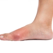
Genetic bombshell shatters gout myths: Lifestyle choices not to blame, landmark study reveals
New research challenges long-held beliefs about the causes of gout, highlighting the significant impact of genetics on the chronic inflammatory condition.

New research challenges long-held beliefs about the causes of gout, highlighting the significant impact of genetics on the chronic inflammatory condition.

A new study suggests that medical interns working on float schedules with concentrated night shifts experience better sleep regularity, mood, and cognitive performance compared to those on traditional call schedules with extended shifts.

Researchers at the Medical Center – University of Freiburg demonstrate that artificial intelligence can effectively produce medical documentation, potentially reducing physicians’ administrative burden.
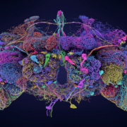
Researchers unveil the most comprehensive neural map of an adult animal brain to date, offering unprecedented insights into brain circuitry and function.
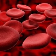
A single-dose gene therapy has demonstrated significant reduction in bleeding episodes for adults with haemophilia B, potentially offering a long-term solution to this genetic disorder.
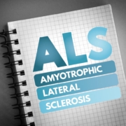
Researchers have developed a highly accurate blood test for amyotrophic lateral sclerosis (ALS), potentially reducing diagnosis times and improving patient outcomes.
Scientists at Weill Cornell Medicine in Qatar have developed a comprehensive molecular map of the human body, integrating data from multiple ‘omics’ platforms to create a powerful new research tool.
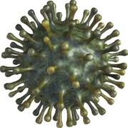
A broadly neutralising antibody capable of protecting against all known SARS-CoV-2 variants and related coronaviruses has been discovered by researchers at the University of Texas at Austin.

New research reveals that patients with stable coronary artery disease who quit smoking at any point after diagnosis can reduce their risk of major cardiovascular events by nearly 50%. The study, presented at ESC Congress 2024, also found that merely reducing smoking habits had minimal impact on cardiovascular risk.

Researchers at Weill Cornell Medicine have utilised advanced machine learning techniques to identify three distinct subtypes of Parkinson’s disease, potentially paving the way for more personalised treatment approaches.
Prins Hendrikstraat 1
5611HH Eindhoven
The Netherlands
info@interhospi.com
PanGlobal Media IS not responsible for any error or omission that might occur in the electronic display of product or company data.
 © Suhre/Halama Labs