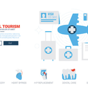
Beukenlaan 137
5616 VD Eindhoven
The Netherlands
+31 85064 55 82
info@interhospi.com
PanGlobal Media IS not responsible for any error or omission that might occur in the electronic display of product or company data.
Medical tourism refers to people who travel overseas for obtaining treatment. In the past, it referred to (wealthy/privileged) patients from developing countries who visited medical centres in industrialized countries to get treatment not available at home.
However, the situation has since reversed, in certain cases dramatically. Medical tourism now typically refers to patients from industrialized countries who travel to poorer countries for lower priced, or more quickly available (and in some cases, superior) treatment. Top medical tourist destinations in this respect include India and Thailand as well as Costa Rica, Mexico and the Gulf.
Medical tourism and health tourism
Some studies do not consider medical tourism to include cosmetic and wellness tourism – with dental treatment also viewed as a cosmetic procedure. This, larger group is often referred to as health tourism.
Market revenues for a major medical tourism destination, Thailand, include a relatively large number of cosmetic surgery procedures. This segment also includes a multitude of places in south America, with the industry’s maturity fed by bustling local demand. For example, according to the Sociedad Boliviana de Cirugia Plastica y Reconstructiva’, over 70percent of middle and upper class women in the country have had plastic surgery.
India leads in higher-end procedures
Conversely, at the other end, if only surgical procedures for overseas patients are included, India leads the global medical tourism market.
Consultants McKinsey estimated 180,000 medical tourists were treated at Indian facilities in 2004 (up from 10,000 just five years earlier). Arrivals have since been rising sharply and are estimated to have reached 250,000 in 2012, contributing 3 billion USD in revenues. This is effectively about 30percent of the global market, estimated for the year at 10.5 billion USD by Transparency Market Research.
India has proven to be a preferred destination for US and UK patients, in particular, because of the use of English in most professional interactions, as well as the fact that both countries have a large number of Indian-origin physicians. Indeed, the US government’s top medic, the Surgeon General, is Vivek Hallegere Murthy, a 40-year old Indian.
India stands out as an interesting destination in another respect. Its massive generic drugs industry provides post-operative medicinal treatment at prices well below the West.
Market drivers: cost, waiting times, accreditation
Key factors driving medical tourism from the West to developing countries include the high cost of healthcare and increasing waiting times for certain procedures. Insurance in several countries often does not cover 100percent of the costs of common age-related requirements such as a knee or hip replacement, or limits the choice of the prosthetics, or the surgeon and facility.
Accreditation of top hospitals in medical tourism destinations has also fuelled demand. The oldest international accrediting body is Accreditation Canada, which has accredited hospitals in about a dozen countries.
The best known accreditation group, however, is Joint Commission International (JCI) in the US. JCI was set up in 1994 to provide international clients education and consulting services, and several international hospitals now see accreditation as a way to attract American patients. JCI is an independent private, not-for-profit organization that seeks to develop nationally and internationally recognized procedures to help improve patient care and safety. It advises hospitals to meet standards for patient care and then accredits hospitals meeting the standards.
A British scheme, QHA Trent Accreditation, is an active independent holistic accreditation scheme. Another is GCR.org, which monitors success metrics and standards of almost 500,000 medical clinics worldwide.
These schemes vary in quality, size and cost to hospitals making use of them.
Increasingly, hospitals are looking towards dual international accreditation, perhaps having both JCI to cover potential US clientele, and Accreditation Canada or QHA Trent for Canadian and British patients.
Indian price advantage boosted by quality, innovations
Practically all surgery procedures performed in medical tourism destinations cost a fraction of what they do in industrialized countries. For example, while a liver transplant in the US costs about 300,000 USD ( Euro 280,000), the figure in India is 50,000 USD ( Euro 47,000). Open heart surgery in India costs between 3,000 ( Euro 2,800) and 10,000 USD ( Euro 9,400), compared to 70,000 USD ( Euro 65,500) in the UK and 150,000 USD ( Euro 140,000) in the US.
Such figures acquire added value when one reviews the conclusions of a Harvard Business School (HBS) study in November 2013, comparing data on angioplasty in the US versus India. The study found that one in 200 US angioplasty patients required emergency surgery, with half of them dying, while only two of 40,000 angioplasty patients at India’s CARE Hospitals required emergency surgery, with just one death in the OR since the hospital’s inception in 1997.
The HBS study also studied other Indian hospitals and interventions, finding them to be on par or better than their US counterparts – for example, Apollo Hospitals with knee, coronary and prostate surgery as well as for infections related to the operating theatre and catheters, Narayana for coronary artery bypass procedures, Deccan for peritoneal dialysis and Aravind for ophthalmology.
The HBS review noted India was not simply an improver but an innovator too, for example Indian doctors pioneered the beating-heart method of surgery, where they operate without shutting patients’ hearts down via a heart-lung machine, leading to fewer complications, shorter hospital stays and quicker recovery.
New segment of intra-Third World medical tourism
While much attention remains on Western medical tourists, one of the fastest growing market segments consists of patients within the Third World, who travel to more advanced developing countries. India again is at the top of the list. In early Dec 2016 / Jan 2017, for instance, it was announced that Iman Abdulati, a 36-year old woman weighing half a tonne, was to be flown to India from her home in Egypt for bariatric surgery.
Such cases have drawn considerable attention for other, political reasons.
Pakistani patients with severe conditions requiring top-notch treatment are a routine media fixture in India. For example, in September 2016, Pakistan’s Express Tribune’ featured the case of Abdul Basit, an 11-year old boy who had been suffering from the rare condition known as Crigler-Najjar syndrome and went for a liver transplant to India. Two years previously, after complications, the wife of former Afghan President Hamid Karzai gave birth to a girl at Fortis Hospital in New Delhi.
In August 2016, The Diplomat’ reported India had emerged as one of the fastest growing global healthcare destinations, particularly for patients from conflict countries like Afghanistan, Iraq, Yemen, Sudan, the Democratic Republic of Congo (DRC), and Somalia, attracting close to 400,000 foreign patients a year, half from war-ravaged countries.
The selection of India as preferred medical tourism destination is being officially sanctioned. In 2004, BBC News’ reported that ‘India was chosen as the place’ for sending sick patients from Tanzania unable to be treated at home, after the Tanzanian government ‘did a comparative analysis of health facilities in South Africa, India and western European countries.’ In 2007, Companion Global Healthcare teamed up with hospitals in India (as well as Thailand and Singapore).
China lags India due to egalitarian healthcare model
Due to a variety of reasons, the other Asian behemoth, China, has a much less mature medical tourism sector. WHO figures show hospital bed densities far lower in India, at just 9 per 10,000 people (making a total of roughly 1 million beds) compared to 42 in China (about 5 million). However, the higher share of private beds in India (40percent against 6.5percent in China) means that India has slightly more private beds – about 400,000, against China’s 325,000.
More than anything, India’s lead over China in medical tourism symbolises its top-down approach to healthcare, in contrast to China’s bottom-up one which first aims at providing top quality healthcare to local Chinese. As a result, China does not provide good healthcare for its middle and upper class. For Britain’s Guardian’, poor rural Chinese were curiously’ better off than their city cousins.’ The Guardian’ contrasted this with India, where ‘many city-based healthcare facilities are excellent….’ This higher-end focus provides India with more medical tourists than China.
Hospital budgets: the sky’s the limit
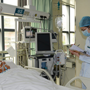
There are now at least a dozen major private hospital groups in India. Leading groups (with 20-50 facilities, and 2,500-8,000 beds) include Apollo, Max Healthcare, Fortis, Escorts Healthcare, Wockhardt and the Manipal Group. Many of the above (as well as newcomers from cash-rich Indian conglomerates such as Reliance, the Hindujas, Sahara and ITC) are also pursuing the new concept of Medicities, involving suburban developments dedicated wholly to integrated hospital facilities.
The procurement budget of such groups is not insignificant. Apollo’s annual spending on medical equipment, for example, has been close to 200 million USD in recent years. Such budgets allows cash-rich Indian hospitals to procure state-of-the-art equipment – from Da Vinci robots and stereotactic laser surgery to wide-bore 3T Silent Scan MRIs.
Nevertheless, as far as spending is concerned, Indian hospital groups face several challenges in the coming years, especially from the cash-flush Gulf.
US hospitals lead Gulf partnerships
US hospitals are at the forefront of partnerships in the Gulf. One of the key reasons was the difficulty for medical tourists from the region to obtain US visas after 9/11, according to the American Hospital Association.
Key US partners of Gulf hospitals include Johns Hopkins Medicine, which has an agreement since 2006 to partner the General Health Authority for Health Services in the UAE. It also manages the 400-plus bed Tawam Hospital in Abu Dhabi and an affiliated centre offering state-of-the-art molecular imaging services. Johns Hopkins also has alliances with King Khaled Eye Specialist Hospital in Saudi Arabia.
Another example is the Cleveland Clinic, which is affiliated with the International Medical Centre in Jeddah, Saudi Arabia, and is a strategic partner at the 360-bed, multi-specialty Cleveland Clinic Abu Dhabi Hospital in the UAE.
Elsewhere in the UAE, Methodist International manages the operations of Burj Dubai Medical Centre as well as clinics in Dubai, while Partners Harvard Medical International is a key strategic collaborator with Dubai Healthcare City (which explicitly seeks to attract foreign medical tourists).
Education and training focus in Gulf
Many of these alliances are increasing their focus on education and training. For example, the Partners-Dubai Healthcare City has added a high profile unit called Harvard Medical School Dubai Center Institute for Postgraduate Education and Research, while in 2014 Johns Hopkins signed a partnership with oil major Aramco to provide medical education and training in Saudi Arabia.
Qatar, too, has sought US partners. The Weill Cornell Medical College was in fact one of the earliest ventures, established in 2001 as a partnership between Cornell University and the Qatar Foundation for Education, Science and Community Development. It aims to provide medical education and cutting-edge research.
Ironically, the focus on training might hit a traditional source of physicians in the Gulf especially hard, namely Indians who would be replaced by skilled locals.
Medicine is in a state of continual progression as new therapies and interventions are developed and technological advances facilitate resuscitation and prolonged organ support. In addition, patients are living longer with increased numbers of comorbidities and complex disease processes. As one result of these demographic changes and medical advances, intensive care units (ICUs) are admitting more patients with a high risk of death, patients who would previously have died before reaching the ICU. As a consequence, the need for end-of-life decisions has become more common than in the past. There has also been a move from an emphasis on survival at all costs to a recognition that the quality of life of survivors must also be taken into account, as well as the quality of dying for those who will not survive.
by Prof Jean-Louis Vincent
With patients who are not considered to have any reasonable chance of benefiting from new or continued intensive care treatments, physicians are faced with four possible options, ranging from continuing with full treatment to support life through to increasing the doses of sedatives to hasten the dying process (Table). In some patients, where withdrawing therapy is permitted, an ICU trial’ can be considered, giving the patient the chance to benefit from a possible intervention. The target of such a test and the time-limit must be set in advance and adhered to; good communication with the family is essential to ensure that these factors are clear. It should be remembered that in some patients, death is actually in their best interest, preventing unnecessary and prolonged suffering.
Recent data suggest that some 40% of ICU non-survivors will have a decision to withhold/withdraw life-sustaining therapy during their ICU stay. Perhaps not surprisingly, there are marked differences in end-of-life decisions and the decision-making practice around the globe. For example, data show that patients are more likely to receive a decision to withhold or withdraw life-sustaining therapy in Oceania, North America, and northern Europe and less commonly in the Middle East, Asia, southern Europe and South America. Although withdrawing and withholding are seen as ethically equivalent in many countries, in others, withholding life-sustaining therapy is considered acceptable but not withdrawing. In Israel, because withdrawal of life-support measures is forbidden, the authorities even passed a law whereby timers can be put on respirators, which then stop by themselves after a preprogrammed time period. The use of sedatives/analgesia at the end-of-life to shorten the dying process also varies considerably among countries and individuals. Some people justify the administration of large doses of sedatives/analgesics in this situation by calling on the double effect’ principle, wherein giving analgesic agents for comfort has the unavoidable effect of hastening death, but this view is rather hypocritical. There is little official guidance available for intensivists regarding this issue and it is perhaps the area of end-of-life management that creates the greatest concern among physicians with fear of possible litigation. The Belgian Society of Intensive Care recently published a statement that ‘Shortening the dying process with use of medication, such as analgesics/sedatives, may sometimes be appropriate, even in the absence of discomfort, and can actually improve the quality of dying’.
The degree of involvement of family members in end-of-life decision making also varies, with families more frequently involved in Northern Europe and the US than in southern European countries. This is in part related to the traditional paternal approach to medical practice still widespread in many southern European countries. Family-centered decision making is also common in East Asian countries, such as Japan, China and South Korea.
The reasons for these international differences are complex. Many are related to the marked cultural and religious diversity among countries. Lack of available resources and financial constraints can also influence end-of-life decision making, particularly in lower income countries. There are also differences among ICUs within a country and among individual intensivists, related again to the cultural and religious backgrounds of the physicians, but also to local legislation, peer and family pressure, and ICU casemix and organization amongst others. The key ethical principles of autonomy, beneficence, non-maleficence and distributive justice must always be used as the basis for any end-of-life decision, but the ways in which these are interpreted and their relative importance may vary according to local factors. It is therefore inappropriate to try and develop a universal consensus on end-of-life decisions as some have suggested, although local guidelines may be useful. Open discussion of these difficult issues must be encouraged within the ICU team and good communication with the family is essential. The aim must always be to provide compassionate end-of-life care, appropriate for the individual patient and his/her particular circumstances.
Suggested reading
Curtis JR and Vincent JL Ethics and end-of-life care for adults in the intensive care unit. Lancet 2010;376:1347-53.
Myburgh J, et al End-of-life care in the intensive care unit: Report from the Task Force of World Federation of Societies of Intensive and Critical Care Medicine. J Crit Care 2016; 34:125-30
Vincent JL, et al ‘Piece’ of mind: end of life in the intensive care unit statement of the Belgian Society of Intensive Care Medicine. J Crit Care 2014;29:174-175
The author
Jean-Louis Vincent, MD, PhD
Dept of Intensive Care,
Erasme University Hospital,
Universite libre de Bruxelles,
Route de Lennik 808,
1070 Brussels,
Belgium
Tel. +32-2-555-3380
Fax +32-2-555-4555
E-mail: jlvincent@intensive.org
4D cardiac imaging, which generates a three-dimensional motion picture of a beating’ heart, offers cardiologists a revolutionary new tool. Indeed, the ability to acquire images across all phases of a heartbeat cycle is the only way to meaningfully visualizing morphological anomalies and make an authentic assessment of cardiac function.
Traditionally, ultrasound has been a preferred modality for 4D cardiac imaging. However, 4D cardiac MRI (known formally as cardiovascular’ MRI) has been gaining ground. Coupled with MRA (magnetic resonance angiography), it enables cardiologists to view images of the heart, major blood vessels and blood flow.
The novelty of 4D
4D cardiac imaging is a recent technique. Its novelty is best illustrated by an editorial in The Journal of the American College of Cardiology’. The editorial, published as recently as 2009, observed the role of ‘2- and 3-dimensional coronary mapping’ in high-resolution digital imaging.
Major imaging vendors now offer real-time 3D/4D imaging products – across all modalities, PET/CT, MRI and ultrasound. However, the bulk of 4D applications so far have involved ultrasound – especially for cardiac imaging. This may be changing, with increased attention, above all, to MRI.
Ultrasound’s longer legacy
One reason for ultrasound’s pole position in 4D consists of a longer legacy. In the early 1980s, researchers from Duke University in the US reported that though MRI was faster, ultrasound offered the closest achievement of ‘3D real-time acquisition,’ or what is now called 4D.
Technical standardization bodies also moved quickly to endorse and drive the take-up of 4D ultrasound. In 2008, the DICOM (Digital Imaging and Communications in Medicine) initiative approved Supplement 43 which addressed the exchange of real time 3D ultrasound datasets between different vendors. In 2011, IHE (Integrating the Health Enterprise) published a White Paper on 3D/4D imaging workflow.
Early adoption of 4D ultrasound by cardiologists
On their part, cardiologists were enthusiastic early adopters of 3D (and later 4D) ultrasound. The IHE’s White Paper mentioned above was written by its Cardiology Technical Committee. Another factor strongly favouring ultrasound was mobility, since small ultrasound devices could be transported to the patient.
During this period, competing imaging modalities seemed to stand little chance as far as cardiology was concerned.
Computerized tomography (CT) was dismissed since it required cardiologists to use complex post-processing techniques in order to visualize the bearing heart. Cardiac magnetic resonance imaging (MRI) was considered relatively expensive, with limited availability and requiring specialized training.
GE’s cSound: industry seizes the ultrasound opportunity
Industry was quick to seize the ultrasound opportunity. In 2015, healthcare technology giant GE released new software for its ultrasound machines called cSound. cSound-equipped machines intelligently process data being returned by an ultrasound signal, analysing almost 5 gigabytes of data every second, and then filtering it on a pixel-by-pixel basis via algorithms which produced real-time 4D views. This allowed cardiologists to observe how blood swirls around clots in arteries, measure blood leakage around the valves and assess damage. cSound reinforced GE’s presence at the cutting edge of ultrasound, reinforcing a technique patented by the company in the early 2000s and known as Spatial Temporal Image Correlation (STIC). STIC allowed for the quick capture of a full fetal heart cycle beating in real-time.
4D PET/CT and MRI turn to diagnostic oncology
Proponents of 4D PET (positron emission tomography)/CT and MRI were however not sitting by idly. Rather than cardiology, they turned their attention to other specialities, above all oncology where 4D offered huge potential in diagnostics.
4D PET, for example, seemed unmatched in characterizing solitary pulmonary nodules, while 4D CT offered a revolutionary approach in oncology – such as gating tumours and determining treatment margins. On its part, 4D MRI demonstrated a superiority to CT in soft-tissue imaging and in cases where radiation exposure was a concern.
From 4D to 5D imaging
As of now, the focus in diagnostics is to combine the anatomical with functional or molecular imaging, in order to make precise assessments of biological and metabolic pathways. Key modalities include PET with radio-labelled tracers for molecular imaging, and MRI using molecular markers for functional imaging. The molecular/functional enhancement is often referred to as 5D, and to its proponents, offers hope in increasing the specificity and sensibility of diagnostics.
At some stage in the future, it is inevitable that cardiologists will see the virtues of 5D imaging for diagnostics.
The challenge from multi-detector ultrasound scanners
Meanwhile, cardiac ultrasound faces competition in certain applications from other imaging modalities.
In recent years, multi-detector CT scanners seem to offer considerable promise, particularly for non-invasive detection of coronary artery disease and higher flexibility for analysis and visualization of individual vessels. These images, nevertheless, continue to require special processing and rendering tools for assessment of segmental narrowing or occlusions.
The growing promise of 4D cardiac MRI
Rather than CT, cardiac (or cardiovascular) MRI in 4D seems to have rapidly become the principal technology paradigm challenger to ultrasound.
Cardiac MRI scanners do not use open’ magnets which face serious limitations in the case of moving objects – such as a beating heart. The magnet strengths most widely used for cardiac MRI are 1.5T and 3T – although the latter, in some conditions, require software to cancel artifacts. Higher strength magnets are, however, the technology of choice in studying conditions such as aortic construction.
What is also a key advantage of cardiac MRI compared to CT is its lack of ionizing radiation, high spatial resolution and the ability to provide a functional cardiac assessment in one scan.
The technique of 4D cardiac MRI is closely based on traditional MRI. However, it is optimized for use in the cardiovascular system in real time, principally via ECG gating and rapid imaging sequences. This results in acquisition of images at each stage of a sequence of cardiac cycles, and functional assessment of the heart. Blood, in such sequences (technically known as balanced steady state free precession or bSSFP), appears bright due to contrast with blood flow. As a result, 4D cardiac MRI makes it possible to discriminate in a relatively easy fashion between the myocardium and blood.
With and without contrast agents
Cardiac MRI typically uses several approaches to make a comprehensive assessment of the heart and cardiovascular system. Some of the most promising applications include the ability to visualize heart muscle fat or scar in high resolution without the need for a contrast agent. This is based on a technique called spin echo’, which shows blood as black, and identifies myocardium abnormalities through differences in intrinsic contrast.
On the other hand, contrast agents like gadolinium-DTPA can be used for applications such as infarct imaging – where healthy heart muscle appears dark, and infarction areas show in bright white. Contrast agents in cardiac MRI have also proven their worth for treatment of coronary artery narrowing, which starves the heart muscle of oxygen. The contrast agent reveals any transient perfusion defects from artery constriction. Knowing about the presence of such a defect assists in guiding interventional procedures.
Image quality, superior access to anatomical structures
Cardiac MRI provides images of superior quality, accuracy and versatility, alongside access to anatomical structures which are tough to achieve with ultrasound. Examples of these include congenital heart anomalies as well as anatomical changes after surgical interventions.
The latest generation of MRI scanners allow for acquiring high-resolution isotropic data with detailed anatomical information and identical resolution in all three dimensions. Frontier areas of research for 4D MRI include qualitative and quantitative flow pattern analysis in mice with aortic constriction.
Detecting hemodynamic alterations with 4D MRI
At present, one of the most promising cardiac applications for 4D MRI consists of the detection of haemodynamic alterations. The incorporation of pharmacological stress procedures allows for enhanced detection of alterations in heart function during stress-induced ischemia.
In April 2014, a team at Northwestern University reported that 4D flow MRI would help better understand altered hemodynamics in patients with cardiovascular diseases and improve patient management and monitoring of therapeutic response. Their study, published in Cardiovascular Diagnosis and Therapy’, noted that these hemodynamic insights could also lead to new risk stratification metrics in patients and impact upon individualized treatment decisions in order to optimize patient outcomes.
Diagnostics and prognosis of heart events
Cardiac MRI is also being seen as a diagnostic tool to predict heart events. In May 2016, a study led by John P. Greenwood from the University of Leeds in Britain noted that it was ‘a better prognosticator of risk for serious cardiovascular events than SPECT, regardless of a person’s risk factors, angiography results, or initial treatment, and that it would be a powerful tool for ‘the diagnosis and management of patients with suspected coronary heart disease.’ The serious events, assessed over a 5-year period, included death, myocardial infarction/acute coronary syndrome, unscheduled coronary revascularization, or hospitalization for stroke, transient ischemic attack, heart failure, or arrhythmia.
The study was based on a multi-parametric cardiovascular MRI protocol, and performed on a 1.5T MRI scanner and published in the Annals of Internal Medicine’. It was formally known as the Clinical Evaluation of Magnetic Resonance Imaging in Coronary Heart Disease (CE-MARC), and billed as ‘the largest prospective comparison of cardiovascular MRI and nuclear myocardial perfusion imaging (MPI) with SPECT’ with X-ray angiography used as the reference standard.
Genotoxicity poses calls for caution
There have, nevertheless, been some calls for caution due to the chance of genotoxic effects of cardiac MRI scanning.
In October 2011, a study by researchers at Seoul National University in South Korea, assessed high-field intensity 3T clinical MRI scans in cultured human lymphocytes in vitro and ‘observed a significant increase in the frequency of single-strand DNA breaks following exposure to a 3T MRI.’
In June 2013, another study on cardiac MRI in European Heart Journal’ reported similar conclusions, this time in vivo. The study, by researchers from University Hospital Zurich, prospectively enrolled 20 patients, and found a ‘significant increase in median numbers of DNA DSBs in lymphocytes induced by routine 1.5T’ MR scanners. The study also made a recommendation, urging cardiac MRI to ‘be used with caution and that similar restrictions may apply as for X-ray-based and nuclear imaging techniques in order to avoid unnecessary damage of DNA integrity with potential carcinogenic effect.’
Finns call for further studies
Nevertheless, there has been no study so far on the genotoxic effects of MRI compared with those of CT scans. In addition, cardiac MRI risk research has been based entirely on cell level experiments with no conclusive and definitive evidence of actual cancer risk. This is in direct contrast to the link between ionizing radiation and cancer risk.
MRI is therefore still considered by its proponents as the safest alternative.
Indeed, weeks after the University Hospital Zurich study, Finnish researchers published a riposte, again in the European Heart Journal”, arguing that the ‘cellular mechanism’ of how cardiac MRI induced DNA damage was unknown ‘and may be different from that of radiation.’ They concluded that it was ‘obvious that further larger studies are warranted before any restrictions’ were imposed on the use of cardiac MRI.
CardioConfirm is Mortara Instrument’s latest tool for connectivity and IT. CardioConfirm has been launched almost a decade after Mortara Instrument started a successful path leading to the adoption of the DICOM standard in all its ELITM series cardiographs, Stress Testing, Holter and Monitoring equipment.
The DICOM standard allows users to seamlessly integrate reports from Mortara devices with existing information systems available in hospitals. CardioConfirm takes connectivity a step further: in addition to traditional viewing options, its user-friendly interface is designed to provide full editing capabilities for all DICOM-enabled systems. Besides opening, editing and storing resting ECGs, physicians may now use dedicated tools for zooming in or measuring ECG waveforms, and may take advantage of a library of statements that conveniently appear with just a few key-strokes, on the basis of those normally used for reports.
CardioConfirm also offers the possibility of editing final reports of stress and Holter tests. Preliminary exports generated by DICOM-friendly systems can be edited by physicians from the main system workstation, a feature that makes the workflow smoother and reduces the time needed to review and edit these types of reports. This new OEM software allows any hospital or clinic to leverage their existing system by simply embedding CardioConfirm into it, thus eliminating the need to spend significant capital investment on an entirely new operation system.
CardioConfirm allows medical professionals to concentrate on patient care with its unique ability to integrate high quality diagnostic display of tests together with all patient information – including test results, vitals, and personal and family history – in one convenient location so that cardiologists do not need to look for additional test results that a technician may have recorded elsewhere.
As the health care landscape continues to grow and change, Mortara’s CardioConfirm is making a big impact on the continued transformation. Hospitals and health care professionals will have a more streamlined workflow, increased efficiency and the ability to focus more attention on patient care.
Mortara Instrument supplies CardioConfirm to all PACS and EMR providers and hospitals that want to expand or complete their PACS/EMR systems to include diagnostic cardiology workflow. It is available in a variety of versions that meet your need for a seamless integration with third party systems.
For further information, click here
DICOM is the registered trademark of the National Electrical Manufacturers Association for its standard publications relating to digital communications of medical information.
The safe use of health technology-from basic infusion pumps to large, complex imaging systems-requires identifying possible sources of danger or difficulty with those technologies and taking steps to minimize the likelihood that adverse events will occur. This list will help healthcare facilities do that.
Produced each year by ECRI Institute’s Health Devices Group, the Top 10 Health Technology Hazards list identifies the potential sources of danger that it believes warrant the greatest attention for the coming year. The list does not enumerate the most frequently reported problems or the ones associated with the most severe consequences-although such information is certainly considered in the analysis. Rather, the list reflects the Health Devices Group’s judgment about which risks should receive priority now.
All the items on the list represent problems that can be avoided or risks that can be minimized through the careful management of technologies. Additional content provided with the full article, which is available separately to members of certain ECRI Institute programmes, provides guidance to help manage the risks. In this way, the list serves as a tool that healthcare facilities can use to prioritize their patient safety efforts.
International Hospital presents here the abridged version of ECRI Institute’s 2017 Top 10 list of health technology hazards which is available as a free public service to inform healthcare facilities about important safety issues involving the use of medical devices and systems.
1. Infusion errors can be deadly if simple safety steps are overlooked
Most large-volume infusion pumps incorporate safety mechanisms for reducing the risks of potentially deadly intravenous (IV) infusion errors. These mechanisms have greatly improved infusion safety, but can’t eliminate all potential errors. And the mechanisms themselves have been known to fail.
ECRI Institute continues to learn about and investigate incidents of infusion errors involving pump or administration set failures, staff unknowingly defeating a safety mechanism, or incorrect infusion programming. Such errors- particularly those that result in the uncontrolled flow of medication to the patient, known as ‘IV free flow’-can lead to patient harm and even death.
In many of these incidents, harm could have been averted if staff had:
Once commonplace, these simple practices are now often overlooked-perhaps because staff implicitly trust the pump’s advanced safety features.
2. Inadequate cleaning of complex reusable instruments can lead to infections
The use of contaminated medical instruments can lead to disabling or deadly patient infections or instrument malfunctions.
Outbreaks associated with the use of contaminated duodenoscopes-such as those that caused headlines in recent years-illustrate the severity of this issue. But duodenoscopes are not the only devices that warrant attention. ECRI Institute has received reports involving a variety of contaminated medical instruments that have been used, or almost used, on patients.
Complex, reusable instruments-such as endoscopes, cannulated drills, and arthroscopic shavers-are of particular concern. They can be difficult to clean and then disinfect or sterilize (i.e., reprocess) between uses, and the presence of any lingering contamination on, or in, the instrument can be difficult to detect.
Often, we find that inattention to the cleaning steps within the reprocessing protocol is a contributing factor. Healthcare facilities should verify that comprehensive reprocessing instructions are available to staff and that all steps are consistently followed, including precleaning of the device at the point of use.
3. Missed ventilator alarms can lead to patient harm
Ventilator alarm management challenges complicate efforts to prevent patient harm resulting from missed alarms. Ventilators deliver life-sustaining therapy, and a missed alarm could be deadly. Concerns include:
These concerns, and the ways to manage them, are similar to those that exist with physiologic monitoring systems, which we have addressed in previous Top 10 Health Technology Hazards lists. Ventilators, however, pose some unique challenges. For example: Collecting and analysing ventilator alarm data can be difficult, making it harder for hospitals to identify where their vulnerabilities lie. And the options for supplementing a ventilator’s alarms-so that the alarm can be noticed outside the patient’s room, for example-are limited.
As a result, ventilators will require different methods for studying the problem and different strategies for addressing it.
4. Undetected opioid-induced respiratory depression
Patients receiving opioids-such as morphine, hydromorphone, or fentanyl-are at risk for drug-induced respiratory depression. If not detected, this condition can quickly lead to anoxic brain injury or death. Thus, spot checks every few hours of a patient’s oxygenation and ventilation are inadequate.
Drug-induced respiratory depression is of particular concern for patients receiving parenteral and neuraxial opioids in medical-surgical and general care areas. However, it is also of concern for hospital or ambulatory surgery/endoscopy facility patients receiving opioids during procedural sedation and while in the postanesthesia care unit (PACU).
Even if they are otherwise healthy, such patients can be at risk if, for example:
ECRI Institute recommends that healthcare facilities implement measures to continuously monitor the adequacy of ventilation of these patients and has recently tested and rated monitoring devices for this application.
5. Infection risks with heater-cooler devices used in cardiothoracic surgery
Heater-cooler systems have been identified as a potential source of nontuberculous mycobacteria (NTM) infections in heart surgery. The likelihood of infection during surgery is not fully understood. However, these infections can be life-threatening and have resulted in patient deaths.
Heater-cooler systems are used in cardiothoracic surgeries to warm or cool the patient by extracorporeal heat exchange with the patient’s blood during heart-lung bypass procedures. These devices circulate warm or cold water through a closed circuit. Water in the circuit is not intended to come into direct contact with the patient or the patient’s circulating blood. However, aerosolized water carried by air from the exhaust vents of contaminated heater-coolers has been suggested as a cause of NTM infections.
Initial reports focused on one specific model of heater-cooler, but models from other suppliers could likewise become contaminated under certain circumstances and if appropriate precautions are not taken.
The U.S. Food and Drug Administration has issued recommendations for all heater-cooler devices; they are intended to help prevent and manage device contamination risks and to minimize patient exposure to heater-cooler exhaust air, which may contain aerosolized contaminated water.
6. Software management gaps put patients, and patient data, at risk
Inadequate medical device software management can delay a facility’s responses to safety alerts, allow cybersecurity vulnerabilities to be exploited, and impact patient safety.
Maintaining a central repository of up-to-date and easily retrievable information about the software versions used in a healthcare facility’s medical devices is challenging. But failure to do so leaves the facility ill-prepared to effectively manage software updates and alerts.
Mismanagement of software updates and alerts can adversely affect patient care or impact patient/staff safety- for example, by:
To address the hazard, a healthcare facility should verify that its computerized maintenance management system (CMMS) provides the capabilities needed to effectively track software versions for its medical devices and systems. In addition, the facility should establish practices for keeping the software version information in the CMMS current and complete.
7. Occupational radiation hazards in hybrid ORs
Clinicians working in hybrid ORs-operating suites that include built-in x-ray imaging systems-are at risk of unnecessary occupational exposures to ionizing radiation if appropriate precautions are not consistently followed.
Particular concern exists in this environment because hybrid OR staff may be less knowledgeable than radiology and interventional radiology staff about the risks of radiation exposure, and they may be less experienced at taking appropriate precautions.
In addition, with the increasing reliance on X-ray imaging systems during complex OR procedures, an increasing number of specialists and staff members who previously would have had little exposure to ionizing radiation during surgeries are now participating in these procedures.
Because long-term exposure to radiation increases the risk of cancer, it is imperative that hybrid OR staff obtain OR-specific radiation protection training, that they put this training into action, and that available tools and methods be used to minimize radiation exposures.
8. Automated dispensing cabinet setup and use errors may cause medication mishaps
Poor choices made when setting up automated dispensing cabinets (ADCs), as well as mistakes made during use, can lead to harmful medication errors.
Medication errors and near misses associated with ADCs have been traced to insufficient planning when setting up medication drawers, as well as errors made when stocking them. Incidents reported to ECRI Institute include: the presence of the wrong drug or dose in an ADC pocket, the availability of high-alert drugs in unsecured areas of the cabinet, and the unavailability of needed drugs.
Problems such as these have resulted in delays in patient care and the administration of incorrect drugs or drug concentrations, leading in some cases to severe patient injury.
Careful planning is required to determine:
9. Surgical stapler misuse and malfunctions
Problems associated with the use and functioning of surgical staplers can lead to intraoperative hemorrhaging, tissue damage, unexpected postoperative bleeding, failed anastomoses, and other forms of patient harm.
Surgical staplers require meticulous technique to operate, and problems during use are not uncommon. The U.S. Food and Drug Administration receives thousands of adverse event reports related to surgical staplers each year, and ECRI Institute likewise consistently receives reports of surgical stapler problems. Although severe injuries are infrequent, they do occur: We have investigated fatalities and other cases of serious patient harm.
Commonly reported problems include: misfiring or difficulty in firing, misapplied staples, unusual sounds during firing (which can indicate a damaged or malfunctioning mechanism), and tissue becoming ‘jammed’ in the mechanism.
To prevent patient harm, users must be familiar with device operation, they must carefully select the appropriate staple size for the patient and tissue type, and they must be alert to the signs that the stapler may not be functioning as intended.
10. Device failures caused by cleaning products and practices
The use of cleaning agents or cleaning practices that are incompatible with the materials used in a medical device’s construction, or that are otherwise inappropriate for the device’s design, can cause the device to malfunction or to fail prematurely, possibly affecting patient care. Specifically:
Because there is no single cleaner or cleaning process that will work with all devices, hospitals must stock and use multiple cleaning products and familiarize staff with device-specific cleaning methods-tasks that pose a significant burden. Nevertheless, failure to do so can lead to ineffective cleaning (a potentially deadly circumstance), as well as excessive component breakage and premature equipment failures (which can affect patient care and be a significant financial burden).
www.ecri.org.uk www.ecri.orgApril 2024
The medical devices information portal connecting healthcare professionals to global vendors
Beukenlaan 137
5616 VD Eindhoven
The Netherlands
+31 85064 55 82
info@interhospi.com
PanGlobal Media IS not responsible for any error or omission that might occur in the electronic display of product or company data.
This site uses cookies. By continuing to browse the site, you are agreeing to our use of cookies.
Accept settingsHide notification onlyCookie settingsWe may ask you to place cookies on your device. We use cookies to let us know when you visit our websites, how you interact with us, to enrich your user experience and to customise your relationship with our website.
Click on the different sections for more information. You can also change some of your preferences. Please note that blocking some types of cookies may affect your experience on our websites and the services we can provide.
These cookies are strictly necessary to provide you with services available through our website and to use some of its features.
Because these cookies are strictly necessary to provide the website, refusing them will affect the functioning of our site. You can always block or delete cookies by changing your browser settings and block all cookies on this website forcibly. But this will always ask you to accept/refuse cookies when you visit our site again.
We fully respect if you want to refuse cookies, but to avoid asking you each time again to kindly allow us to store a cookie for that purpose. You are always free to unsubscribe or other cookies to get a better experience. If you refuse cookies, we will delete all cookies set in our domain.
We provide you with a list of cookies stored on your computer in our domain, so that you can check what we have stored. For security reasons, we cannot display or modify cookies from other domains. You can check these in your browser's security settings.
.These cookies collect information that is used in aggregate form to help us understand how our website is used or how effective our marketing campaigns are, or to help us customise our website and application for you to improve your experience.
If you do not want us to track your visit to our site, you can disable this in your browser here:
.
We also use various external services such as Google Webfonts, Google Maps and external video providers. Since these providers may collect personal data such as your IP address, you can block them here. Please note that this may significantly reduce the functionality and appearance of our site. Changes will only be effective once you reload the page
Google Webfont Settings:
Google Maps Settings:
Google reCaptcha settings:
Vimeo and Youtube videos embedding:
.U kunt meer lezen over onze cookies en privacy-instellingen op onze Privacybeleid-pagina.
Privacy policy