• News
    • Featured Articles
    • Product News
    • E-News
  • Magazine
    • About us
    • Digital edition
    • Archived issues
    • Media kit
    • Submit Press Release
  • White Papers
  • Events
  • Suppliers
  • E-Alert
  • Contact us
  • FREE newsletter subscription
  • Search
  • Menu Menu
International Hospital
  • AI
  • Cardiology
  • Oncology
  • Neurology
  • Genetics
  • Orthopaedics
  • Research
  • Surgery
  • Innovation
  • Medical Imaging
  • MedTech
  • Obs-Gyn
  • Paediatrics

Archive for category: Featured Articles

Featured Articles

Mortara Instrument, Inc. Announces Expansion of ECG Warehouse Contract Award with the U.S. FDA

, 26 August 2020/in Featured Articles /by 3wmedia

Mortara has been awarded a multi-year contract for ongoing maintenance and support of the FDA ECG Warehouse including continuous ECG studies analyzed by VERITASTM.

Mortara collaborated with the FDA to develop the ECG Warehouse which was initially deployed in 2005. The ECG Warehouse acts as a repository for annotated electrocardiograph (‘ECG’) studies provided to the FDA in support of new drug applications. With the ECG Warehouse, the FDA uses Mortara’s VERITAS ECG algorithms and viewing technologies to review ECG data submitted as part of new drug applications.

Since inception of the ECG Warehouse, more than 9 million resting ECGs have been analyzed with Mortara’s VERITAS algorithms, making this one of the largest cloud-based clinical data repositories in the world. The ECG Warehouse has subsequently been expanded to also include continuous 12-lead recordings, which now number nearly 800 in total. The warehouse tools include web-based upload, navigation of continuous data, arrhythmia identification and waveform morphology comparison.

Under this expanded ECG Warehouse contract, Mortara will continue to support Sponsor and ECG Central Laboratory upload of ECG studies, provide support to FDA personnel and provide on-going basic development enhancements to the ECG Warehouse including advances in the VERITAS ECG algorithms.

‘Mortara is pleased to continue its longstanding relationship with the FDA in providing the ECG Warehouse solution,’ said Dr. Justin Mortara, CEO of Mortara. ‘This award is testimony to our leadership role in ECG acquisition and algorithm technologies. We are honored to be chosen by the FDA and to play our part in the cardiac safety evaluation of new drugs.’

About Mortara
For over 30 years, Mortara Instrument, Inc. has served as a leading designer, developer, and manufacturer of diagnostic cardiology and, most recently, patient monitoring technologies. Mortara is focused on delivering world-class medical devices, as evidenced by its innovative portfolio of solutions designed to serve throughout the continuum of clinical care. The company’s comprehensive range of products spans modalities including resting ECG, cardiac stress exercise, Holter monitoring, cardiac and pulmonary rehabilitation, and ambulatory blood pressure and multi-parameter patient monitoring. Mortara’s global headquarters is located in Milwaukee, Wisconsin with direct operations in Australia, Germany, Italy, the Netherlands, and the United Kingdom. While Mortara distributes its products and technologies globally, it remains dedicated to manufacturing in the United States in order to consistently deliver the quality products for which it is known.
Mortara’s approach to innovation has a global reach that impacts both mature and emerging healthcare systems. To learn more about Mortara and its expanding product portfolio, including the Burdick and Quinton brands, visit www.mortara.com.

https://interhospi.com/wp-content/uploads/sites/3/2020/06/logo-footer.png 44 200 3wmedia https://interhospi.com/wp-content/uploads/sites/3/2020/06/Component-6-–-1.png 3wmedia2020-08-26 14:18:122021-01-08 12:30:54Mortara Instrument, Inc. Announces Expansion of ECG Warehouse Contract Award with the U.S. FDA

Automated breast ultrasound detects significantly more invasive cancers in dense breasts

, 26 August 2020/in Featured Articles /by 3wmedia

While mammography remains the gold standard for detecting breast cancer, research has shown it is not equally effective in all women. In the 40percent of with dense breast tissue, mammography can miss up to one third of breast cancers. This may lead to a delay in diagnosis and a worse prognosis for women with dense breast tissue. Mammography has been shown to miss 30percent of cancer in dense breasts. Using screening ultrasound for women with dense breasts is helping address this challenge. However, the limitations of traditional hand-held ultrasound (HHUS), which include operator dependency, variability and long acquisition times, make it inefficient for broad-scale breast cancer screening. With the introduction of ABUS (automated breast ultrasound), clinicians are able to address these variables and shorten both exam and read times, while increasing sensitivity with a multi-modality approach.

New findings from a Swedish study show a 57percent relative increase in breast cancer detection in women with dense breast tissue when ABUS was used together with mammography.

The system is found to have significantly improved cancer detection in women with dense breast tissue when used together with mammography.

The European Asymptomatic Screening Study (EASY) aimed to evaluate the impact of ABUS in conjunction with full field digital screening mammography (FFDSM) in 1,668 women aged 40-74 with dense breasts. The study showed a 57 percent relative increase in breast cancer detection in dense breast tissue, compared with mammography alone.

‘If ABUS would be a part of national screening programmes in dense breasts, more cancers could be detected at an earlier stage. Many countries are working to try to optimize screening so that each woman can get examinations according to her assessed risk,’ said Dr Brigitte Wilczek, lead researcher on the EASY study.

Dense breast tissue is linked with an increase in the risk of developing cancer. It also makes detecting cancer more difficult. This is because both masses and breast tissue appear white in the mammogram, which makes the search for masses like a search for a snowball in a snowstorm. By contrast, masses appear dark against white tissue with ultrasound technology.

Dense breasts are particularly common in younger women and seems to reduce with age, as on average 74percent of women in their 40s, 57percent of women in their 50s, 44percent of women in their 60s and 36percent of women in their 70s have dense breast tissue.

In the study, published in the European Journal of Radiology, FFDSM was first used in the examination followed by a 3D ABUS exam which took 15 minutes to complete per patient. The inclusion criteria for the women in the study was that they be 40 years or older, asymptomatic, and have heterogeneously dense parenchyma or extremely dense breast on assessment by the radiographer in the screening.

‘The study shows that it is feasible to implement 3D ABUS into a high volume mammography center and increase the cancer detection rate while maintaining an acceptable low recall rate,’ said Dr Wilczek.

The recall rate for ABUS and FFDSM combined was only +0.9percent compared to FFDSM alone. This is an acceptable low recall rate well within the recommendations of the European guidelines for quality assurance in breast cancer screening.

www.gehealthcare.com
https://interhospi.com/wp-content/uploads/sites/3/2020/06/logo-footer.png 44 200 3wmedia https://interhospi.com/wp-content/uploads/sites/3/2020/06/Component-6-–-1.png 3wmedia2020-08-26 14:18:122021-01-08 12:30:36Automated breast ultrasound detects significantly more invasive cancers in dense breasts

How to reduce hospital noise at neonatal units

, 26 August 2020/in Featured Articles /by 3wmedia

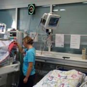
Hospital noise is an issue for babies born prematurely who are at high risk when it comes to external influences such as noise. Their systems are underdeveloped and they need as much sleep and rest as possible to recuperate. This is one of the reasons why the Neonatal Unit at the highly specialized Rigshospitalet hospital in Denmark, began cooperating with the noise measurement company SoundEar this year.

Not all sound is noise
Staff at the Neonatal Unit stress that there is a difference between what they call good sound’ and noise. The point being that not all sound is noise, and not all sound should be eliminated. It is important for the development of the newborns that they hear sound such as their parents and siblings talking and singing to them. It is also important that staff can communicate audibly in critical situations, and it is inevitable that some medical equipment, such as respirators, are noisy. What they do want to reduce is unnecessary sound stemming from alarms, furniture, work flow and talk.

Keeping parents in the loop
An important part of the project is to keep parents informed about why the noise meters are installed and not only focus on reducing noise, but also inform them that they should still talk to their newborns and that some medical care routines will result in a certain level of noise. One way of going about this has been to develop a flyer to hand out to new parents in the neonatal intensive care unit (NICU).

Reducing hospital noise through awareness
The aim is to bring down hospital noise levels at the NICU through installing noise meters in all rooms. Half of the noise meters are anonymous white boxes which solely measure and collect noise levels. The other half of the noise meters also have a display with an ear, that lights up green, yellow or red, indicating the current level of noise in the room.
Reduction in noise levels at the NICU is expected to be achieved through different layers of nudging:

  • The noise meters with displays should help staff and families become aware of their own noise levels and change their noisy behaviour.
  • The software helps staff become aware of when and where noise levels are critical and something should be done differently.
  • The software sends out noise reports on a weekly basis via email to key staff members who use these reports as a basis for further discussion about noise at staff meetings.

Changing routines
After having the SoundEar devices hanging in the NICU for a few months, staff was asked to fill out a questionnaire about the perceived hospital noise levels, and whether the SoundEar devices seemed to have changed anything. 14 staff members, primarily nurses, answered the questionnaire and 78.6 percent reported that the SoundEar devices had made them more attentive to noise levels. The same amount reported to have changed some of their behaviour because of the SoundEar devices.
The change that most staff members reported to have made, was to unpack syringes and other types of medical equipment outside of patient rooms, because they had noticed that the ripping of plastic made an unnecessary amount of noise around the children. Others reported lowering their voices and lowering the noise level of alarms as changes they had made after the installation of the SoundEar devices.
Several staff members also reported to have seen an increase in parents’ attention to noise levels, and that they commented on noise to other visitors and siblings, thereby spreading the attention to noise.

Custom-made software
All the noise meters transfer noise measurement data wirelessly to a central computer, where it is accessible to staff through a piece of software, developed specifically for hospital use by SoundEar in cooperation with staff at the NICU.

Jointly creating a noise measurement system
An important part of the project was to create a system that would help reduce hospital noise and become part of the daily routine at hospitals. For SoundEar, this meant focusing on what staff needed and what their everyday work life looked like and adjusting to that.
In the early days of the project, SoundEar viewed the software platform as the main component of the system and something that staff should be able to interact with daily. They should monitor noise levels just as they were used to monitoring the health levels of the newborns.
To make the software as useful as possible, SoundEar conducted several interviews with staff members. Very soon, it became clear that even though nurses viewed reducing hospital noise as important, their focus was on the critical medical care for the newborns and keeping them alive and well. Their time was limited and they would not be able to prioritize time from their busy schedules to consult a piece of software that did not have immediate medical importance for the children.
Instead, they suggested that a few members of the staff should be responsible for driving the hospital noise reduction, checking the software and gathering insights for the rest of the staff to discuss at weekly meetings. Along the way, the procedure evolved into auto-generated noise reports being sent to key staff members to be discussed at staff meetings on a weekly basis.
www.soundear.dk

https://interhospi.com/wp-content/uploads/sites/3/2020/08/IH207_Soundear_image1blog.jpg 225 300 3wmedia https://interhospi.com/wp-content/uploads/sites/3/2020/06/Component-6-–-1.png 3wmedia2020-08-26 14:18:122021-01-08 12:30:41How to reduce hospital noise at neonatal units

Making medical equipment safer for technicians, manufacturers and patients

, 26 August 2020/in Featured Articles /by 3wmedia
https://interhospi.com/wp-content/uploads/sites/3/2020/08/47084_6006488A_EN_Covered_Ad_Medica2015_IHE_PRINT.jpg 989 700 3wmedia https://interhospi.com/wp-content/uploads/sites/3/2020/06/Component-6-–-1.png 3wmedia2020-08-26 14:18:122021-01-08 12:30:46Making medical equipment safer for technicians, manufacturers and patients

Shear wave elastography – reducing need for invasive biopsy

, 26 August 2020/in Featured Articles /by 3wmedia

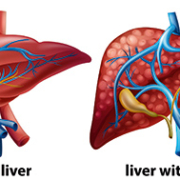
Liver disease is a growing problem across the world. It includes a large range of disorders, such as fatty liver disease (both alcoholic and non-alcoholic), drug-induced liver damage, primary biliary cirrhosis and hepatitis (viral and autoimmune).

Biopsy is gold standard for liver disease
Fibrosis is a relatively common consequence of chronic liver diseases, and its staging, alongside exclusion or confirmation of early compensated cirrhosis, are considered to be vital for surveillance and treatment decisions.
The gold standard for the confirmation of hepatic fibrosis is biopsy. However, biopsy of the liver has several disadvantages. First of all, it is invasive. It is also associated with rare but serious complications. Finally, it can sample only a small portion of the parenchyma (functional rather than connective tissue). This makes it vulnerable to sampling errors.

Non-invasive tests becoming norm
To overcome such constraints, a variety of non-invasive imaging and serological methodologies have been researched and developed for assessing fibrosis. Aside from staging, an ever-growing corpus of data from non-invasive liver tests is also yielding considerable insights for prognostic patient care.
Liver biopsy is now largely restricted to patients showing unexplained discordances in non-invasive testing or those where hepatologists suspect additional etiologies of the disease.
Indeed, non-invasive tests are fast becoming the norm in much of the world, outside the US, although there are several exceptions. The reasons for the lower penetration of non-invasive tests in the US are discussed later.

Ultrasound at forefront
New non-invasive methods for assessing liver fibrosis consists of ultrasound elastography, a diagnostic methodology to evaluate stiffness of tissue, magnetic resonance elastography and serologic testing.
To some of its proponents, elastography is simply a form of the centuries-old systems of diagnosing and assessing diseases via palpation, now extending beyond the scope of physical touch.
While a biopsy is invasive and carries bleeding and infection risks, elastography is seen as a way to get the data needed by clinicians to diagnose and stage liver diseases without the associated complications.

Ultrasound-based elastography is not only used as an alternative to liver biopsy for measuring fibrosis, but also to predict complications in patients with cirrhosis. Another advantage is that elastography, like other non-invasive imaging modalities, can be repeated as often as required to monitor disease progression. Due to their risks, this is simply not feasible with biopsy.

Strain elastography and shear wave elastography
The best-known commercial ultrasound-based techniques for assessing fibrosis include strain elastography and shear wave elastography (SWE). SWE is a real-time two-dimensional elastography technique which enables making quantitative estimates of tissue stiffness in kilopascals (kPa) by virtue of the shear wave speed.
Technologically, even though strain elastography predates SWE, the latter is more easily reproducible than strain elastography, and has rapidly gained interest as the preferred technique. The two are quite different, and outside the hepatology area, seem to have significant complementarities.
Broadly speaking, strain imaging is a qualitative/semi-quantitative method influenced by histotype and lesion size. The use of semi-quantitative indices does not improve performance. Neither does it reduce interoperator variability.

SWE provides accuracy, comparability
Shear wave, on the other hand, is a quantitative method which provides a more accurate and easily comparable assessment of spatial distribution of tissue stiffness.
Most practitioners see SWE as quick and easy to perform, and easily repeated to monitor liver disease progression and measure the effect of a particular treatment. An ultrasound shear wave propagates like ripples of water, as it spreads across tissue. A coherent pattern indicates that a pulse has been applied properly and that there are no artifacts (e.g. from vessels) that would provide erroneous results.
SWE systems provide variable depth of measurement. A depth of 5-6 cms may make it difficult to scan the liver in a large or obese patient, but depths of up to 8 cms are available in certain SWE systems. However, results are not reproducible at such depths, across commercial SWE vendors.

Ease of use not universally accepted
Nevertheless, not everyone agrees that the procedure is easy, especially if SWE results need to be matched against reproducible serological tests. The Society of Radiologists in Ultrasound notes the considerable training required for precision. SWE begins with the positioning of a patient in a left posterior oblique position with the arm raised. Patients need to also breathe slowly, and when asked, suspend breathing, since movement of the liver can reduce accuracy in measurement.

Liver is principal application for SWE
So far, SWE has been used to evaluate and quantify liver fibrosis/cirrhosis of multiple etiologies or with complicating co-morbidities, including chronic hepatitis, liver cancer, steatohepatitis, and biliary atresia. The two-dimensional shear wave elastographic technique offers better performance for assessing liver fibrosis as compared to conventional transient elastography, according to a May 2016 study in the Chinese publication, World Journal of Gastroenterology’.

SWE and hepatitis C

SWE practitioners see it as a tool to assist in earlier detection of conditions such as hepatitis C, and both fatty liver and alcoholic liver disease. Alongside lab studies, SWE offers a means to closely monitor the impact of treatment and assess if the liver will normalize. For many hepatologists, fighting a liver condition before Stage 4 cirrhosis provides a good chance of reversibility.
SWE can also provide information on which hepatitis C patients might benefit from viral therapy. There are numerous reports of patients who would not have been suspected of severe fibrosis or cirrhosis, based on traditional ultrasound grey scaling. At best, the latter provides indicators such as anomalies in the liver contour. However, it does not show signs of cirrhosis such as surface nodularity which are immediately apparent in elastography.

Guiding biopsies
Some clinicians have sought to use SWE to guide liver biopsies and in certain cases, avoid or postpone biopsy. As part of this process, they have addressed one of the major limitations of biopsy, namely restrictions to choice of affected areas, erroneous samples, or inadequacy in sample size enough for interpretation. SWE allows multiple sampling across the liver and generating a mean value. This reduces what in the past would have been a large number of unnecessary biopsies, and minimizes the morbidity of liver biopsy.

SWE in children
SWE has shown specific advantages in pediatric patients. Cincinnati Children’s Hospital Medical Center is gathering data on normal’ stiffness values in children, and on rates of progression, given that published data is almost wholly based on adults.
The study groups cover children with liver transplants, metabolic disorders, cystic fibrosis and those on prolonged intravenous feeding (TPN). One specific area for attention is biliary atresia, a rare but life-threatening condition where the bile ducts in an infant’s liver lack normal openings. The bile builds up and causes damage to the liver.
The pediatric data collection for SWE on newborns with jaundice or cholestasis makes ten measurements. This adds just 5 minutes to a typical ultrasound exam.
Nevertheless, pediatric SWE also has its limitations. According to Dr. Sara O’Hara, who heads the Ultrasound Department at Cincinnati Children’s Hospital, SWE can give variable results in areas such as children with non alcoholic steatohepatitis (NASH) and fatty liver disease.

Breast applications benefit from SWE-plus-strain elastography

In adults, aside from the liver, SWE is seen as a useful technique for evaluation of breast lesions and prostate imaging. In both cases, the technique seems to provide best results in combination with another elastography mode.
For instance, a literature review published in the Journal of Ultrasound’ in 2012 reported that SWE and strain elastography complement each other and overcome mutual limitations in the evaluation of breast lesions.
Clearly, when both types of elastography provide similar results, there is a greater degree of confidence – especially in terms of a near-total elimination of false negatives, which sharply cuts the need for breast biopsies which later prove unnecessary.
There are however some limitations which have been reported in measuring shear wave velocity in the stiffest of breast lesions. Here, rather than propagating through the tumour, the shear wave tends to bounce back. Nevertheless, ongoing improvements in SWE, which have been further reducing examination time and enhancing field of view, means that at some point it could be a tool for breast cancer screening.

Prostate applications benefit from SWE-plus-MR elastography
The use of SWE in prostate cancer, too, shows similar potential for benefits as with breast screening. The first factor is a reduction in biopsies, which prove to have been unnecessary post facto. Studies are under way which seek to correlate stiffness with abnormalities (as well as aggressiveness of tumours) and to assist urologists determine when patients with low-grade prostate cancer must start treatment.
As with SWE and strain elastography in the breast, best results in terms of the prostate are obtained by complementing SWE with another imaging modality – magnetic resonance (MR) elastography. Some findings reveal SWE significantly superior in detecting prostate cancer in the peripheral zone – which is where most tumours occur. However, MR seems to show greater promise in the anterior gland and transitional zone.
Again, as with the breast, the fusion of two modalities permits multiple sampling and tackles a major limitation of prostate biopsy, namely inconvenience and risk, as well as limited choice of affected areas. A few experimental procedures have also targeted fusing MR and SWE images to help guide biopsies.

Using SWE in other organs
SWE has also demonstrated considerable (if still early-stage) promise for evaluating thyroid nodules, indeterminate lymph nodes and uterine fibroids. Another area for investigating SWE include kidney transplants, in order to to avoid excessive biopsies. However, limitations to shear wave captured depth remains a technology challenge for manufacturers to address.

US remains laggard in ultrasound elastography
While most of the world’s regions (Europe, Asia and Latin America) are seeing growth in the use of ultrasound elastography (both SWE and strain), in the US neither is eligible for reimbursement, even in the largest application area – the liver. This is unlike transient elastography, although critics allege it is a blind methodology which neither directly measure fibrosis and often over-estimates it.
Currently, studies in both the US and other parts of the world are seeking to establish the clinical and economic benefits of SWE and strain elastography, including unnecessary invasive biopsies with their associated costs and complications. Eventually, the results of ongoing trials are expected to produce the data which will make ultrasound elastography eligible for reimbursement.
The most self-evident advantage of ultrasound elastography is its non-invasive nature. Unlike a biopsy, it is clearly more feasible to use SWE to screen for patients at greatest risk of chronic liver disease and in need of referral or treatment.

https://interhospi.com/wp-content/uploads/sites/3/2020/08/IH115_shear-wave_Tosh_thematic_crop.jpg 189 400 3wmedia https://interhospi.com/wp-content/uploads/sites/3/2020/06/Component-6-–-1.png 3wmedia2020-08-26 14:18:122021-01-08 12:30:50Shear wave elastography – reducing need for invasive biopsy

Mortara’s WiFi based Telemetry Monitoring: one more success story

, 26 August 2020/in Featured Articles /by 3wmedia

Mortara Instrument’s new family of Surveyor WiFi telemetry solutions is designed to offer diagnostic-quality ECG acquisition and to work on the existing WiFi network, with no need for a dedicated network infrastructure. Its outstanding features have been the key decision factors for Policlinico San Donato (Milan, Italy), one of the top-ranking centers for the study and treatment of cardiovascular diseases, to select Mortara telemetry system.

Mortara designed the Surveyor S4 solution based on three main criteria: cost saving, coverage and clinical excellence.

Cost is a major priority of today’s healthcare professionals and also one of Mortara’s top concerns. The Surveyor S4, thanks to its advanced design, can operate on existing WiFi infrastructure to broadcast physiological signals. It eliminates the cost of a proprietary antenna network, which is required by traditional telemetry systems. Removable, rechargeable batteries allow a lower ecological footprint than disposable batteries, while also reducing running costs.

Coverage (i.e. the areas where the patients can be monitored) is also revolutionized with the Surveyor S4; the use of WiFi technology allows patients to be monitored virtually wherever the WiFi signal is available throughout the facility. This means more freedom for the patient, but also extends patient monitoring to more departments; the ability to clinically monitor and evaluate patients is enhanced without additional beds being added to the traditional telemetry area.

Mortara takes pride in delivering clinical excellence. VERITASTM is the suite of algorithms created by Mortara to analyze ECG signals. The Surveyor S4 family includes the latest algorithms that provide clinicians with absolute reliable data. From basic to lethal arrhythmias, VERITAS is the ideal companion for clinicians. In addition, all Surveyor S4 mobile monitors offer diagnostic quality acquisition; combined with the true 12-lead ECG amplifier, they offer best-in-class 12-lead ST segment analysis. True 12-lead ECG monitoring allows physicians to detect early ST segment changes and obtain a complete evaluation of the cardiac profile of the patient, without additional tests.

Founded in 1969, IRCCS Policlinico San Donato is part of an 18-hospital network that provides over 5,000 beds, and is also host to the Medicine School of the University of Milan. The clinical arrhythmology and electrophysiology ward, run by Professor Carlo Pappone, is one of the international excellence centers for the treatment of all types of cardiac arrhythmias.

Atrial fibrillation, Brugada syndrome, Wolff-Parkinson-White (WPW) syndrome, and cardiac electro-stimulation are among the main research fields. In particular, the research on, and treatment of, supraventricular arrhythmias is a primary focus and area of expertise for this group of clinicians, as testified by the number of publications on international top-ranking journals, and directly witnessed by the large population of patients who have already successfully undergone trans-catheter ablation procedures.

Given the outstanding reputation of his center, Professor Pappone has chosen Mortara as the best-in-class partner in order to deliver excellent diagnosis and treatment.

Policlinico San Donato is one of the many centers where the Mortara monitoring solutions have been adopted and that every day helps to improve healthcare throughout the world.

https://interhospi.com/wp-content/uploads/sites/3/2020/06/logo-footer.png 44 200 3wmedia https://interhospi.com/wp-content/uploads/sites/3/2020/06/Component-6-–-1.png 3wmedia2020-08-26 14:18:122021-01-08 12:30:54Mortara’s WiFi based Telemetry Monitoring: one more success story

Reviews of new Hologic Affirm prone biopsy system are very positive

, 26 August 2020/in Featured Articles /by 3wmedia

Breast tomosynthesis exams, Hologic calls the exams 3D MAMMOGRAPHYTM, have shown to be an advance over digital mammography, with higher cancer detection rates and fewer patient recalls for additional testing.

The new Affirm prone system, which was installed for the first time in Europe earlier last year, is widely considered one of the most significant advance in biopsy technology since the first prone biopsy system was introduced more than 20 years ago. It uses the same proven detector technology as the Hologic Selenia Dimensions breast tomosynthesis system, a top selling breast cancer screening and diagnostic system in the U.S. and in many other countries around the world. With a significantly larger field of view than the MultiCare Platinum system, along with its translucent paddles, the new prone system is designed to deliver exceptional 2D and 3DTM images and better target lesions found during 3D MAMMOGRAPHYTM exams, as well as other screening modalities. In addition, the Affirm prone system allows full 360degree Celsius access to the breast to accommodate most lesion locations. Users can go from a standard to lateral needle approach in seconds to accelerate procedures and ensure reaching targeted lesions.

Doctors in Spain report handling complex biopsies that they were only able to see with breast tomosynthesis imaging with the new system
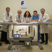
As Doctor Tejerina, a radiologist with the Centro de Patologia de la Mama, Tejerina Foundation, in Madrid, Spain, reports, feedback from the first wave of patients is very positive. ‘We have been suffering to handle complex biopsies of subtle lesions like faint calcifications or distortions that we were only able to see on 3D images,’ Dr. Tejerina says. ‘Older breast biopsy systems are restricted to 2D imaging with a narrow window for targeting the lesions. Often they require multiple X-ray exposures to find and position the suspect tumour for the biopsy needle. With tomosynthesis imaging on the new Affirm prone system, there is a much wider field of view. So the biopsy device can be positioned anywhere in a 360-degree circle, and areas of suspicion seen only with 3D imaging can be easily biopsied.’
Dr. Tejerina also notes that with the previous Hologic biopsy table, the tube head of the biopsy device had to be positioned manually. ‘The new system does this for us automatically, which saves time,’ he says. ‘The software really streamlines our workflow, so the procedure goes faster.’ And he adds, the Affirm Prone table, with its translucent paddles and wider detector, ‘helps us see lesions in the first scout and significantly reduce the number of images needed to get to the lesion.’
The Centro de Patologia de la Mama, Tejerina Foundation has been leading the way in women’s breast health for over 40 years. In 1997, the Centre was first centerein Spain to install a stereotactic guided prone biopsy table. In 2010 the Centre installed a Hologic Selenia Dimensions breast tomosynthesis system, the first site in Spain to use the innovative technology. In 2010 the Centre was also the first site in Spain to combine the Hologic AffirmTM upright biopsy system with the Hologic tomosynthesis system. The Centre was also one of the first sites in the world to offer prone biopsies on the new Affirm system from Hologic.

Doctors in the Netherlands say Affirm system is fast and comfortable for patients
Dr. Henebiens, a radiologist at Spaarne Gasthuis Hospital in Hoofddorp-the first Affirm prone user in the Netherlands-commented on how fast doctors can do a procedure on the Affirm system and how comfortable the new system is for patients.
‘We make fewer exposures on the new Affirm prone system, compared to the older MultiCare Platinum table,’ she notes. ‘And because the table uses 3DTM technology, we use fewer steps getting to the target and getting biopsies.’
Dr. Henebiens also likes how easy it was to get up to speed on the table. ‘The learning curve for the new table was very fast. Training was scheduled for two days, but in one day, the staff knew how to use it.’
The Spaarne Gasthuis Hospital staff had completed over 60 procedures on the table in their first 7 months of use.

Doctors in Italy report faster and lower patient dose biopsies with the new system
Doctor Gianfranco Scaperrotta, Chief of the Breast Imaging and Interventional Radiology at Fondazione IRCCS Istituto Nazionale dei Tumori (INT) in Milano, Italy was an early adopter of the Affirm prone system.
‘The Affirm prone system is a quick, effective and easy to use system,’ he says. ‘The image quality is high, comparable to the Hologic Selenia Dimensions digital mammography system. Workflow is quick thanks to a dedicated workstation and the system’s fully integrated C-arm and automated tube-head. Procedures are faster and safer with the new system thanks to the programmed needle parameters and automated calculations such as the display of safety margins and relative distance in real time.’
After 73 procedures on the new system, INT has seen a 20percent reduction in the time needed for performing a biopsy (patient time under compression) and approximately a 50percent drop in the mean glandular patient dose when they compare the new system to the older Hologic system.
The Fondazione IRCCS Istituto Nazionale dei Tumori is the largest oncology site in Lombardia, the most populous region in Italy. The research and cancer treatment site draws patients from throughout Italy.
In sum, doctors at the first three European Affirm prone install sites reported that the new system offers significant benefits to the patient, the doctor and the technologist.
So what’s next from Hologic in 2D and 3DTM Breast Biopsy after The AffirmTM Prone system? Hologic will show at ECR an all-integrated breast biopsy system that combines tissue acquisition, real-time imagining, and tissue handling. The new system is designed to work in synergy with imaging guidance systems like the AffirmTM Prone table and provide actionable real-time information in the procedure room and improve biopsy workflow.

For specific information on what products are available for sale in a particular country, please contact your local Hologic representative or write to iims@hologic.com

https://interhospi.com/wp-content/uploads/sites/3/2020/08/IH135_Hologic_image1.jpg 217 300 3wmedia https://interhospi.com/wp-content/uploads/sites/3/2020/06/Component-6-–-1.png 3wmedia2020-08-26 14:18:122021-01-08 12:30:37Reviews of new Hologic Affirm prone biopsy system are very positive

Medical tourism – reversing traditional trends, quality and innovation in developing countries

, 26 August 2020/in Featured Articles /by 3wmedia

Medical tourism refers to people who travel overseas for obtaining treatment. In the past, it referred to (wealthy/privileged) patients from developing countries who visited medical centres in industrialized countries to get treatment not available at home.
However, the situation has since reversed, in certain cases dramatically. Medical tourism now typically refers to patients from industrialized countries who travel to poorer countries for lower priced, or more quickly available (and in some cases, superior) treatment. Top medical tourist destinations in this respect include India and Thailand as well as Costa Rica, Mexico and the Gulf.

Medical tourism and health tourism
Some studies do not consider medical tourism to include cosmetic and wellness tourism – with dental treatment also viewed as a cosmetic procedure. This, larger group is often referred to as health tourism.
Market revenues for a major medical tourism destination, Thailand, include a relatively large number of cosmetic surgery procedures. This segment also includes a multitude of places in south America, with the industry’s maturity fed by bustling local demand. For example, according to the Sociedad Boliviana de Cirugia Plastica y Reconstructiva’, over 70percent of middle and upper class women in the country have had plastic surgery.

India leads in higher-end procedures
Conversely, at the other end, if only surgical procedures for overseas patients are included, India leads the global medical tourism market.
Consultants McKinsey estimated 180,000 medical tourists were treated at Indian facilities in 2004 (up from 10,000 just five years earlier). Arrivals have since been rising sharply and are estimated to have reached 250,000 in 2012, contributing 3 billion USD in revenues. This is effectively about 30percent of the global market, estimated for the year at 10.5 billion USD by Transparency Market Research.
India has proven to be a preferred destination for US and UK patients, in particular, because of the use of English in most professional interactions, as well as the fact that both countries have a large number of Indian-origin physicians. Indeed, the US government’s top medic, the Surgeon General, is Vivek Hallegere Murthy, a 40-year old Indian.
India stands out as an interesting destination in another respect. Its massive generic drugs industry provides post-operative medicinal treatment at prices well below the West.

Market drivers: cost, waiting times, accreditation
Key factors driving medical tourism from the West to developing countries include the high cost of healthcare and increasing waiting times for certain procedures. Insurance in several countries often does not cover 100percent of the costs of common age-related requirements such as a knee or hip replacement, or limits the choice of the prosthetics, or the surgeon and facility.
Accreditation of top hospitals in medical tourism destinations has also fuelled demand. The oldest international accrediting body is Accreditation Canada, which has accredited hospitals in about a dozen countries.
The best known accreditation group, however, is Joint Commission International (JCI) in the US. JCI was set up in 1994 to provide international clients education and consulting services, and several international hospitals now see accreditation as a way to attract American patients. JCI is an independent private, not-for-profit organization that seeks to develop nationally and internationally recognized procedures to help improve patient care and safety. It advises hospitals to meet standards for patient care and then accredits hospitals meeting the standards.
A British scheme, QHA Trent Accreditation, is an active independent holistic accreditation scheme. Another is GCR.org, which monitors success metrics and standards of almost 500,000 medical clinics worldwide.
These schemes vary in quality, size and cost to hospitals making use of them.
Increasingly, hospitals are looking towards dual international accreditation, perhaps having both JCI to cover potential US clientele, and Accreditation Canada or QHA Trent for Canadian and British patients.

Indian price advantage boosted by quality, innovations
Practically all surgery procedures performed in medical tourism destinations cost a fraction of what they do in industrialized countries. For example, while a liver transplant in the US costs about 300,000 USD ( Euro 280,000), the figure in India is 50,000 USD ( Euro 47,000). Open heart surgery in India costs between 3,000 ( Euro 2,800) and 10,000 USD ( Euro 9,400), compared to 70,000 USD ( Euro 65,500) in the UK and 150,000 USD ( Euro 140,000) in the US.
Such figures acquire added value when one reviews the conclusions of a Harvard Business School (HBS) study in November 2013, comparing data on angioplasty in the US versus India. The study found that one in 200 US angioplasty patients required emergency surgery, with half of them dying, while only two of 40,000 angioplasty patients at India’s CARE Hospitals required emergency surgery, with just one death in the OR since the hospital’s inception in 1997.
The HBS study also studied other Indian hospitals and interventions, finding them to be on par or better than their US counterparts – for example, Apollo Hospitals with knee, coronary and prostate surgery as well as for infections related to the operating theatre and catheters, Narayana for coronary artery bypass procedures, Deccan for peritoneal dialysis and Aravind for ophthalmology.
The HBS review noted India was not simply an improver but an innovator too, for example Indian doctors pioneered the beating-heart method of surgery, where they operate without shutting patients’ hearts down via a heart-lung machine, leading to fewer complications, shorter hospital stays and quicker recovery.

New segment of intra-Third World medical tourism
While much attention remains on Western medical tourists, one of the fastest growing market segments consists of patients within the Third World, who travel to more advanced developing countries. India again is at the top of the list. In early Dec 2016 / Jan 2017, for instance, it was announced that Iman Abdulati, a 36-year old woman weighing half a tonne, was to be flown to India from her home in Egypt for bariatric surgery.
Such cases have drawn considerable attention for other, political reasons.
Pakistani patients with severe conditions requiring top-notch treatment are a routine media fixture in India. For example, in September 2016, Pakistan’s Express Tribune’ featured the case of Abdul Basit, an 11-year old boy who had been suffering from the rare condition known as Crigler-Najjar syndrome and went for a liver transplant to India. Two years previously, after complications, the wife of former Afghan President Hamid Karzai gave birth to a girl at Fortis Hospital in New Delhi.
In August 2016, The Diplomat’ reported India had emerged as one of the fastest growing global healthcare destinations, particularly for patients from conflict countries like Afghanistan, Iraq, Yemen, Sudan, the Democratic Republic of Congo (DRC), and Somalia, attracting close to 400,000 foreign patients a year, half from war-ravaged countries.
The selection of India as preferred medical tourism destination is being officially sanctioned. In 2004, BBC News’ reported that ‘India was chosen as the place’ for sending sick patients from Tanzania unable to be treated at home, after the Tanzanian government ‘did a comparative analysis of health facilities in South Africa, India and western European countries.’ In 2007, Companion Global Healthcare teamed up with hospitals in India (as well as Thailand and Singapore).

China lags India due to egalitarian healthcare model
Due to a variety of reasons, the other Asian behemoth, China, has a much less mature medical tourism sector. WHO figures show hospital bed densities far lower in India, at just 9 per 10,000 people (making a total of roughly 1 million beds) compared to 42 in China (about 5 million). However, the higher share of private beds in India (40percent against 6.5percent in China) means that India has slightly more private beds – about 400,000, against China’s 325,000.

More than anything, India’s lead over China in medical tourism symbolises its top-down approach to healthcare, in contrast to China’s bottom-up one which first aims at providing top quality healthcare to local Chinese. As a result, China does not provide good healthcare for its middle and upper class. For Britain’s Guardian’, poor rural Chinese were curiously’ better off than their city cousins.’ The Guardian’ contrasted this with India, where ‘many city-based healthcare facilities are excellent….’ This higher-end focus provides India with more medical tourists than China.

Hospital budgets: the sky’s the limit
There are now at least a dozen major private hospital groups in India. Leading groups (with 20-50 facilities, and 2,500-8,000 beds) include Apollo, Max Healthcare, Fortis, Escorts Healthcare, Wockhardt and the Manipal Group. Many of the above (as well as newcomers from cash-rich Indian conglomerates such as Reliance, the Hindujas, Sahara and ITC) are also pursuing the new concept of Medicities, involving suburban developments dedicated wholly to integrated hospital facilities.
The procurement budget of such groups is not insignificant. Apollo’s annual spending on medical equipment, for example, has been close to 200 million USD in recent years. Such budgets allows cash-rich Indian hospitals to procure state-of-the-art equipment – from Da Vinci robots and stereotactic laser surgery to wide-bore 3T Silent Scan MRIs.
Nevertheless, as far as spending is concerned, Indian hospital groups face several challenges in the coming years, especially from the cash-flush Gulf.

US hospitals lead Gulf partnerships
US hospitals are at the forefront of partnerships in the Gulf. One of the key reasons was the difficulty for medical tourists from the region to obtain US visas after 9/11, according to the American Hospital Association.
Key US partners of Gulf hospitals include Johns Hopkins Medicine, which has an agreement since 2006 to partner the General Health Authority for Health Services in the UAE. It also manages the 400-plus bed Tawam Hospital in Abu Dhabi and an affiliated centre offering state-of-the-art molecular imaging services. Johns Hopkins also has alliances with King Khaled Eye Specialist Hospital in Saudi Arabia.
Another example is the Cleveland Clinic, which is affiliated with the International Medical Centre in Jeddah, Saudi Arabia, and is a strategic partner at the 360-bed, multi-specialty Cleveland Clinic Abu Dhabi Hospital in the UAE.
Elsewhere in the UAE, Methodist International manages the operations of Burj Dubai Medical Centre as well as clinics in Dubai, while Partners Harvard Medical International is a key strategic collaborator with Dubai Healthcare City (which explicitly seeks to attract foreign medical tourists).

Education and training focus in Gulf

Many of these alliances are increasing their focus on education and training. For example, the Partners-Dubai Healthcare City has added a high profile unit called Harvard Medical School Dubai Center Institute for Postgraduate Education and Research, while in 2014 Johns Hopkins signed a partnership with oil major Aramco to provide medical education and training in Saudi Arabia.
Qatar, too, has sought US partners. The Weill Cornell Medical College was in fact one of the earliest ventures, established in 2001 as a partnership between Cornell University and the Qatar Foundation for Education, Science and Community Development. It aims to provide medical education and cutting-edge research.
Ironically, the focus on training might hit a traditional source of physicians in the Gulf especially hard, namely Indians who would be replaced by skilled locals.


https://interhospi.com/wp-content/uploads/sites/3/2020/08/IH129_Tosh_med-tourism_thematic.jpg 333 500 3wmedia https://interhospi.com/wp-content/uploads/sites/3/2020/06/Component-6-–-1.png 3wmedia2020-08-26 14:18:122021-01-08 12:30:41Medical tourism – reversing traditional trends, quality and innovation in developing countries

Immunotherapy – promise of dissolving and melting tumours

, 26 August 2020/in Featured Articles /by 3wmedia

In spring 2015, the New England Journal of Medicine’ reported the case of a patient with Stage IV metastatic melanoma – a disease considered close to untreatable. Although three growths in the skin had been surgically removed, one tumour under her left breast had grown deep into her chest wall. The 49-year old woman received a single treatment of an experimental combination of two drugs.
When she returned in three weeks for a second dose, the tumour had ‘kind of just dissolved’, according to Paul Chapman, the physician treating her at Memorial Sloan Kettering Cancer Center in the US.

Over one-fifth patients show complete response
The results were not an exception. 22percent of the 142 patients enrolled in the Memorial Sloan Kettering trial showed a complete response (with their cancer melting’ away), while 53percent had at least 80percent tumour shrinkage. However, there were downsides, too. Half the patients had side effects that were severe or life-threatening.
The drugs used in the trial were Yervoy (ipilimumab) and Opdivo (nivolumab). Approved by the Food and Drug Administration (FDA) for melanoma, the two belong to a small, new arsenal of drugs which supercharge the immune system to attack tumours. The process is known as immunotherapy, and brings together experts from several fields, ranging from oncology and immunology to cell biology and genomics.
Another well-known immunotherapy medication is Keytruda, sometimes called the Jimmy Carter drug’. Combined with surgery and radiotherapy, Keytruda has halted recurrence of melanoma in the former US president, although the disease had spread to his liver and brain.

Single drugs work too
One analysis of 4,846 advanced melanoma patients treated with Yervoy alone found 21percent still alive after three years. Patients who make it to three years ‘do not die of melanoma,’ according to James Allison of the MD Anderson Cancer Center in Houston, Texas, who is widely credited with pioneering modern immunotherapy.
Meanwhile, beyond melanoma, the combination of Yervoy and Opdivo has also shown extraordinary potential in bringing about remission in advanced stages of non-small-cell lung cancer, a leading cause of cancer-related mortality.

Leveraging the immune system

Leveraging the immune system to fight cancer, once little more than a medical dream, is becoming real. Using gene sequencing technologies to classify tumours, the immune system is now becoming primed with drugs and genetically-engineered cells.
The immune system itself consists of a biochemical network which defends the body against viruses, bacteria and other invaders. Cancer, however, finds ways to hide from the immune system, or block its ability to fight.
Immunotherapy seeks to help the immune system recognize cancer as a threat, and attack it.

The medical equivalent of atomic fission
At the moment, there are hundreds of immunotherapy clinical trials under way for almost all types of cancer, individually or combined with other treatments. Eventually, researchers hope to develop blood tests that allow for the early detection of cancer, determine which medicines can be effective and monitor the response in real time.
For some oncologists, immunotherapy is the medical equivalent of splitting the atom. John Heymach, a lung cancer specialist at MD Anderson, has described immunotherapy as a ‘complete game-changer.’ Several others concur. At an AACR press conference in 2015, Louis Weiner of Georgetown University observed: ‘We are in the middle of a revolution,’ and added that ‘I don’t think that is hyperbolic.’
The media too has leaped into the fray, latching on to the enticing concept of dissolving the tumours that physically embody one of humanity’s most intractable struggles against disease. Forbes’, for example, headlines an article: ‘Immune System Drugs Melt Tumours In New Study, Leading A Cancer Revolution.’

Checkpoint inhibitors
In practical terms, there are two contemporary approaches to immunotherapy.
The first (and more-widely used) method involves the use of drugs that block a so-called checkpoint’ mechanism used by cancers to shut down the immune system. This type of drug, known as a checkpoint inhibitor, is used to treat advanced melanoma, Hodgkins lymphoma and cancers of the lung, kidney and bladder.
The drugs work in 20-40percent of patients. In many such cases, the results are nothing short of spectacular, with prolonged remissions that persist, even after treatment is halted.

Checkpoint inhibitors harness T-cells, the white blood cells which could be described as the special force soldiers of the immune system. The T-cells can, however, run out of control and attack normal, healthy tissue, leading to autoimmune disorders like rheumatoid arthritis, Crohns disease and lupus. To avoid this, built-in brakes or checkpoints’ slow or shut down T-cells.
One type of checkpoint inhibitor stops T-cells from multiplying. Another weakens them and shortens their life span. The two drugs in the Yervoy-Opdivo study reported by the New England Journal of Medicine’ were both checkpoint inhibitors. Yervoy (ipilimumab) interferes with a molecule which switches off T-cells. Opdivo (nivolumab) prevents the death of T-cells.

Limitations with checkpoint inhibitors

Nevertheless, for the bulk of patients, checkpoint inhibitors do not show any results, or work for a while and then stop. In the Yervoy-Opdivo study, 126 of 142 patients did not see their cancer vanish entirely. One of the theories being researched to explain this setback is that other, to-be-discovered checkpoints are playing a role, and these would lead to new drugs that increase the scope of their effectiveness.
Meanwhile, harnessing an immune system in overdrive can also be very risky. As mentioned earlier, one out of two patients in the Yervoy-Opdivo study had side effects that were severe or life-threatening. In many cases, treatment for such patients needs to be discontinued.
Conversely, checkpoint inhibitors can also slow down vital glands such as the pituitary and thyroid, thus creating a lifelong need for hormone treatment. This can have an impact in other areas. For example, kidney transplant patients have suffered rejection after taking checkpoint inhibitors since the latter spurred their immune system to attack the grafted organ.
Checkpoint inhibitors can also take months to begin working, and sometimes cause inflammation that make scanner data show what may, confusingly, look like a growing tumour.

CART: personalized immunotherapy

The second approach involves highly personalized treatments known as CART, with the abbreviation arising from the use of a protein chimeric antigen receptor (CAR) to modify a T-cell, which are first removed from a patient, genetically altered to kill cancer, and then re-infused.
CARTs effectively synergize antibodies, which provide precision recognition of disease targets, with the power of T-cells. Unlike antibodies, however, the modified T-cells continue to multiply, serving as a living therapy.
In autumn 2013, researchers at Fred Hutchinson Cancer Research Center in Seattle launched a (preliminary) safety trial with a CART on a lymphoma patient, who had failed to respond to elevated doses of chemotherapy. It was the first trial of its kind to be conducted on a human. At the end of a fortnight, the patient was reported telling his physicians that the lymph nodes in his neck felt like ‘ice cubes melting.’

Beyond leukemia and lymphoma
CARTs have largely worked so far in cases of leukemia or lymphoma, albeit dramatically. However, Fred Hutchinson is also working on several other cancer types, including Merkel cell carcinoma, melanoma and several sarcoma subtypes.
Elsewhere, researchers at the University of Pennsylvania are working on a CART which targets mesothelin, a protein often encountered on the surface of tumour cells. Trials involve patients with serious ovarian cancer, epithelial mesothelioma, and pancreatic cancer.

Challenges with CART
Nevertheless, many practical challenges remain to be overcome with CARTs too. They require extensive research and refinement in the lab before patient trials. Production is also labour-intensive, requiring isolation of specific T-cells from a blood sample, followed by multiplication in an incubator and the use of a hemacytometer for counting, and then concentration in a centrifuge. Apart from fine-tuning their therapeutic effects, means to cost-effectively scale the technology will also be required to bring CARTs to market.
Like checkpoint inhibitors, CART therapy also has clinical limitations, even in its mainstay application in leukemia. 20-30percent of patients are not helped, and are likely to die.

Some way to go
In the final analysis, immunotherapy still has some way to go.
In spite of their often near-miraculous performance, immunotherapy drugs have worked in what is still a minority of patients.
Researchers are clearly aware that immunotherapy is unique, potent and extraordinary, but they cannot fully understand why – or yet control it adequately.

The risks of hype
Physicians also urge caution. Media-hype has led many patients to believe the age of chemotherapy is past. There are cases of unresponsive immunotherapy patients (or those suffering from unacceptable side effects) being switched back to chemotherapy, successfully. Though this may be due to a delayed effect of immunotherapy, it is too early to tell. Indeed, one explanation is that chemotherapy and immunotherapy may be working synergistically in such cases.

Industry too may need a reality check. Asset management companies like Piper Jaffray have forecast immunotherapy boosting the cancer treatment market to half a trillion dollars a year. This may of course face a collision with reality.
Yervoy costs over USD 120,000 ( Euro 110,000) for a four-course treatment, while Keytruda is billed at about USD 150,000 ( Euro 138,000) for a year. At current prices, the combination of Opdivo and Yervoy would result in an annual cost of USD 270,000 ( Euro 248,000). On their part, CART therapies may cost even more. How exactly these sums will be financed is indeed the trillion dollar question.

https://interhospi.com/wp-content/uploads/sites/3/2020/08/IH124_Tosh_melting-tumours_thematic.jpg 186 300 3wmedia https://interhospi.com/wp-content/uploads/sites/3/2020/06/Component-6-–-1.png 3wmedia2020-08-26 14:18:122021-01-08 12:30:46Immunotherapy – promise of dissolving and melting tumours

International differences in end-of-life issues in the ICU

, 26 August 2020/in Featured Articles /by 3wmedia

Medicine is in a state of continual progression as new therapies and interventions are developed and technological advances facilitate resuscitation and prolonged organ support. In addition, patients are living longer with increased numbers of comorbidities and complex disease processes. As one result of these demographic changes and medical advances, intensive care units (ICUs) are admitting more patients with a high risk of death, patients who would previously have died before reaching the ICU. As a consequence, the need for end-of-life decisions has become more common than in the past. There has also been a move from an emphasis on survival at all costs to a recognition that the quality of life of survivors must also be taken into account, as well as the quality of dying for those who will not survive.

by Prof Jean-Louis Vincent

With patients who are not considered to have any reasonable chance of benefiting from new or continued intensive care treatments, physicians are faced with four possible options, ranging from continuing with full treatment to support life through to increasing the doses of sedatives to hasten the dying process (Table). In some patients, where withdrawing therapy is permitted, an ICU trial’ can be considered, giving the patient the chance to benefit from a possible intervention. The target of such a test and the time-limit must be set in advance and adhered to; good communication with the family is essential to ensure that these factors are clear. It should be remembered that in some patients, death is actually in their best interest, preventing unnecessary and prolonged suffering.

Recent data suggest that some 40% of ICU non-survivors will have a decision to withhold/withdraw life-sustaining therapy during their ICU stay. Perhaps not surprisingly, there are marked differences in end-of-life decisions and the decision-making practice around the globe. For example, data show that patients are more likely to receive a decision to withhold or withdraw life-sustaining therapy in Oceania, North America, and northern Europe and less commonly in the Middle East, Asia, southern Europe and South America. Although withdrawing and withholding are seen as ethically equivalent in many countries, in others, withholding life-sustaining therapy is considered acceptable but not withdrawing. In Israel, because withdrawal of life-support measures is forbidden, the authorities even passed a law whereby timers can be put on respirators, which then stop by themselves after a preprogrammed time period. The use of sedatives/analgesia at the end-of-life to shorten the dying process also varies considerably among countries and individuals. Some people justify the administration of large doses of sedatives/analgesics in this situation by calling on the double effect’ principle, wherein giving analgesic agents for comfort has the unavoidable effect of hastening death, but this view is rather hypocritical. There is little official guidance available for intensivists regarding this issue and it is perhaps the area of end-of-life management that creates the greatest concern among physicians with fear of possible litigation. The Belgian Society of Intensive Care recently published a statement that ‘Shortening the dying process with use of medication, such as analgesics/sedatives, may sometimes be appropriate, even in the absence of discomfort, and can actually improve the quality of dying’.

The degree of involvement of family members in end-of-life decision making also varies, with families more frequently involved in Northern Europe and the US than in southern European countries. This is in part related to the traditional paternal approach to medical practice still widespread in many southern European countries. Family-centered decision making is also common in East Asian countries, such as Japan, China and South Korea.

The reasons for these international differences are complex. Many are related to the marked cultural and religious diversity among countries. Lack of available resources and financial constraints can also influence end-of-life decision making, particularly in lower income countries. There are also differences among ICUs within a country and among individual intensivists, related again to the cultural and religious backgrounds of the physicians, but also to local legislation, peer and family pressure, and ICU casemix and organization amongst others. The key ethical principles of autonomy, beneficence, non-maleficence and distributive justice must always be used as the basis for any end-of-life decision, but the ways in which these are interpreted and their relative importance may vary according to local factors. It is therefore inappropriate to try and develop a universal consensus on end-of-life decisions as some have suggested, although local guidelines may be useful. Open discussion of these difficult issues must be encouraged within the ICU team and good communication with the family is essential. The aim must always be to provide compassionate end-of-life care, appropriate for the individual patient and his/her particular circumstances.

Suggested reading
Curtis JR and Vincent JL Ethics and end-of-life care for adults in the intensive care unit. Lancet 2010;376:1347-53.
Myburgh J, et al End-of-life care in the intensive care unit: Report from the Task Force of World Federation of Societies of Intensive and Critical Care Medicine. J Crit Care 2016; 34:125-30
Vincent JL, et al ‘Piece’ of mind: end of life in the intensive care unit statement of the Belgian Society of Intensive Care Medicine. J Crit Care 2014;29:174-175

The author
Jean-Louis Vincent, MD, PhD
Dept of Intensive Care,
Erasme University Hospital,
Universite libre de Bruxelles,
Route de Lennik 808,
1070 Brussels,
Belgium
Tel. +32-2-555-3380
Fax +32-2-555-4555
E-mail: jlvincent@intensive.org

https://interhospi.com/wp-content/uploads/sites/3/2020/08/IH118_Vincent_thematic_crop.jpg 244 300 3wmedia https://interhospi.com/wp-content/uploads/sites/3/2020/06/Component-6-–-1.png 3wmedia2020-08-26 14:18:122021-01-08 12:30:50International differences in end-of-life issues in the ICU
Page 81 of 103«‹7980818283›»

Latest issue of International Hospital

April 2024

2 June 2026

DeepHealth achieves multiple regulatory milestones for Neuro, Prostate and LumbarMR

7 January 2026

Gulf Aorta Summit 2026 Returns to Dubai with a Global Lineup of Aortic Experts

17 December 2025

GE HealthCare receives CE mark for 128cm total body PET/CT

Digital edition
All articles Archived issues

Free subscription

View more product news

Get our e-alert

The medical devices information portal connecting healthcare professionals to global vendors

Sign in for our newsletter
  • News
    • Featured Articles
    • Product News
    • E-News
  • Magazine
    • About us
    • Archived issues
    • Media kit
    • Submit Press Release

Prins Hendrikstraat 1
5611HH Eindhoven
The Netherlands
info@interhospi.com

PanGlobal Media IS not responsible for any error or omission that might occur in the electronic display of product or company data.

Scroll to top

This site uses cookies. By continuing to browse the site, you are agreeing to our use of cookies.

Accept settingsHide notification onlyCookie settings

Cookie and Privacy Settings


How we use cookies

We may ask you to place cookies on your device. We use cookies to let us know when you visit our websites, how you interact with us, to enrich your user experience and to customise your relationship with our website.

Click on the different sections for more information. You can also change some of your preferences. Please note that blocking some types of cookies may affect your experience on our websites and the services we can provide.

Essential Website Cookies

These cookies are strictly necessary to provide you with services available through our website and to use some of its features.

Because these cookies are strictly necessary to provide the website, refusing them will affect the functioning of our site. You can always block or delete cookies by changing your browser settings and block all cookies on this website forcibly. But this will always ask you to accept/refuse cookies when you visit our site again.

We fully respect if you want to refuse cookies, but to avoid asking you each time again to kindly allow us to store a cookie for that purpose. You are always free to unsubscribe or other cookies to get a better experience. If you refuse cookies, we will delete all cookies set in our domain.

We provide you with a list of cookies stored on your computer in our domain, so that you can check what we have stored. For security reasons, we cannot display or modify cookies from other domains. You can check these in your browser's security settings.

.

Google Analytics Cookies

These cookies collect information that is used in aggregate form to help us understand how our website is used or how effective our marketing campaigns are, or to help us customise our website and application for you to improve your experience.

If you do not want us to track your visit to our site, you can disable this in your browser here:

.

Other external services

We also use various external services such as Google Webfonts, Google Maps and external video providers. Since these providers may collect personal data such as your IP address, you can block them here. Please note that this may significantly reduce the functionality and appearance of our site. Changes will only be effective once you reload the page

Google Webfont Settings:

Google Maps Settings:

Google reCaptcha settings:

Vimeo and Youtube videos embedding:

.

Privacy Beleid

U kunt meer lezen over onze cookies en privacy-instellingen op onze Privacybeleid-pagina.

Privacy policy
Accept settingsHide notification only

Sign in for our newsletter

Free subscription