
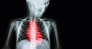
Cardiac imaging – strengthening case for real-time MR
4D cardiac imaging, which generates a three-dimensional motion picture of a beating’ heart, offers cardiologists a revolutionary new tool. Indeed, the ability to acquire images across all phases of a heartbeat cycle is the only way to meaningfully visualizing morphological anomalies and make an authentic assessment of cardiac function.
Traditionally, ultrasound has been a preferred modality for 4D cardiac imaging. However, 4D cardiac MRI (known formally as cardiovascular’ MRI) has been gaining ground. Coupled with MRA (magnetic resonance angiography), it enables cardiologists to view images of the heart, major blood vessels and blood flow.
The novelty of 4D
4D cardiac imaging is a recent technique. Its novelty is best illustrated by an editorial in The Journal of the American College of Cardiology’. The editorial, published as recently as 2009, observed the role of ‘2- and 3-dimensional coronary mapping’ in high-resolution digital imaging.
Major imaging vendors now offer real-time 3D/4D imaging products – across all modalities, PET/CT, MRI and ultrasound. However, the bulk of 4D applications so far have involved ultrasound – especially for cardiac imaging. This may be changing, with increased attention, above all, to MRI.
Ultrasound’s longer legacy
One reason for ultrasound’s pole position in 4D consists of a longer legacy. In the early 1980s, researchers from Duke University in the US reported that though MRI was faster, ultrasound offered the closest achievement of ‘3D real-time acquisition,’ or what is now called 4D.
Technical standardization bodies also moved quickly to endorse and drive the take-up of 4D ultrasound. In 2008, the DICOM (Digital Imaging and Communications in Medicine) initiative approved Supplement 43 which addressed the exchange of real time 3D ultrasound datasets between different vendors. In 2011, IHE (Integrating the Health Enterprise) published a White Paper on 3D/4D imaging workflow.
Early adoption of 4D ultrasound by cardiologists
On their part, cardiologists were enthusiastic early adopters of 3D (and later 4D) ultrasound. The IHE’s White Paper mentioned above was written by its Cardiology Technical Committee. Another factor strongly favouring ultrasound was mobility, since small ultrasound devices could be transported to the patient.
During this period, competing imaging modalities seemed to stand little chance as far as cardiology was concerned.
Computerized tomography (CT) was dismissed since it required cardiologists to use complex post-processing techniques in order to visualize the bearing heart. Cardiac magnetic resonance imaging (MRI) was considered relatively expensive, with limited availability and requiring specialized training.
GE’s cSound: industry seizes the ultrasound opportunity
Industry was quick to seize the ultrasound opportunity. In 2015, healthcare technology giant GE released new software for its ultrasound machines called cSound. cSound-equipped machines intelligently process data being returned by an ultrasound signal, analysing almost 5 gigabytes of data every second, and then filtering it on a pixel-by-pixel basis via algorithms which produced real-time 4D views. This allowed cardiologists to observe how blood swirls around clots in arteries, measure blood leakage around the valves and assess damage. cSound reinforced GE’s presence at the cutting edge of ultrasound, reinforcing a technique patented by the company in the early 2000s and known as Spatial Temporal Image Correlation (STIC). STIC allowed for the quick capture of a full fetal heart cycle beating in real-time.
4D PET/CT and MRI turn to diagnostic oncology
Proponents of 4D PET (positron emission tomography)/CT and MRI were however not sitting by idly. Rather than cardiology, they turned their attention to other specialities, above all oncology where 4D offered huge potential in diagnostics.
4D PET, for example, seemed unmatched in characterizing solitary pulmonary nodules, while 4D CT offered a revolutionary approach in oncology – such as gating tumours and determining treatment margins. On its part, 4D MRI demonstrated a superiority to CT in soft-tissue imaging and in cases where radiation exposure was a concern.
From 4D to 5D imaging
As of now, the focus in diagnostics is to combine the anatomical with functional or molecular imaging, in order to make precise assessments of biological and metabolic pathways. Key modalities include PET with radio-labelled tracers for molecular imaging, and MRI using molecular markers for functional imaging. The molecular/functional enhancement is often referred to as 5D, and to its proponents, offers hope in increasing the specificity and sensibility of diagnostics.
At some stage in the future, it is inevitable that cardiologists will see the virtues of 5D imaging for diagnostics.
The challenge from multi-detector ultrasound scanners
Meanwhile, cardiac ultrasound faces competition in certain applications from other imaging modalities.
In recent years, multi-detector CT scanners seem to offer considerable promise, particularly for non-invasive detection of coronary artery disease and higher flexibility for analysis and visualization of individual vessels. These images, nevertheless, continue to require special processing and rendering tools for assessment of segmental narrowing or occlusions.
The growing promise of 4D cardiac MRI
Rather than CT, cardiac (or cardiovascular) MRI in 4D seems to have rapidly become the principal technology paradigm challenger to ultrasound.
Cardiac MRI scanners do not use open’ magnets which face serious limitations in the case of moving objects – such as a beating heart. The magnet strengths most widely used for cardiac MRI are 1.5T and 3T – although the latter, in some conditions, require software to cancel artifacts. Higher strength magnets are, however, the technology of choice in studying conditions such as aortic construction.
What is also a key advantage of cardiac MRI compared to CT is its lack of ionizing radiation, high spatial resolution and the ability to provide a functional cardiac assessment in one scan.
The technique of 4D cardiac MRI is closely based on traditional MRI. However, it is optimized for use in the cardiovascular system in real time, principally via ECG gating and rapid imaging sequences. This results in acquisition of images at each stage of a sequence of cardiac cycles, and functional assessment of the heart. Blood, in such sequences (technically known as balanced steady state free precession or bSSFP), appears bright due to contrast with blood flow. As a result, 4D cardiac MRI makes it possible to discriminate in a relatively easy fashion between the myocardium and blood.
With and without contrast agents
Cardiac MRI typically uses several approaches to make a comprehensive assessment of the heart and cardiovascular system. Some of the most promising applications include the ability to visualize heart muscle fat or scar in high resolution without the need for a contrast agent. This is based on a technique called spin echo’, which shows blood as black, and identifies myocardium abnormalities through differences in intrinsic contrast.
On the other hand, contrast agents like gadolinium-DTPA can be used for applications such as infarct imaging – where healthy heart muscle appears dark, and infarction areas show in bright white. Contrast agents in cardiac MRI have also proven their worth for treatment of coronary artery narrowing, which starves the heart muscle of oxygen. The contrast agent reveals any transient perfusion defects from artery constriction. Knowing about the presence of such a defect assists in guiding interventional procedures.
Image quality, superior access to anatomical structures
Cardiac MRI provides images of superior quality, accuracy and versatility, alongside access to anatomical structures which are tough to achieve with ultrasound. Examples of these include congenital heart anomalies as well as anatomical changes after surgical interventions.
The latest generation of MRI scanners allow for acquiring high-resolution isotropic data with detailed anatomical information and identical resolution in all three dimensions. Frontier areas of research for 4D MRI include qualitative and quantitative flow pattern analysis in mice with aortic constriction.
Detecting hemodynamic alterations with 4D MRI
At present, one of the most promising cardiac applications for 4D MRI consists of the detection of haemodynamic alterations. The incorporation of pharmacological stress procedures allows for enhanced detection of alterations in heart function during stress-induced ischemia.
In April 2014, a team at Northwestern University reported that 4D flow MRI would help better understand altered hemodynamics in patients with cardiovascular diseases and improve patient management and monitoring of therapeutic response. Their study, published in Cardiovascular Diagnosis and Therapy’, noted that these hemodynamic insights could also lead to new risk stratification metrics in patients and impact upon individualized treatment decisions in order to optimize patient outcomes.
Diagnostics and prognosis of heart events
Cardiac MRI is also being seen as a diagnostic tool to predict heart events. In May 2016, a study led by John P. Greenwood from the University of Leeds in Britain noted that it was ‘a better prognosticator of risk for serious cardiovascular events than SPECT, regardless of a person’s risk factors, angiography results, or initial treatment, and that it would be a powerful tool for ‘the diagnosis and management of patients with suspected coronary heart disease.’ The serious events, assessed over a 5-year period, included death, myocardial infarction/acute coronary syndrome, unscheduled coronary revascularization, or hospitalization for stroke, transient ischemic attack, heart failure, or arrhythmia.
The study was based on a multi-parametric cardiovascular MRI protocol, and performed on a 1.5T MRI scanner and published in the Annals of Internal Medicine’. It was formally known as the Clinical Evaluation of Magnetic Resonance Imaging in Coronary Heart Disease (CE-MARC), and billed as ‘the largest prospective comparison of cardiovascular MRI and nuclear myocardial perfusion imaging (MPI) with SPECT’ with X-ray angiography used as the reference standard.
Genotoxicity poses calls for caution
There have, nevertheless, been some calls for caution due to the chance of genotoxic effects of cardiac MRI scanning.
In October 2011, a study by researchers at Seoul National University in South Korea, assessed high-field intensity 3T clinical MRI scans in cultured human lymphocytes in vitro and ‘observed a significant increase in the frequency of single-strand DNA breaks following exposure to a 3T MRI.’
In June 2013, another study on cardiac MRI in European Heart Journal’ reported similar conclusions, this time in vivo. The study, by researchers from University Hospital Zurich, prospectively enrolled 20 patients, and found a ‘significant increase in median numbers of DNA DSBs in lymphocytes induced by routine 1.5T’ MR scanners. The study also made a recommendation, urging cardiac MRI to ‘be used with caution and that similar restrictions may apply as for X-ray-based and nuclear imaging techniques in order to avoid unnecessary damage of DNA integrity with potential carcinogenic effect.’
Finns call for further studies
Nevertheless, there has been no study so far on the genotoxic effects of MRI compared with those of CT scans. In addition, cardiac MRI risk research has been based entirely on cell level experiments with no conclusive and definitive evidence of actual cancer risk. This is in direct contrast to the link between ionizing radiation and cancer risk.
MRI is therefore still considered by its proponents as the safest alternative.
Indeed, weeks after the University Hospital Zurich study, Finnish researchers published a riposte, again in the European Heart Journal”, arguing that the ‘cellular mechanism’ of how cardiac MRI induced DNA damage was unknown ‘and may be different from that of radiation.’ They concluded that it was ‘obvious that further larger studies are warranted before any restrictions’ were imposed on the use of cardiac MRI.
