

Advances in in-vitro diagnostics (IVD) point-of-care (POC) technology have made it possible to bring the diagnostic power of the central laboratory to the patient, reducing waiting time and in turn improving outcomes [1]. A good starting point and significant pathological area for the use of IVD POC systems is cardiovascular disease. The World Health Organization predicts the number of deaths from cardiovascular disease to increase from 17 million to 23 million people per year by 2030 [2].
Innovations which accelerate diagnostic process have a key role to play in global efforts to reduce these numbers. For example, at Philips, we have enlisted the power of magnetic nanobeads to deliver a next generation of stable and rapid cardiac markers blood testing for suspected acute cardiac patients on the Minicare I-20 handheld immunoassay device, launched last year. With Minicare I-20, the emergency department (ED) can now run a cTnI test next to the patient, and obtain the result within 10 minutes.
The advantages of robust, accurate POC tests are particularly relevant to clinicians working in the ED and ambulance setting where having access to shortened assay turnaround time may improve outcomes. With near-patient testing, it is no longer necessary to send the blood sample to the hospital laboratory and wait up to 60 minutes for the results to come back.
Reduces crowding and patient waiting times
When patients present with symptoms of a heart attack, there is a critical need to make rapid yet precise decisions. However, only about 10% of patients can be accurately diagnosed as AMI based on an ECG [3]. Most patients presenting with suspected heart attack require blood tests, predominately the gold standard troponin biomarker. Serial testing of cTn is part of the recommended diagnostic protocol that aids in ruling in, or ruling out, Myocardial Infarction (MI). The availability of a sensitive and accurate point-of-care test for cardiac troponin could allow clinicians to reduce the standard serial testing of cTn at presentation and six hours after to a safe zero-three hour rule out protocol.
The use of point-of-care testing (POCT) in the ED and ambulance setting to reduce turnaround time for assay results has the potential to improve overall efficiency, by reducing crowding and the length of stay in acute care. Further, for the patient, it can reduce the stress of waiting for their results, and the time to diagnosis and initiation of therapy.
To make the most efficient use of hospital resources, near-patient testing protocols need to be integrated into the acute care workflow and the patient care pathway reorganized, with the full support of the clinical teams and their managers [4]. We are already seeing closer cooperation between clinical teams and the central laboratory, as they recognize the need to help reduce crowding in the ED by supporting the use of POC testing to speed up the delivery of certain blood test results.
BNP assays for rapid ruling out of acute heart failure
Critical cardiovascular disease also covers acute heart failure (AHF), the most common cause of hospitalization in patients aged over 65 years. A brain natriuretic peptide (BNP) test measures the amount of the BNP hormone in the blood. Acute heart failure is a serious condition that accounts for 5% of all emergency admissions in Europe and USA and patients presenting with AHF require immediate treatment [5]. International guidelines recommend the use of the BNP biomarker to rule-out acute heart failure (AHF) in patients presenting with acute dyspnea.
The ED clinician needs to be able to distinguish AHF as quickly as possible. Minicare BNP is the second cardiac marker assay to be introduced on the Philips Minicare I-20 handheld analyser. It provides the ED clinician with access to a fast and accurate BNP marker test to help rule out acute heart failure patients more quickly. Like the first Philips Minicare cTnI assay, Minicare BNP provides clinicians with lab comparable results, and clinically significant information within 10 minutes. It is expected to be commercially available later this year.
The Minicare I-20 platform and both cardiac marker assays are simple and easy to use by non-laboratory POC staff. Its integrated calibration and fail-safe functionalities ensure the robustness and accuracy needed for confident, on-the-spot decision making for better outcomes.
POC test streamlines workflow
The use of POC tests, however, is not limited to the ED or hospital and there is increasing demand, for example, from clinicians to use POC testing systems for both acute and chronic conditions [6].
Two more extensions to the Minicare family are expected to be available in the second half of 2017:
Minicare H-300* point-of-care thromboelastography system:
to aid in the diagnosis and monitoring of hemostasis abnormalities. In critical care situations, such as a heavy blood loss, trauma or before, during and after surgery, understanding a patient’s hemostatic status is critical. Philips will offer a point-of-care hemostasis system that delivers real-time insights in the whole blood hemostasis status of the patient. This novel, small footprint, portable system delivers full results within 15 minutes, with the first results already visible within five. Unlike current hemostasis analysers which are complex to operate, this device is easy to use with minimal training. It is suitable for both the operating room and the ED.
Minicare C-300 clinical chemistry system with an extensive range of chemistry parameters:
Clinical chemistry testing can now be done near-patient with this small benchtop, point-of-care clinical chemistry system for rapid and efficient near-patient testing and diagnosis. Now there’s no need to send blood samples to the central lab and wait for them to return. Shorter waiting time for blood test results is likely to improve workflow and the overall patient experience. Within 15 minutes, the Minicare C-300 will deliver results for an extensive range of clinical chemistry parameters, with a good correlation to the central laboratory instruments. It is easy to operate with limited sample preparatory work required.
Improving patient care
In-vitro diagnostics tests at the point of care provide clinically significant information faster than is possible from the central laboratory. Near-patient testing offers the potential to improve levels of patient’ satisfaction with their treatment, while making more efficient use of healthcare resources [1]. As a global leader in health technology, Philips is expanding its Minicare family of IVD near-patient testing systems for a range of clinical care settings – from critical care in (pre) hospital acute care to primary care. The Philips message is to develop IVD POC solutions ‘ready where you are’, enabling near-patient testing to play a key part in improving patient’s experience.
References
1. Laurence, Caroline O et al. “Patient Satisfaction with Point-of-Care Testing in General Practice.” The British Journal of General Practice 60.572 (2010): e98–e104. PMC. Web. 17 Aug. 2017.
2. World Health Organisation. The Global Burden of Disease. Updated 2004. Available at: www.who.int/healthinfo/global_burden_disease/GBD_report_2004update_full.pdf accessed July 2017
3. European Society of Cardiology. ESC Guidelines. Updated 2016. Available at: www.escardio.org/Guidelines/Clinical-Practice-Guidelines accessed July 2017
4. Bingisser R, Cairns C, Christ M, Hausfater P, Lindahl B, Mair J, Panteghini M, Price C, Venge P. Cardiac troponin: a critical review of the case for point-of-care testing in the ED. Am J Emerg Med. 2012 Oct;30 1.
5. Cowie M. R., et al. (2014) Improving care for patients with acute heart failure: before, during and after hospitalization, ESC Heart Failure, 1, 110–145, doi: 10.1002/ehf2.12021.
6. Howick J, et al. (2014). Current and future use of point-of-care tests in primary care: an international survey in Australia, Belgium, The Netherlands, the UK and the USA. BMJ Open. 4:8. (8):1639-49. doi: 10.1016/j.ajem.2012.03.004. Epub 2012 May 23.
Philips Medical Systemswww.healthcare.philips.com
*Philips is distributor and Entergrion is legal manufacturer
The march of healthcare technology is not always even. Benefits on one front can often be outweighed by problems on another. Radiology is no exception to this rule.
Like other medical professionals, radiologists have begun using portals and social media to connect to patients and join the move towards personal healthcare.
Websites and radiology
Today, websites staffed by imaging professionals seek to directly address the public about radiology. Such a trend is especially pronounced in the US. Examples include radiology Q&A portals at the University of Texas’ John P. and Kathrine G. McGovern Medical School, Northwest Radiology Consultants in Atlanta, Georgia, and a host of others. One of the best known is the RSNA/ACR public information website, RadiologyInfo.org, which offers a library of resources for patients including information on how various imaging procedures are performed. In Europe, the ESR has a Website page dedicated to ‘Radiation and Patients’ and an ‘Ask EuroSafe Imaging’ Q&A page, split into three sections (CT, interventional radiology and pediatric imaging), with answers provided by radiologists from across the continent.
Radiologists and social media
Radiologists have also sought to use social media to build and continuously strengthen interactive relationships with patients outside a formal hospital or physician office setting. Such approaches have spilt over into tackling concerns after widespread reports in the media about the ‘over-use’ of medical radiation. In the US, for example, the Health Physics Society has a site dedicated largely to addressing such risk perceptions in the general public. The UK too has seen such a step with the British Institute of Radiology and the Institute of Physics and Engineering in Medicine endorsing Ask for Evidence, as part of which a panel of radiologists and medical physicists respond to questions from the public on radiation safety.
Technology versus patient downtime
These are clearly significant and laudable developments. Informed patients are increasingly regarded to be better patients by several physicians. However, other recent developments in technology, above all electronic medical/health records (EMR/EHR), are placing a growing burden on clinicians to update medical documentation, in order to facilitate real-time sharing and reduce errors. This results in less time for patient care. A key driver here, in the US, consists of federal government meaningful use (MU) requirements, which provides physicians with financial incentives to use EHRs.
For radiologists, these incentives are hardly negligible and range from 44,000 to 63,750 dollars (39,000 to 56,735 Euros) over a 5-or 6-year period via Medicare and Medicaid, respectively.
On the other hand, MU also requires 10% of patients viewing, downloading or transmitting their electronic health information, with over 40% of all imaging scans to be made accessible via certified EHR technology.
The above requirements are hardly a testimonial to efficiency. One study on MU published by the Radiological Society of North America (RSNA) in 2012 found that medical residents reported having to spend the bulk of their time updating charts and documentation, and that EHR adoption correlated directly to reduced time for direct patient care.
Radiology strives to remain at technology cutting edge
This is a profound challenge. Radiology has traditionally been the medical speciality at the cutting edge of technical advancement. It was radiology which first moved away from paper to digital technology. As a result, radiologists and industry are currently seeking to fast track solutions for increasing patient downtime and improving workflow.
Data use
In the first stage, the focus was on enhancing use of available data. Ironically, illustrating the unevenness and asynchronicity in the progress of technology, efforts were concentrated on getting more usable data out of electronic records, which did not always trickle down to radiologists. One reason was the lack of skills. Referring physicians often left responsibility to get approval for imaging to office staff, many of of who lacked the clinical knowledge required to seek such approval.
Automation: From CPOE to CDS
Soon after, the effort shifted to automation, especially in the shape of decision support (and so-called assistant clinical reasoning) tools. Such a process continues, with evolution from static to dynamic, patient-centred tools. A good example of this is the computerized order entry (CPOE) system. In 2012, a study in the ‘Journal of the American College of Radiology’ proved the clinical viability of combining radiology CPOE with imaging decision support, including pathways and algorithms, as well as classification for actionable findings.
One of the longest-used clinical decision support systems is ACR Assist from the American College of Radiology, which is designed to blend in seamlessly with radiology workflow. Clinical data is encoded in vendor-neutral ways, in order to quickly build commercial applications. The ACR has since created guidelines for radiologists and referring physicians to proceed after clinical findings. Others are also stepping in with new initiatives to enhance automation and decision support. Massachusetts General Hospital, for example, has developed Procedure Order Entry (PrOE), a surgical appropriateness system to help identify whether a procedure is necessary, and the implications of this for radiology are under active investigation. By utilizing evidence-based guidelines rather than have a less-informed entity authorize diagnostic imaging, CPOE in radiology not only enhances efficiency, but also the quality of care.
IPads and speed
One unexpected finding cited in the 2012 RSNA study on meaningful use was that residents using iPads were able to enter and update data more rapidly. Indeed, a majority of those surveyed found that iPads led to significant increases in work efficiency.
This was an opportune moment, given that a year previously, the US Food and Drug Administration had cleared the first mobile app to allow physicians to make diagnoses using iPads or iPhones.
Currently, radiology imaging applications for mobile platforms allow remote monitoring and control for a PACS administrator. Fuelled by standard tools such as DICOM viewers, these impact directly on quality control, data management and workflow efficiency.
The implications of teleradiology connectivity are especially dramatic in emergency settings. About five years ago, Mayo Clinic physicians deployed smartphones in order to assess their utility in a telemedicine stroke-management network which connected radiologists to neurologists and emergency physicians at a remote facility. The findings were encouraging, with over 90% of agreement on the key radiological findings. In the future, smartphone-based teleradiology systems are likely to become commonplace among first responders.
Image management and automation
Image management is also being used as a means to automate processes. The fast growth of technology has also necessitated unprecedented collaborations between specialists. Oncologists, for example, have been working with radiologists to analyse datasets for tumour detection and monitoring, and some studies report sharp reduction in the time required to study suspicious tissue.
On its part, Massachusetts General has also developed QPID (Queriable Patient Interface Dossier) to integrate electronic records and streamline providers’ abilities to access details in a patient’s medical history.
Other areas for attention include voice-enabled documentation, accompanied by structured reporting and data sets that pre-populate a radiology report. These not only reduce human error when inputting data but also enables radiologists to interpret and diagnose a study when a referring physician is most in need of the information – while meeting a patient.
From automation to deep machine learning
The greatest benefit of automation is to maximize the use of available data. This enhances the ability to provide not just personal but precision medicine, too. When interfaced to an appropriate radiology-focused IT platform, individual radiologists and the broader radiology (as well as clinical) community will be empowered to benefit from feedback loops that reinforce positive lessons, de-emphasize negative ones and continuously build appropriateness and best-practice guidelines. Based on the templated information in a report, colleagues (real and virtual) would be able to rapidly offer second opinions and perspectives on how to best serve a specific patient-case.
Further down the road are deep machine learning tools which will provide sophisticated, structured and in-depth data on a patient, to enable increasingly informed decisions in the context of specific and individual challenges – influenced by factors ranging from pharmacogenomics to disease staging, age and lifestyle. Such knowledge, which would create highly actionable reports, are expected to dramatically impact upon patient outcomes.
Radiology and public perception
It is no secret that professional radiological societies strongly believe there is a need to improve patient (and public) perception of the role played by radiologists in healthcare, and that this necessitates closer contact with patients. Patients after all seldom choose a radiologist. This choice is made by a referring physician or health plan.
Though radiology is essential to patient care, radiological services often seem inconvenient, a threat to privacy, sometimes mysterious and scary. The connect between a radiologist and patient is intermediated by nurses and assistants (e.g. for injecting contrast material or preparing them for the imaging procedure), or by technologists seen as managers of machines. Various studies have shown that radiologists are not always present during performance of a study and seldom introduce themselves to a patient.
As a result, patients increasingly consider radiologists to be supervisors of a technological process. The clinician requesting the examination and receiving the radiology report is considered to be the one interpreting the study and making the decision.
Patient at the core
To sum up, the core value proposition in transforming and keeping radiology up to date involves the patient. Although the growing digitization of healthcare pushes radiologists away from patients, there is a need to make these interactions more prominent. Some radiologists warn that otherwise, there is a risk of their services becoming commoditized. For such a process, there is clearly a need to draw more patient data into decision-making. One of the most ambitious efforts on this count was launched at the turn of the decade by RSNA, with funding from the National Institute of Biomedical Imaging and Bioengineering. The project, which promotes patient access to self-management tools, is known as Image Share, and consists of a secure network based on open-standards architecture. Images are exchanged between servers at radiology departments and imaging centres via the Cloud. A two-year pilot began in 2011 at Mount Sinai Medical Center in New York, followed by university hospitals in several states. In 2016, RSNA introduced a validation programme for the project, to test vendor system compliance with standards for exchange of medical images. To date, results have been satisfying.
Although initiatives like this will continue to grow in importance, they are unlikely to do more than enhance the efficiency of radiologists – and their professional judgement – in improving patient care.


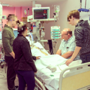
Visiting hours for hospitalized patients have traditionally been restricted to set periods during the day and limited in duration. However, the situation is slowly changing towards a more open approach to family visits, even in wards where visits are often most restricted, such as intensive care units (ICUs). As just a few examples of this general change in attitudes towards visiting, many American hospitals have now completely removed restricted visiting hours; a campaign of extended visiting hours was launched in France a few months ago; and a bill is currently being discussed in Italy to expand hospital visits.
by Prof Jean-Louis Vincent
Why restrict hospital visiting?
The reasons behind restrictive visiting are not very clear or, in today’s context, very credible. The fear of transmission of infection was perhaps the earliest reason for restricting visits, but with improved infection control measures, this concern is generally unfounded. Other suggested reasons include the need for patient to have adequate rest periods and the belief that visitors interfere negatively with medical and nursing care.
Because sick patients need rest?
It was widely believed that having periods of the day without visiting would ensure that patients had sufficient periods of rest, without disturbance from visitors. However, the need for sick patients to rest is often exaggerated. Indeed, this idea is now rather out-of-date, even for the sickest of patients. Although patients must clearly not be exhausted by their visitors, too much rest can encourage muscle weakness and prolong convalescence. When a family member says “doesn’t he/she need to rest Doctor?”, I often reply “certainly not; in fact you should wake him/her up!”. The current trend is to encourage physical and intellectual stimulation for all patients.
Of course patients need some time to sleep and rest, as we all do, but this can be determined on an individual basis, preferably after discussion with the patient, rather than being enforced at fixed times by restricted visiting hours. Moreover, the presence of a loved one in the room does not necessarily prevent restorative sleep. Rest is also important for family members and it is sometimes necessary to remind them to take a break, particularly at night. In any case, access to hospitals is generally limited during the night, for security reasons.
Because visitors interfere with patient care?
The presence of visitors was often believed to interfere negatively with medical care. Visiting hours were therefore concentrated on periods of the day during which patients were least likely to be undergoing medical consults or examinations. However, hospitals of today function almost continuously or at least with considerably more extensive hours than in the past, notably for laboratory and radiological investigations, making it difficult to predict when examinations and rounds are most likely to take place.
The presence of visitors was also often believed to hinder good nursing care, and perhaps much restricted visiting was devised for the benefit of nurses, rather than the patient. Nurses often complained that they were unable to perform the necessary care in the best possible way, because they were bothered by the presence of relatives, sometimes numerous and noisy, who asked a lot of questions, and were even critical of the care being provided!
However, it is now widely believed that extended visiting hours can be beneficial not only for the patient and visitors, but also for the staff. Staff members, especially nurses, are often initially reluctant to the proposed change to more extensive or unlimited visiting, concerned that it will increase their workload. But this is not necessarily true, and is in fact often the reverse. Allowing visitors to be present at different times during the day enables them to understand better the work of the nurses, doctors and other healthcare personnel. When visiting hours are restricted, nurses often make use of the visiting periods to have a small break, to catch up or even have a joke with their colleagues. This can sometimes give visitors the impression that nurses have nothing to do, or are not really concerned about looking after the patients under their care. By arriving at different times of the day and staying for longer periods, family members can better appreciate hospital life and realize that nurses also need some time for relaxation and distraction, thus reducing the risk of conflicts between family members and staff. Extending visiting hours also reduces the number of telephone calls from relatives asking after their loved one, thus freeing up nursing time.
Let’s welcome visitors
Importantly, fixed visiting hours can discourage relatives from visiting a patient. For example, it can be difficult for family members who are working to request time off during the day to be able to observe the fixed visiting hours; sometimes family members simply forget (or are unaware of) the specified times, especially when units have different hours on different days of the week, and have to go home having missed the allocated slot; similarly, visitors who have to travel some distance to visit their loved one may be put off by the risk of being late and missing the fixed visiting period. Finally it is sometimes just easier to say, “I’ll visit when they’re better and out of hospital…”
Rather than being made to feel that they are the enemy and not welcome, relatives should be encouraged to visit and be involved. We must not talk about “them” and “us”. The patient must be at the centre of our preoccupations at all times and we must all work together to ensure he/she has the best possible chances of a good recovery without complications. Family members and loved ones form part of the patient’s immediate supportive environment and can form a useful bridge between the patient and hospital staff. They can also play an active role in patient surveillance, for example by indicating to staff if there is a problem that has not been noticed or that the patient may not want to report. In certain American hospitals, pamphlets are now available to explain how relatives can identify and report important signs of deterioration, for example, confusion that wasn’t there before or a small change in respiration that has gone unnoticed.
Family members can even sometimes contribute directly to some aspects of patient care, for example helping with feeding, washing or dressing. Indeed, these practices are commonplace in countries with limited resources, where family members never leave the bedside. In western society, however, patient care has been completely transferred from the family to professional carers, which can sometimes lead to the patient feeling patronized or being treated like a child.
The hospital structure is also changing to be more welcoming for visitors. Instead of a few folding seats at the end of the corridor for relatives waiting while the patient is examined or comes back from an examination, many hospitals have now introduced reception rooms where relatives can stay as long as they wish, in comfortable conditions. In the United States in particular, hospitals have set up small kitchen-lounges where families can rest, prepare a meal in the microwave or watch television… and why not socialize, chat, share experiences with relatives of other patients.
Indeed, the hospital is no longer a detached world, which we are somewhat hesitant or even scared to enter. Hospitals are increasingly user friendly and should be seen as somewhere positive and welcoming. After all, many hospitals now have a cafeteria (if not a restaurant), small shops, a bank, a post-office, pleasant gardens… creating the idea that hospitals can be part of everyday life, and indeed are for the many patients and visitors that pass through the doors daily. Visitors can make use of these areas when their relative is undergoing an examination or receiving nursing care.
Family presence during interventions?
As families spend more time visiting their loved ones in hospital, the chances that they will be present when an intervention is needed are increasing, perhaps particularly on high acuity wards. But should they be allowed to stay in the room? Perhaps yes for a simple blood test or changing of a dressing, but what about during cardiopulmonary resuscitation (CPR)? This issue continues to raise considerable debate, not least because the patient needing CPR cannot be asked if they mind. Although some staff members find having family members present adds stress to an already complex situation, studies have suggested that the presence of a relative can help a surviving patient understand what has happened and, if the patient dies, having been present can reassure the family member that everything possible was done. This is an area where attitudes are changing and, if a family member wishes to be present during CPR, this request should not be refused.
The rights and responsibilities of visitors ….
Clearly, although visitors have the right to see their loved ones in hospital, they must also abide by certain rules. They must leave the room when asked to do so by the hospital staff and should not interfere with patient care. They should not slow the work of the nursing or medical staff by asking repetitive, unnecessary questions or by engaging in prolonged conversation. Importantly, too, visitors are there to visit only their relative/loved one and must not look, even surreptitiously, into the rooms of other patients!
… and the rights of the patient
On reflection, rather than asking whether visiting the sick patient is allowed, the question should rather be the reverse, whether the patient is allowed to see his/her relatives? Limiting hospital visits is generally harmful for the patient and opening up visiting is reported to improve patient satisfaction. By bringing news from the outside world, family, friends, pets, … visitors can stimulate a patient’s intellect and interest, helping promote a quick recovery. There is nothing worse than lying in bed all day just looking at the ceiling… But, it is important to consider the patient’s viewpoint when considering visitor access. For example, some patients may prefer to have only close family members visit, feeling embarrassed about less well-known friends and relatives seeing them unwell, and others may prefer not to discuss their condition when family members are present for fear of upsetting them. Patients have the right to see visitors whenever they wish, but should not have visiting forced upon them.
Conclusion
It is not so long ago that, when visiting a patient in hospital, an often rather officious nurse would announce the end of visiting hours and insist you leave your loved one. Such strict practices have become less common and there is much more flexibility, particularly on general hospital wards. We need to go further and extend open visiting to all areas of the hospital, including ICUs, where visiting still remains, in general, more restricted. In many cases, we should be actively inviting relatives to visit more and to stay longer, especially when the patient has few visitors and feels isolated. Visiting is humane and good for the patient.
If you still have restricted visiting hours at your hospital, I am sure this will change in the near future. I am not convinced that there should be a law on this subject, whether in Belgium, Italy or elsewhere, but rather a collective effort needs to be made to change our mentality related to visiting hours and thus improve the quality of care for our patients.
Suggested reading
Giannini A, et al. What’s new in ICU visiting policies: can we continue to keep the doors closed? Intensive Care Med 2014; 40: 730-33
Jabre P, et al. Family presence during cardiopulmonary resuscitation. N Engl J Med 2013; 368: 1008–18.
McAdam JL & Puntillo KA. Open visitation policies and practices in US ICUs: can we ever get there? Crit Care 2013; 17: 171
Shulkin D, et al. Eliminating visiting hour restrictions in hospitals. J Healthc Qual 2014; 36: 54-7
The author
Jean-Louis Vincent, MD, PhD
Dept of Intensive Care, Erasme University Hospital, Université libre de Bruxelles,
Route de Lennik 808, 1070 Brussels,
Belgium
jlvincent@intensive.org
Rupture of the coronary plaque surface, accompanied by the exposure of thrombogenic, red cell-rich necrotic core material, is one of the most important underlying mechanisms in acute coronary syndrome (ACS). After decades of idling, such plaques sometimes suddenly burst into this life-threatening condition. The rupturing occurs during the evolution of coronary atherosclerotic lesions, and is often accompanied by super-imposed thrombosis.
To date, the precise mechanisms involved in plaque erosion remain generally unknown. Coronary spasm is simply a universal suspect.
Prevention is therefore considered to be the only effective means for reducing the mortality and morbidity of coronary heart disease.
Coronary lesions that are prone to rupture have a distinct morphology compared with stable plaques, and provide a unique opportunity for non-invasive imaging to identify vulnerable plaques, before they lead to clinical events.
Plethora of terminology
The severity and prognosis of plaque rupture is characterized by a plethora of terminology. Plaque vulnerability describes the risk of symptomatic thrombosis in the short term, whereas plaque ‘activity’ remains ambiguous (referring to one of a wide variety of processes associated with progression).
‘Plaque burden’ is accepted to denote extent of disease. It is a measure of the extent of atherosclerosis, regardless of the cellular composition or activity of plaques. There are various ways to measure the burden: plaque volume, lesion-coverage of arterial surface – sometimes based on using computed tomography (CT) to measure coronary calcium score, or ultrasound to assess plaque area in the carotid bed. Given that atherosclerosis is multi-focal (and impacts upon the entire vasculature), a high plaque burden in one region (e.g. the lower limbs) may be a marker for advanced disease elsewhere. The highest concern on the latter consists of the coronary arteries due to their high degree of susceptibility.
Size is not everything
Rather than plaque size alone, the risk of rupture depends more on the composition and type of plaque, inter alia, richness in soft extracellular lipids – and macrophages. Indeed, structurally what is required for plaque rupture is an extremely thin fibrous cap. As a result, ruptures are usually minuscule and occur mainly at the periphery of the cap covering the lipid-rich core – among lesions clinically defined as thin-cap fibroatheromas. They have reduced tensile strength and are more extensible than intact caps, while the presence of collagen and smooth muscle cells is lower. In effect, as extracellular lipid accumulation progresses (usually due to external stress/triggers), the fibrous cap weakens and predisposition of a plaque to rupture increases.
Several other factors are also believed to play a concurrent role, among them inflammatory cell recruitment, macrophage formation, necrosis, matrix synthesis, calcification, arterial remodelling, etc.
The interaction between these factors is not only complex but variable too, as far as the development of plaque is concerned. This leads to unpredictable rates of progression and variable clinical outcomes.
Not all ruptures lead to ACS
Nevertheless, there are, once again, certain other issues in play with regard to the clinical relevance of vulnerable plaque detection. Most plaques remain subclinical and asymptomatic. Others elicit acute thrombosis and may lead to an acute coronary syndrome (ACS).
However, not all plaque ruptures cause ACS. Some develop obstructively (stable angina). Indeed, on its own, stable angina pectoris derived from atherosclerosis is rarely fatal without scarring of the myocardium – the latter can provoke an arrhythmia presenting as sudden cardiac death.
Confusion arises in other contexts too. For example, the presence of thrombosis is not the same as the occurrence of ACS. Indeed, some physicians believe that the majority of ruptures and erosions are asymptomatic in the short term, although they may sometimes lead to gradual coronary narrowing.
Nightmare for prognosis
This lack of clarity has proven to be a nightmare. ACS occurs only when vulnerable plaque, platelet activation and impaired fibrinolysis occur alongside inflammatory states. Such vulnerability may change with time, and it is these changing dynamics vis-a-vis stress/triggers which determines the exact moment and point of rupture. As a result, the non-invasive detection of vulnerable plaques is considered to be of great clinical relevance, especially in ultra-high risk patients.
At the cutting-edge
Currently, a host of new, non-invasive techniques are being harnessed to assess and predict the likelihood of coronary plaque rupture. Leading the way are computational fluid dynamics (CFD) and fractional flow reserve (FFR) methodologies. They are based on harnessing supercomputing capability to the analysis of CT angiography.
The high quality imaging and sub-millimetre resolution of modern computed tomography (CT) scanners allows characterization and quantification of lesions at accuracies unimaginable barely a decade ago. CFD supplements the functional information of CT-based plaque assessment by calculating lesion-specific endothelial shear stress and FFR. Such supplementation of functional information by quantified morphologic data about coronary plaques is considered to be one of the best means to detect vulnerable plaques.
FFR guided therapy
For patients with coronary calcification and hemodynamically significant obstructive disease, FFR has long been considered the best solution for guiding re-vascularization of lesions and improving outcomes. FFR provides an index of atherosclerosis and lesion significance, as measured with a pressure-sensitive angioplasty guidewire. FFR-guided therapy has improved patient outcomes, reduced stent insertions. However, it is used in less than one-tenth of cases due to procedural and operator related factors – above all, patient discomfort due to time and motion artifact as well as cost.
Coupling FFR to CT angiography
More recently, due to the developments in non-invasive CT imaging and the application of CFD modelling to CT angiography datasets, FFR can be derived non-invasively without requiring modification of standard CT angiography acquisition protocols or inducing hyperemia.
Such non-invasive FFR, moreover, has been shown to demonstrate excellent correlation with invasive FFR.
PLATFORM Study
One of the key studies investigating the impact of combining FFR and CT was called PLATFORM (the Prospective LongitudinAl trial of FFRCT: Outcome and Resource Impacts).
PLATFORM, which ran from the end of 2013 to 2015 at centres in the US and Europe, demonstrated improved patient selection for invasive angiography using a combination of coronary CT angiography (CCTA) along with fractional flow reserve CT (FFRCT). The so-called CCTA-FFRCT approach increased the chance of identifying obstructive coronary artery disease among those intended for invasive testing and held forth the promise of serving as an efficacious gatekeeper to invasive coronary angiography (ICA).
The findings were conclusive, with numbers presented by researchers at the European Society of Cardiology at London in 2015. The use of FFRCT in patients with planned invasive catheterization, they noted, was associated with a reduction in the rate of finding no obstructive CAD at ICA, from 73% to 12%. It also resulted in cancellation of 61% of ICAs.
Computational fluid dynamics
In effect, the adoption and translation of CFD modelling may be considered to have revolutionized cardiovascular medicine.
CFD is a specialist IT discipline bringing together advanced mathematics and fluid mechanics. Its roots lie in mission-critical/high-performance engineering systems. Much of its history is intimately connected to the aerospace industry, to enhance the accuracy of complex simulation scenarios such as transonic or turbulent air flows.
In medicine, the first-ever CFD investigations began in cardiovascular research, to clarify the characteristics of aortic flow in a degree of detail below the threshold of experimental measurements. Computer-aided design (CAD) models of the human vascular system were built using modern imaging techniques, coupled to rapid, economical, low-risk 3-D prototyping. The ensuing models precisely computed factors such as blood flow and tissue behaviour and response, taking close consideration of boundary conditions such as complex systemic/physiological pressure and ‘virtualized’ metrics such as wall shear stress.
CFD modelling has already revolutionized the development of devices such as stents, valve prostheses, and ventricular assist devices.
CFD is currently being translated into cardiovascular clinical tools for minimally-invasive application to a wide spectrum of coronary, valvular, myocardial and peripheral vascular diseases. One of the biggest advantages offered by combining high-resolution imaging with CFD is that unique patient-specific data can be juxtaposed into multi-scale, variable duration models to make individualized risk prediction and planning possible. This is directly opposed to registry-based, population-averaged data.
In the future, it is expected that the trend to ‘digital patient’ representation, combined with population-scale numerical models, will reduce cost, time and risk associated with clinical trials.
The massive processing power brought to play by CFD quickly led to the understanding that mechanistic forces of arterial wall shear stress (WSS) and axial plaque force acting on coronary plaques might be responsible for both the development of coronary plaque and its vulnerability to rupture.
For example, it is difficult to measure WSS, a key factor in the development of atherosclerosis and in-stent restenosis, without invasive procedures – with all the latters’ attendant risks and frequent futility. One study demonstrated that less than a third of patients with suspected obstructive coronary artery disease (CAD) showed its presence after invasive coronary angiography (ICA), while an even-smaller number had flow-limiting obstructive disease based on invasive fractional flow reserve (FFR).
In contrast, CFD models can both compute and map the spatial distribution of WSS, establishing links between haemodynamic disturbance and atherogenesis and explaining why atherosclerotic plaque tends to be deposited at arterial bends or bifurcations.
CFD modelling has also been central to comprehending the role of WSS in endothelial homoeostasis. While turbulent blood flow reduces WSS and stimulates adverse vessel remodelling, non-disturbed laminar blood flow seems to be associated with higher WSS – which reduces endothelial cell activation. In a March 2012 issue of ‘Circulation’, researchers from Johns Hopkins University School of Medicine, CVPath Institute at Maryland and the Mount Sinai School of Medicine in New York established that a complex series of WSS-related signalling pathways and interactions underlie the above phenomenon.
Though much more remains to be understood before such pathways can be exploited to their full extent to yield new anti-atherosclerotic therapies, few doubt that the way forward lies in further CFD models that combine dynamic fluid behaviour analysis with cellular response.
Other emerging techniques
Apart from CFD, other methodologies under consideration to quantify measurement of coronary plaque and lesions include the use of radio-frequency (RF) backscatter intravascular ultrasound. A prospective study in 2011 in the US known as ATLANTA sought to make the first-ever assessment of the accuracy of 3-dimensional, quantitative measurements of coronary plaque by computed tomography angiography (CTA) against intravascular ultrasound with radiofrequency backscatter analysis (IVUS/VH).
For the ATLANTA study, 60 patients underwent coronary X-ray angiography, IVUS/VH and coronary CTA. Plaque geometry and composition was quantified after spatial co-registration on segmental and slice-by-slice bases. The researchers found significant correlation for all pre-specified parameters by segmental and slice-by-slice analyses. Compositional analysis suggested that high-density non calcified plaque on CTA best correlated with fibrous tissue and low-density non calcified plaque correlated with necrotic core plus fibrofatty tissue by IVUS/VH.


Prins Hendrikstraat 1
5611HH Eindhoven
The Netherlands
info@interhospi.com
PanGlobal Media IS not responsible for any error or omission that might occur in the electronic display of product or company data.