

Data sharing between healthcare organizations can improve clinical efficiency and enable better decision-making. This is essential at a time when health services in Europe are facing perhaps their greatest challenge to date. Providers must accommodate increasing demand for services due to ageing populations, the rising prevalence of chronic diseases and budgets being constrained. The ability to compare costs to benchmarks will be crucial for healthcare providers needing to maintain or improve standards of care and achieve cost reductions and efficiency targets without cutting frontline staff. However, it will be necessary to look at key performance indicators (KPIs) more from a clinical achievement perspective than an operational cost or activity-based perspective. Data collection and dissemination will also be central to achieving these aims, as hospitals and providers adopt a more collaborative and integrated approach to healthcare.
by Peter Osborne
Growth in healthcare spending per capita has slowed or fallen sharply in real terms in almost all European countries since 2010 [see Figure 1]. Previous to the economic crisis, it had been rising faster than the rest of the economy, according to the European Commission (EC) and Organization for Economic Co-operation and Development (OECD).
As a result, there is a pressing need to change the way healthcare services are commissioned and delivered. Although national health systems in Europe are diverse, with models varying from predominantly single payer health services, to systems of competing insurers and providers, there is a growing recognition that advanced analytics employed at every point of care will play a key role in advising on how to move forward.
According to analysts at Frost & Sullivan, hospitals and other healthcare providers will need to invest in advanced data analytics solutions to monitor end-to-end care delivery across a variety of settings, as well as provide comprehensive reporting on performance and quality measures to a variety of stakeholders.
Up until recently however, the healthcare sector has suffered from disjointed approaches to collecting data, with highly-fragmented systems used by various payers, providers, and government agencies. Furthermore, many have yet to adopt any form of analytical approach to the clinical, financial and administrative data they collate.
A recent Accenture survey of 3,700 physicians across eight countries globally found that the main barriers to health information exchange were: IT systems that are unable to
Although cardiovascular disease, predominantly coronary heart disease (CHD) and stroke, is still the leading cause of mortality in Europe (around 47% of deaths), deaths from CHD have more than halved since the 1980s. There have been steady reductions in mortality from the disease in both genders and in most European countries, largely due to tests that allow timely diagnosis, as well as treatment with the new anti-thrombotic drugs. The major concern now, though, is that this steady reduction in mortality is beginning to plateau in many EU countries, particularly in younger people, and even more alarming is that an actual rise in mortality has recently been demonstrated in populations in Romanian, Greece and Lithuania. The danger is that such increases will be sustained and will begin to occur in other European countries, fuelled by the diabetes and obesity

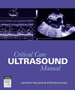
This is a concise step-by-step guide on the assessment of ultrasounds. Its aim is to train critical care physicians in applying Rapid Assessment by Cardiac Echo (RACE) and Focused Assessment with Sonography in Trauma (FAST) to sonography principles. The focus is on helping readers to obtain rapid practical information to assist management decisions. The user-friendly layout is further enhanced by explanatory diagrams and ultrasound images which help with the learning experience. Also included is a DVD showing video clips of procedures cross-referenced in the book assisting the reader in comprehending the content covered in the manual. Practical tips and cautions stand out in highlighted boxes. Multiple choice questions at the end of each chapter allow readers to analyse what they have learnt. The appendices provide a checklist to assist interpretation of transthoracic echocardiogram in a systematic way. A chapter on Doppler principles help those who wish to prepare the way for Doppler measurements.
ELSEVIERwww.elsevier.com
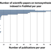
X-ray based imaging techniques include a variety of different implementations and applications: computed radiography (CR), digital radiography (DR) and variations of computed tomography (CT): clinical CT, C-arm, tomosynthesis, micro-CT, industrial CT. These X-ray based methods are widely used not only for diagnostics and assistance in clinical practice but also for screening in security applications and for non-destructive material testing in industry, archeology and material science. The aim of this review paper is to give an introduction into a modern X-ray based tomographic imaging technique, called Digital Tomosynthesis (DT). DT is known as an attractive low-dose alternative to CT in medical (and non-medical) imaging applications.
by Yulia M. Levakhina, Thorsten M. Buzug
Historical overview: from radiostereoscopy to digital tomosynthesis
The history of X-ray imaging starts in 1985 when Wilhelm Conrad Roentgen discovered a new kind of radiation which he called X-rays. It was a breakthrough invention that allowed visualization of inner structures of the human body without surgical intervention.
Analogue imaging
A simple radiographic image contains the superposition of all three-dimensional structures in an object as a two-dimensional image. This means that is it impossible to recover the depth of information of any particular feature (e.g. tumour). At the beginning of the 1920s there were many attempts to erase superimposed shadows from X-ray images and to benefit from the use of X-rays for imaging of the human body. Owing to the fact that the communication between researchers from different countries was very limited at that time, many scientists were re-discovering similar imaging concepts. It resulted in a number of patent applications and scientific papers, which all discussed the same imaging technique where the X-ray tube and X-ray receptor move in parallel planes. The result of each acquisition was an analogue image showing sharply the only one plane located in focus while blurring all other planes. This technique was called stratigrafia by A. Vallebona, planigraphy by A. E. M. Bocage and B. G. Ziedses des Plantes or laminography by J. Kieffer. More information can be found e.g. in the historical article written by a curator of the Belgian Museum of Radiology, R. van Tiggelen.
Digital tomosynthesis or computed tomography?
The next step forward was the implementation of a device, where each measured radiographic image can be stored separately and processed after the examination instead of integrating the measurements directly on film. By doing this, it is possible to generate an arbitrary number of planes or laminograms through the object based on the limited number of measured radiographs. The total radiation dose can be reduced because only one examination is needed to produce images of the whole volume. This is essentially the main idea of modern tomosynthesis as it is known today. The word tomosynthesis was introduced by D. G. Grant in 1972. A number of further improvements of tomosynthesis, mainly focused on improving image quality and shortening acquisition time, have been proposed during the 1970s and 1980s. The review papers by Dobbins give a detailed overview of tomosynthesis research during the 1970s and 1980s.
In that same year (1972), there was an another development when Sir Godfrey Hounsfield and James Ambrose gave a talk on

Last March, the 29th edition of the Korean International Medical & Hospital Equipment Show attracted a record number of visitors, over 68,000 in total, which represents a growth of nearly 14% over last year. That
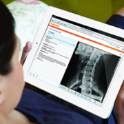
At ECR 2013 Carestream is showcasing a raft of new solutions designed to help radiology professionals improve patient care. These include a new portal that allows patients to electronically access and manage their X-ray exams; new lesion management tools that can help enhance accuracy in assessing changes in cancerous lesions and, as a works-in-progress, a smaller-format digital radiography detector that provides high-quality, dose-sensitive X-ray images for pediatric, orthopedic and general radiology exams.
Healthcare IT
The MyVue patient portal empowers patients to electronically access and manage their X-ray exams—and then share that data with specialists and other healthcare professionals. The Web-based portal is implemented by healthcare providers to allow patients to download information to PCs, laptops, iPads or other devices. It is easy to use and reduces the time and cost of outputting medical exams onto DVD/CDs or other physical storage formats for medical records. My Vue is currently available as an option for Vue PACS and Vue Archive users and is now available for order as a Vue Cloud Service.
New lesion management tools can help enhance accuracy in assessing changes in cancerous lesions as part of diagnosis and treatment for oncology patients. A native follow-up application that can be added to Vue PACS, the new module provides semi-automatic tracking and segmentation of lesions from modalities. Each measurement generates an anatomical bookmark within the exam.
Enhancements to Carestream’s RIS including the storage and tracking of radiation dose information and other capabilities that lay the groundwork to support cumulative dose tracking, which is an important patient care initiative worldwide.
Digital Capture
In the wireless digital radiography market, a smaller-format 25 cm x 30 cm Carestream DRX 2530C detector is demonstrated as a work in progress. The new cesium iodide detector is designed to offer high efficiency for dose sensitive pediatric, orthopedic and general radiology exams. The smaller detector is designed to fit into pediatric incubator trays and offer higher DQE (detective quantum efficiency), which can lead to lower dose requirements than CR cassettes or gadolinium scintillator detectors. The new DRX 2530C detector is intended to be used with Carestream DRX-Revolution or Carestream DRX-Mobile Retrofit Kits for mobile imaging of neonatal or pediatric patients. In orthopedic and general radiology imaging, the smaller detector is designed to aid in positioning for tabletop exams such as knee, elbow, skull and other exams.
Also highlighted will be a new non-motorized option for the Carestream DRX-Evolution, a versatile DR system with modular components. The new DRX-Evolution Standard-Q offers DR capability at an affordable cost. The ergonomically designed wall stand makes standing exams easier with its extensive vertical travel range. In addition, the extra-wide Standard-Q elevating float-top table lowers easily to accommodate stretcher and wheelchair patients and offers increased patient weight capacity.
Mammography
A new module displays digital breast tomosynthesis (DBT) exams from DICOM-compliant acquisition devices on its Carestream Vue Mammo Workstation, where radiologists can also view traditional mammograms, breast ultrasound, breast MRI and general radiology exams from a single desktop. The module streamlines workflow by allowing healthcare providers to store, route, display and query/retrieve DBT exams from DICOM-compliant acquisition devices. Comparison tools enable radiologists to use personalized hanging protocols for DBT exams along with other procedures.
Digital Output
The new DryView 5950 Laser Imaging System produces 508 pixels-per-inch output for general radiology and mammography images. The new imager can support efficient printing and time-saving film cartridges and delivers an enhanced quality control system for mammography images. This innovative internal quality control system includes a built-in densitometer that will produce test prints and display data needed to support mammography quality control charting.
Dental
The CS 9300 System is a high-quality cone beam CT (CBCT – 3D imaging) and true panoramic imaging system for ENT and dental indications. The system can be used for a variety of ENT and dental applications – including sinus, temporal bone and maxillofacial exams; dental implantology; oral surgery; orthodontics; periodontics and endodontics. The CS 9300 is a cost-effective solution to offload ENT and dental CT, delivering up to 94 % less radiation dose than conventional CT units, and images at a much higher resolution, making it ideal for visualizing fine bony structures in the
middle ear and radicular (root) structures.
Carestream Health
www.ihe-online.com & search 46315


Prins Hendrikstraat 1
5611HH Eindhoven
The Netherlands
info@interhospi.com
PanGlobal Media IS not responsible for any error or omission that might occur in the electronic display of product or company data.