
Digital tomosynthesis at a glance: a scientific review
X-ray based imaging techniques include a variety of different implementations and applications: computed radiography (CR), digital radiography (DR) and variations of computed tomography (CT): clinical CT, C-arm, tomosynthesis, micro-CT, industrial CT. These X-ray based methods are widely used not only for diagnostics and assistance in clinical practice but also for screening in security applications and for non-destructive material testing in industry, archeology and material science. The aim of this review paper is to give an introduction into a modern X-ray based tomographic imaging technique, called Digital Tomosynthesis (DT). DT is known as an attractive low-dose alternative to CT in medical (and non-medical) imaging applications.
by Yulia M. Levakhina, Thorsten M. Buzug
Historical overview: from radiostereoscopy to digital tomosynthesis
The history of X-ray imaging starts in 1985 when Wilhelm Conrad Roentgen discovered a new kind of radiation which he called X-rays. It was a breakthrough invention that allowed visualization of inner structures of the human body without surgical intervention.
Analogue imaging
A simple radiographic image contains the superposition of all three-dimensional structures in an object as a two-dimensional image. This means that is it impossible to recover the depth of information of any particular feature (e.g. tumour). At the beginning of the 1920s there were many attempts to erase superimposed shadows from X-ray images and to benefit from the use of X-rays for imaging of the human body. Owing to the fact that the communication between researchers from different countries was very limited at that time, many scientists were re-discovering similar imaging concepts. It resulted in a number of patent applications and scientific papers, which all discussed the same imaging technique where the X-ray tube and X-ray receptor move in parallel planes. The result of each acquisition was an analogue image showing sharply the only one plane located in focus while blurring all other planes. This technique was called stratigrafia by A. Vallebona, planigraphy by A. E. M. Bocage and B. G. Ziedses des Plantes or laminography by J. Kieffer. More information can be found e.g. in the historical article written by a curator of the Belgian Museum of Radiology, R. van Tiggelen.
Digital tomosynthesis or computed tomography?
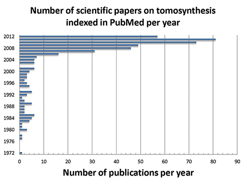
The next step forward was the implementation of a device, where each measured radiographic image can be stored separately and processed after the examination instead of integrating the measurements directly on film. By doing this, it is possible to generate an arbitrary number of planes or laminograms through the object based on the limited number of measured radiographs. The total radiation dose can be reduced because only one examination is needed to produce images of the whole volume. This is essentially the main idea of modern tomosynthesis as it is known today. The word tomosynthesis was introduced by D. G. Grant in 1972. A number of further improvements of tomosynthesis, mainly focused on improving image quality and shortening acquisition time, have been proposed during the 1970s and 1980s. The review papers by Dobbins give a detailed overview of tomosynthesis research during the 1970s and 1980s.
In that same year (1972), there was an another development when Sir Godfrey Hounsfield and James Ambrose gave a talk on
