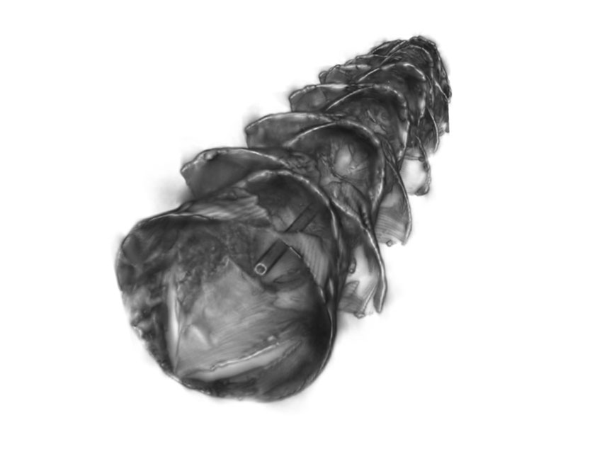
The kirigami-inspired stent
Inspired by kirigami, the Japanese art of folding and cutting paper to create three-dimensional structures, MIT engineers and their collaborators have designed a new type of stent that could be used to deliver drugs to the gastrointestinal tract, respiratory tract, or other tubular organs in the body.
The stents are coated in a smooth layer of plastic etched with small “needles” that pop up when the tube is stretched, allowing the needles to penetrate tissue and deliver a payload of drug-containing microparticles. Those drugs are then released over an extended period of time after the stent is removed.
This kind of localized drug delivery could make it easier to treat inflammatory diseases affecting the GI tract such as inflammatory bowel disease or eosinophilic esophagitis, says Giovanni Traverso, an MIT assistant professor of mechanical engineering, a gastroenterologist at Brigham and Women’s Hospital, and the senior author of the study.
“This technology could be applied in essentially any tubular organ,” Traverso says. “Having the ability to deliver drugs locally, on an infrequent basis, really maximizes the likelihood of helping to resolve patients’ conditions and could be transformative in how we think about patient care by enabling local, prolonged drug delivery following a single treatment.”
Sahab Babaee, an MIT research scientist, is the lead author of the paper, which appears in Nature Materials.
Stretchable stents
Inflammatory diseases of the GI tract, such as IBD, are often treated with drugs that dampen the body’s immune response. These drugs are usually injected, so they can have side effects elsewhere in the body. Traverso and his colleagues wanted to come up with a way to deliver such drugs directly to the affected tissues, reducing the likelihood of side effects.
Stents could offer a way to deliver drugs to a targeted portion of the digestive tract, but inserting any kind of stent into the GI tract can be tricky because digested food is continuously moving through the tract. To make this possibility more feasible, the MIT team came up with the idea of creating a stent that would be inserted temporarily, lodge firmly into the tissue to deliver its payload, and then be easily removed.
The stent they designed has two key elements – a soft, stretchy tube made of silicone-based rubber, and a plastic coating etched with needles that pop up when the tube is stretched.
“The novelty of our approach is that we used tools and concepts from mechanics, combined with bioinspiration from scaly-skinned animals, to develop a new class of drug-releasing systems with the capacity to deposit drug depots directly into luminal walls of tubular organs for extended release,” Babaee says. “The kirigami stents were engineered to provide a reversible shape transformation: from flat, to 3D, buckled-out needles for tissue engagement, and then to the original flat shape for easy and safe removal.”
In this study, the MIT team coated the plastic needles with microparticles that can carry drugs. After the stent is inserted endoscopically, the endoscope is used to inflate a balloon inside the tube, causing the tube to elongate. As the tube stretches, the pulling motion causes the needles in the plastic to pop up and release their cargo.
“It’s a dynamic system where you have a flat surface, and you can create these little needles that pop up and drive into the tissue to do the drug delivery,” Traverso says.
For this study, the researchers created kirigami needles of several different sizes and shapes. By varying those features, as well as the thickness of the plastic sheet, the researchers can control how deeply the needles penetrate into the tissue. “The advantage of our system is that it can be applied to various length scales to be matched with the size of the target tubular compartments of the gastrointestinal tract or any tubular organs,” Babaee says.
GI drug delivery
The researchers tested the stents by endoscopically inserting them into the oesophagus of pigs. Once the stent was in place, the researchers inflated the balloon inside the stent, allowing the needles to pop up. The needles, which penetrated about half a millimetre into the tissue, were coated with microparticles containing a drug called budesonide, a steroid that is used to treat IBD and eosinophilic esophagitis.
Once the drug-containing particles were deposited in the tissue, the researchers deflated the balloon, flattening out the needles so the stent could be endoscopically removed. This process took only a couple of minutes, and the microparticles then stayed in the tissue and gradually released budesonide for about one week.
Depending on the composition of the particles, they could be tuned to release drugs over an even longer period of time, Traverso says. This could make it easier to keep patients on the correct drug schedule, because they would no longer need to take the drug themselves, but would periodically receive their medicine via temporary insertion of the stent. It would also avoid the side effects that can occur with systemic drug administration.
The researchers also showed that they could deliver the stents into blood vessels and the respiratory tract. They are now working on delivering other types of drugs and on scaling up the manufacturing process, with the goal of eventually testing the stents in patients.


Cognoa’s autism spectrum disorder diagnosis aid approved by US FDA
, /in Product News /by panglobalThe U.S. FDA has authorized marketing of a device to help diagnose autism spectrum disorder (ASD). The Cognoa ASD Diagnosis Aid is a machine learning-based software intended to help healthcare providers diagnose ASD in children 18 months through 5 years of age who exhibit potential symptoms of the disorder.
“Autism spectrum disorder can delay a child’s physical, cognitive and social development, including motor skill development, learning, communication and interacting with others. The earlier ASD can be diagnosed, the more quickly intervention strategies and appropriate therapies can begin,” said Jeff Shuren, M.D., J.D., director of the FDA’s Center for Devices and Radiological Health. “Today’s marketing authorization provides a new tool for helping diagnose children with ASD.”
The Centers for Disease Control and Prevention defines ASD as a “developmental disability that can cause significant social, communication and behavioral challenges” and is estimated to affect about 1 in 54 children. Because ASD symptoms can vary greatly, the disorder may be difficult to diagnose. While ASD may be detected as early as 18 months old, many children are not diagnosed until later in childhood, which can delay treatment and early intervention. The average age of diagnosis for ASD is 4.3 years. Some delays in diagnosis are due to the need for children to be referred to specialists with expertise in ASD.
Software as a medical device
The Cognoa ASD Diagnosis Aid is a software as a medical device that uses a machine learning algorithm to receive input from parents or caregivers, video analysts and health care providers to assist physicians evaluate a patient at risk of ASD. The device consists of three main components: a mobile app for caregivers and parents to answer questions about behaviour problems and to upload videos of their child; a video analysis portal that allows manufacturer-trained and certified specialists to view and analyse uploaded videos of patients; and a health care provider portal that is intended for a health care provider to enter answers to pre-loaded questions about behaviour problems, track the information provided by parents or caregivers and review a report of the results. After processing the information provided by parents, caregivers and healthcare providers, the ASD Diagnosis Aid reports a positive or negative diagnosis if there is sufficient information for its algorithm to make a diagnosis. If there is insufficient information to render a “Positive for ASD” or “Negative for ASD” result to help determine a diagnosis, the ASD Diagnosis Aid will report that no result can be generated.
Effectiveness of Cognoa ASD Diagnosis Aid
The FDA assessed the safety and effectiveness of the Cognoa ASD Diagnosis Aid in a study of 425 patients aged 18 months through 5 years in 14 different clinical care sites, with an average age of 2.8 years. The study compared the assessments made by the device directly against the assessments made by a panel of clinical experts who used the current standard ASD diagnostic process. The device provided a “Positive for ASD” or “Negative for ASD” result to aid in making a diagnosis in 32% of patients. For those with a “Positive for ASD” or “Negative for ASD” result, the device results matched the panel’s conclusions for 81% of patients who tested positive for ASD by the device and 98% of patients who tested negative for ASD by the device. In addition, the device made an accurate ASD determination in 98.4% of patients with the condition and in 78.9% of patients without the condition.
The risks associated with the use of the device include misdiagnosis and delayed diagnosis of ASD, based on a false positive result (observed in 15 out of 303 study subjects without ASD), a false negative result (observed in one out of 122 study subjects with ASD) or when no result was generated. Both misdiagnosis or missed diagnosis can result in delayed treatment of ASD and delivery of treatment not appropriate for ASD.
The FDA reviewed the Cognoa ASD Diagnosis Aid through the De Novo premarket review pathway, a regulatory pathway for low- to moderate-risk devices of a new type.
The Cognoa ASD Diagnosis Aid is indicated as an aid in the diagnosis of ASD for patients 18 months through 5 years of age who are at risk of developmental delay based on concerns of a parent, caregiver, or healthcare provider. The device is not indicated for use as a stand-alone diagnostic device but as an adjunct to the diagnostic process.
Stents inspired by paper-cutting art designed to deliver drugs to GI tract
, /in E-News, Featured Articles /by panglobalThe kirigami-inspired stent
Inspired by kirigami, the Japanese art of folding and cutting paper to create three-dimensional structures, MIT engineers and their collaborators have designed a new type of stent that could be used to deliver drugs to the gastrointestinal tract, respiratory tract, or other tubular organs in the body.
The stents are coated in a smooth layer of plastic etched with small “needles” that pop up when the tube is stretched, allowing the needles to penetrate tissue and deliver a payload of drug-containing microparticles. Those drugs are then released over an extended period of time after the stent is removed.
This kind of localized drug delivery could make it easier to treat inflammatory diseases affecting the GI tract such as inflammatory bowel disease or eosinophilic esophagitis, says Giovanni Traverso, an MIT assistant professor of mechanical engineering, a gastroenterologist at Brigham and Women’s Hospital, and the senior author of the study.
“This technology could be applied in essentially any tubular organ,” Traverso says. “Having the ability to deliver drugs locally, on an infrequent basis, really maximizes the likelihood of helping to resolve patients’ conditions and could be transformative in how we think about patient care by enabling local, prolonged drug delivery following a single treatment.”
Sahab Babaee, an MIT research scientist, is the lead author of the paper, which appears in Nature Materials.
Stretchable stents
Inflammatory diseases of the GI tract, such as IBD, are often treated with drugs that dampen the body’s immune response. These drugs are usually injected, so they can have side effects elsewhere in the body. Traverso and his colleagues wanted to come up with a way to deliver such drugs directly to the affected tissues, reducing the likelihood of side effects.
Stents could offer a way to deliver drugs to a targeted portion of the digestive tract, but inserting any kind of stent into the GI tract can be tricky because digested food is continuously moving through the tract. To make this possibility more feasible, the MIT team came up with the idea of creating a stent that would be inserted temporarily, lodge firmly into the tissue to deliver its payload, and then be easily removed.
The stent they designed has two key elements – a soft, stretchy tube made of silicone-based rubber, and a plastic coating etched with needles that pop up when the tube is stretched.
“The novelty of our approach is that we used tools and concepts from mechanics, combined with bioinspiration from scaly-skinned animals, to develop a new class of drug-releasing systems with the capacity to deposit drug depots directly into luminal walls of tubular organs for extended release,” Babaee says. “The kirigami stents were engineered to provide a reversible shape transformation: from flat, to 3D, buckled-out needles for tissue engagement, and then to the original flat shape for easy and safe removal.”
In this study, the MIT team coated the plastic needles with microparticles that can carry drugs. After the stent is inserted endoscopically, the endoscope is used to inflate a balloon inside the tube, causing the tube to elongate. As the tube stretches, the pulling motion causes the needles in the plastic to pop up and release their cargo.
“It’s a dynamic system where you have a flat surface, and you can create these little needles that pop up and drive into the tissue to do the drug delivery,” Traverso says.
For this study, the researchers created kirigami needles of several different sizes and shapes. By varying those features, as well as the thickness of the plastic sheet, the researchers can control how deeply the needles penetrate into the tissue. “The advantage of our system is that it can be applied to various length scales to be matched with the size of the target tubular compartments of the gastrointestinal tract or any tubular organs,” Babaee says.
GI drug delivery
The researchers tested the stents by endoscopically inserting them into the oesophagus of pigs. Once the stent was in place, the researchers inflated the balloon inside the stent, allowing the needles to pop up. The needles, which penetrated about half a millimetre into the tissue, were coated with microparticles containing a drug called budesonide, a steroid that is used to treat IBD and eosinophilic esophagitis.
Once the drug-containing particles were deposited in the tissue, the researchers deflated the balloon, flattening out the needles so the stent could be endoscopically removed. This process took only a couple of minutes, and the microparticles then stayed in the tissue and gradually released budesonide for about one week.
Depending on the composition of the particles, they could be tuned to release drugs over an even longer period of time, Traverso says. This could make it easier to keep patients on the correct drug schedule, because they would no longer need to take the drug themselves, but would periodically receive their medicine via temporary insertion of the stent. It would also avoid the side effects that can occur with systemic drug administration.
The researchers also showed that they could deliver the stents into blood vessels and the respiratory tract. They are now working on delivering other types of drugs and on scaling up the manufacturing process, with the goal of eventually testing the stents in patients.
GammaDelta Therapeutics receives FDA clearance of IND application for GDX012, a novel allogeneic variable delta 1 gamma-delta T cell cancer therapy
, /in E-News /by panglobalFDA also granted orphan drug designation to GDX012 for the treatment of Acute Myeloid Leukaemia
GammaDelta Therapeutics has received US FDA clearance for the Investigational New Drug (IND) application for the Company’s allogeneic variable delta 1 (Vδ1) gamma-delta (γδ) T cell therapy, GDX012, to be investigated as a treatment for haematological malignancies. The FDA also granted orphan drug designation to GDX012 for the treatment of Acute Myeloid Leukaemia (AML).
Dr. Paolo Paoletti, CEO of GammaDelta Therapeutics, commented: “The clearance of our IND application for GDX012 marks an important step for our company in establishing a portfolio of innovative allogeneic cell therapies. The unique properties of Vδ1 γδ T cells will be evaluated for the first time in a clinical study for patients with AML. This important milestone results from our efforts to establish a robust pipeline of cellular immunotherapies derived from our proprietary platforms and processes for the isolation and expansion of Vδ1 γδ T cells from both blood and tissues for targeting haematological malignancies and solid tumours.”
GammaDelta plans to initiate a Phase 1 clinical trial for patients with measurable residual disease (MRD) positive AML. Expected to begin later in 2021 as a multicentre study in the US, the trial will evaluate safety, tolerability and anti-leukemic activity of GDX012.
GammaDelta Therapeutics is advancing its novel T cell platform under an ongoing collaboration with Takeda Pharmaceutical Company formed in 2017.
Changing treatment paradigm for AML
Dr. Michael Koslowski, Head of R&D and Chief Medical Officer of GammaDelta Therapeutics, said: “Although progress has been made in the treatment of AML, the median overall five-year survival rate for patients diagnosed with AML remains under 30 percent. With the development of GDX012 we are aiming to change the treatment paradigm for AML and potentially other haematologic malignancies. The unique biological characteristics of Vδ1 γδ T cells offer a first-in-class Vδ1 γδ T cell therapy for AML, where the development of cell therapies has been historically limited due to the lack of specific targets.”
Dr. Chris Arendt, Oncology Therapeutic Area Unit Head of Takeda, commented: “The progression of GammaDelta Therapeutics’ platform technology underscores the potential of γδ T cells and the power of the innate immune system. Through collaboration with pioneers like GammaDelta Therapeutics, we hope to advance next-generation cell therapies and to maximise off-the-shelf treatments in the battle against hard-to-treat cancers.”
GammaDelta has developed proprietary technologies to generate both blood-and tissue-derived allogeneic immunotherapies based on Vδ1 γδ T cells for the treatment of haematologic malignancies and solid tumours. Both platforms have enabled the creation of highly active and selective non-engineered and genetically engineered allogeneic cell therapies, which demonstrate cellular activity and tumour cell killing capacity. Vδ1 γδ T cells are a unique subset of T cells that specifically recognise and are activated by molecular patterns of dysregulation on cancer cells. The non-MHC-restricted activity of Vδ1 γδ T cells makes them a unique cell type for the development of fully allogeneic, “off-the-shelf” cell therapies.
Carestream advances X-ray image quality with Smart Noise Cancellation
, /in Product News /by panglobalObjective testing demonstrated that SNC processing enables a 2X to 4X noise reduction in flat areas, preserves high frequency sharpness, and improves contrast detail.
Carestream Health has released Smart Noise Cancellation (SNC), a groundbreaking artificial intelligence (AI)-based technology that significantly improves image quality – producing images that are clearer than with standard processing. SNC has received FDA 510(k) Clearance and is available as an optional feature with Carestream’s ImageView Software powered by Eclipse – the intelligent image-processing engine behind the company’s innovative imaging software – on DRX-Evolution and DRX-Evolution Plus systems.
“Carestream is a leader in using AI for noise cancellation with X-ray images. Our team of imaging scientists has been able to separate image noise from sharpness and contrast using AI-based algorithms that result in remarkable image quality,” said Jill Hamman, Worldwide Marketing Manager, Global X-ray Solutions at Carestream. “This technology provides improved anatomical clarity, preservation of fine detail and better contrast-to-noise ratio for images acquired at a broad range of exposures, which can help improve diagnostic confidence and alleviate physician fatigue. It also enables radiology professionals to better optimize radiation dose.” Optimizing radiation dose is especially important with neonatal and paediatric diagnostic imaging, where imaging at the lowest possible dose is crucial for young patients.
Challenge of separating noise from image
Separating noise from an image has been a challenge for medical imaging scientists. Traditional noise reduction introduces blurring, which degrades image sharpness and might remove important anatomical information. Conversely, the more an image is sharpened, the more noise may be enhanced. Noise is often an undesirable by-product of image capture and can obscure critical anatomical data. Carestream’s SNC is able to isolate noise to produce images that are significantly clearer than with standard processing.
As the preferred level of noise on X-ray images is subjective – for example, some radiologists expect to see a certain degree of noise in images, which assures them that the patient was not overexposed – Carestream enables imaging professionals to adjust the amount of noise cancellation and exposure to meet their desired image quality.
In a blinded Clinical Reader study, 89.5% of all study ratings showed a slight to strong preference for SNC processed images.
Objective testing demonstrated that SNC processing enables a 2x–4x noise reduction in flat image areas, preserves high frequency sharpness and improves contrast detail. Additionally, a blind Clinical Reader Study using board-certified radiologists found that 89.5% of all study ratings showed a slight to strong preference for SNC-processed images. Sixty-four percent of the diagnostic quality ratings improved – based on the RadLex rating scale – and 56% of these ratings improved from “limited” or “diagnostic” to “exemplary”.
When combined with SmartGrid software, Smart Noise Cancellation software promises benefits in gridless imaging where the removal of scatter typically leads to an increase in noise appearance.
New material to treat wounds can protect against resistant bacteria
, /in E-News /by panglobalResearchers at Chalmers University of Technology, Sweden, have developed a new material that kills bacteria and could potentially prevent infections in wounds – a specially designed hydrogel, that works against all types of bacteria, including antibiotic-resistant ones. The active substance in the new bactericidal material consists of antimicrobial peptides, small proteins which are found naturally in our immune system. Photo: Anna-Lena Lundqvist/Chalmers
Researchers at Chalmers University of Technology, Sweden, have developed a new material that prevents infections in wounds – a specially designed hydrogel, that works against all types of bacteria, including antibiotic-resistant ones. The new material offers hope for combating a growing global problem antibiotic-resistant bacteria.
The World Health Organization describes antibiotic-resistant bacteria as one of the greatest threats to global health. To deal with the problem, there needs to be a shift in the way we use antibiotics, and new, sustainable medical technologies must be developed.
“After testing our new hydrogel on different types of bacteria, we observed a high level of effectiveness, including against those which have become resistant to antibiotics,” says Martin Andersson, research leader for the study and Professor at the Department of Chemistry and Chemical Engineering at Chalmers University of Technology.
Research and development of the material has been ongoing for many years at Martin Andersson’s group at Chalmers, growing in scope along the way, with a particular focus on the possibilities for wound care. Now, the important results are published as a scientific article in the journal ACS Biomaterials Science & Engineering.
In recent years, foundational research into the antimicrobial peptide hydrogel has run in parallel with commercial development of the innovation through the spin-off company Amferia AB. The company has developed an antibacterial patch, which is expected to be commercialised soon. Photo: Anna-Lena Lundqvist/Chalmers
The main purpose of the studies so far has been to explore new medical technology solutions to help reduce the use of systemic antibiotics. Resistant bacteria cause what is referred to as hospital-acquired infection – a life-threatening condition that is increasing in incidence worldwide.
Mimicking the natural immune system
The active substance in the new bactericidal material consists of antimicrobial peptides, small proteins which are found naturally in our immune system.
“With these types of peptides, there is a very low risk for bacteria to develop resistance against them, since they only affect the outermost membrane of the bacteria. That is perhaps the foremost reason why they are so interesting to work with,” says Martin Andersson.
Researchers have long tried to find ways to use these peptides in medical applications, but so far without much success. The problem is that they break down quickly when they come into contact with bodily fluids such as blood. The current study describes how the researchers managed to overcome the problem through the development of a nanostructured hydrogel, into which the peptides are permanently bound, creating a protective environment.
“The material is very promising. It is harmless to the body’s own cells and gentle on the skin. In our measurements, the protective effect of the hydrogel on the antimicrobial peptides is clear – the peptides degrade much slower when they are bound to it,” says Edvin Blomstrand, Doctoral Student at the Department of Chemistry and Chemical Engineering at Chalmers, and one of the main authors of the article.
“We expected good results, but we were really positively surprised at quite how effective the material has proven,” adds Martin Andersson.
According to the researchers, this new material is the first medical device to make successful use of antimicrobial peptides in a clinically and commercially viable manner. There are many varied and promising opportunities for clinical application.
Start-up company Amferia takes the research from lab to market
In recent years, foundational research into the antimicrobial peptide hydrogel has run in parallel with commercial development of the innovation through the spin-off company Amferia AB.
The material and the idea, which is currently developed as an antibacterial wound patch, has generated interest around the world, attracting significant investment and receiving several awards. The company is working intensively to get the material to market so that it can benefit wider society.
Before the new material can benefit hospitals and patients, clinical studies are needed, which are ongoing. A CE marking of the material is expected to be completed in 2022.
OR Technology unveils motorised X-ray system for efficient, smooth workflow
, /in Product News /by panglobalGerman company OR Technology has released their versatile Amadeo R motorised X-ray system, expanding their product range for the inpatient sector.
The X-ray system, consisting of a bucky table and grid wall stand, has an auto-tracking function suitable for all X-ray exposures in sitting, lying and standing positions.
An intuitive 10″ touchscreen display simplifies operation. Up to 60 pre-set positions of stand height, alignment and SID (Source-Image Distance) speed up the alignment of the unit.
The X-ray tube and bucky tray of the grid wall stand are designed to be lowered to the floor. The X-ray tube automatically follows the bucky tray of the wall stand as long as the column stand is not above the X-ray table. The X-ray table with six-position height-adjustable table top has a high load-bearing capacity. An armrest is attached to the grid drawer of the wall stand to provide support for patients during taxing exposures.
The company says X-ray staff quickly become familiar with the use of the acquisition and reporting software dicomPACS®DX-R.
A wide range of standard features such as motorised auto-tracking, the wireless three-way foot switch for all motorised functions, the integrated safety functions or the removable grid on the table or grid wall stand enable fast and efficient work in daily routine operation.
OR Technology, based in Rostock, Germany, has been a manufacturer of digital X-ray technology and developer of image management systems since 1991. The company’s own solutions are successfully used in practices and clinics in more than 120 countries. Their portfolio ranges from DR retrofits for existing stationary or mobile X-ray systems, to imaging plate systems (X-ray with cassettes), complete X-ray systems and mobile DR detector case solutions for outdoor use.
medK launches needle-free extension lines for vascular access
, /in Product News /by panglobalThe needle-free extension line with triple valve. photo: ©medK GmbH
medK, a German company specialising in interventional products, has launched a needle-free solution for vascular access. The needle-free connection offers secure and fast access to fluid lines eliminating the risk of needle sticks. The extension line can also be connected with a glass syringe. With the multi-way versions there is no need to use a stopcock or a manifold.
medK Needle free Extension Lines are compatible with disinfection with IPA wipes (Isopropanol). This and the smooth valve surface allows easy disinfection and reduces the risk of infections. The leak free disconnection protects against blood contaminations for health workers. Once disconnected it provides reliable closure and can stay up to seven days or two hundred activations with the patient. The fully visible flow path allows visual checking of the fluid.
The needle-free extension line valve. Photo: ©medK GmbH
The needle-free extension lines are also available with a combined check valve that safeguards against back tracking. The needle-free extension lines are compatible with Isopropanol, Chlorohexidine, blood and lipids. The device is suitable for use in MRI procedures and is latex and animal product free.
medK extension lines are available as single, dual and triple access with needle free or combined check valve.
Researchers identify why Covid-19 patients develop life-threatening blood clots
, /in Corona News, E-News /by panglobalScientists have identified how and why some Covid-19 patients can develop life-threatening blood clots, which could lead to targeted therapies that prevent this from happening.
The work, led by researchers from RCSI University of Medicine and Health Sciences, is published in the Journal of Thrombosis and Haemostasis.
Previous research has established that blood clotting is a significant cause of death in patients with Covid-19. To understand why that clotting happens, the researchers analysed blood samples that were taken from patients with Covid-19 in the Beaumont Hospital Intensive Care Unit in Dublin.
They found that the balance between a molecule that causes clotting, called von Willebrand Factor (VWF), and its regulator, called ADAMTS13, is severely disrupted in patients with severe Covid-19.
When compared to control groups, the blood of Covid-19 patients had higher levels of the pro-clotting VWF molecules and lower levels of the anti-clotting ADAMTS13. Furthermore, the researchers identified other changes in proteins that caused the reduction of ADAMTS13.
“Our research helps provide insights into the mechanisms that cause severe blood clots in patients with Covid-19, which is critical to developing more effective treatments,” said Dr Jamie O’Sullivan, the study’s corresponding author and research lecturer within the Irish Centre for Vascular Biology at RCSI.
He added: “While more research is needed to determine whether targets aimed at correcting the levels of ADAMTS13 and VWF may be a successful therapeutic intervention, it is important that we continue to develop therapies for patients with Covid-19. Covid-19 vaccines will continue to be unavailable to many people throughout the world, and it is important that we provide effective treatments to them and to those with breakthrough infections.”
UV Smart aims to provide healthcare facilities with validated UV-C disinfection methods
, /in Product News /by panglobalDisinfection with high-energy UV-C light has been proven safe and effective. UV Smart makes this technology easily applicable for the validated disinfection of medical instruments and equipment. The result: a safer environment for healthcare professionals and their patients.
Study presented at EHA21 looks at Pfizer/BioNTech SARS-COV-2 vaccine impairment in immunosuppressed patients
, /in Corona News, E-News /by panglobalThe European Hematology Association held their virtual congress from 9-17 June – it remains online until 15 August here. Many studies were presented, key among them was one titled: Humoral Response to the Pfizer/BioNTech BNT162b2 Vaccine Is Impaired in Patients Receiving CAR-T or High-Intensity Immunosuppressive Therapy.
In the study the researchers evaluated the efficacy and safety of the Pfizer/BioNTech BNT162b2 vaccine – approved for the prevention of SARS-CoV-2 infection – in patients that underwent hematopoietic cell transplantation (HCT) and chimeric antigen receptor (CAR)-T therapy. They prospectively followed 79 vaccinated patients who were actively treated at the Tel Aviv Sourasky Medical Center and monitored the safety profile and the humoral immune response to the vaccine.
They note that although the vaccine is recommended for immunosuppressed patients, its efficacy and safety in patients undergoing immunologic cell therapy have not been well-documented.
In their study they found that: “Overall, the vaccine was well-tolerated and all adverse events resolved within a few days except for one secondary graft rejection, which is still under investigation. We observed that only 36% of patients who received CAR-T therapy developed a humoral antibody response compared with 81% of patients who underwent allogeneic HCT. In addition, patients with B cell aplasia and those who received the vaccine shortly after infusion of cells were less likely to develop antibodies. Taken together, these data demonstrate that the humoral response to the BNT162b2 vaccine is significantly impaired in patients receiving CAR-T , as opposed to those after allogeneic HCT who had a good response.”
EHA21 Virtual Congress
This study presentation and others can be accessed at the EHA21 Virtual Congress. The education and scientific program of the congress focuses on clinical practice, recent advances, new data and views from different stakeholders and international organizations. Registration is open until 1 August and the sessions are online until 15 August.