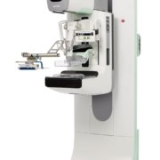
The new 3Dimensions system features Clarity HD high-resolution 3D imaging, which provides the industry’s fastest, highest resolution 3D images. Clarity HD technology’s advanced detector and innovative 3D imaging algorithm work together to deliver exceptional 3D images, regardless of breast size or density, while reducing recalls by up to 40% compared to 2D alone. In addition, the 3Dimensions system offers Intelligent 2D imaging technology, which features smart mapping, enabling radiologists to instantly move from suspicious areas detected on the 2D image to the point of interest on the 3D slice. The 3Dimensions system also includes the new SmartCurve breast stabilization system, which is clinically proven to deliver a more comfortable mammogram without compromising image quality, workflow or dose. The SmartCurve system has been shown to improve comfort in 93% of women surveyed who reported moderate to severe discomfort with standard compression. The system features a curved compression surface that mirrors the shape of a woman’s breast to reduce pinching and allow uniform compression over the entire breast. Advanced processing software, specifically developed for the SmartCurve system, ensures optimal image quality. Another feature is the Quantra 2.2 breast density assessment software, which enables standardization in patient protocols, providing reproducible and consistent breast density assessment.
https://interhospi.com/wp-content/uploads/sites/3/2020/08/IH159_Hologic-_1_.jpg8005213wmediahttps://interhospi.com/wp-content/uploads/sites/3/2020/06/Component-6-–-1.png3wmedia2020-08-26 14:31:322021-01-08 12:10:42Highest resolution breast tomosynthesis system
Xenios AG, a company of the Fresenius Medical Care Group, reports that it has seen a significant growth in demand for its extracorporeal membrane oxygenation (ECMO) devices, which can be used for the treatment of patients who develop severe pneumonia and acute respiratory distress syndrome (ARDS) due to COVID-19 infection.
Jürgen Böhm, CMO of Xenios, explained that for critically ill COVID-19 patients with acute lung failure and refractory hypoxemia – despite use of standard therapy – “our treatment often remains the last therapeutic option and has been a lifesaver for many patients”.
Xenios’s ECMO therapy bypasses the function of the lungs. The patient’s blood is freed from carbon dioxide outside the body and enriched with oxygen. The lungs are thus given time to heal. Because of the increase of critically ill COVID-19 patients, more physicians are opting for ECMO therapy, and thus the increase in demand for Xenios’s ECMO devices.
To meet the demand, the company has increased production of its ECMO devices. “We have put many measures in place to maximize the utilization of our capacity to manufacture ECMO devices as well as patient kits. Our biggest challenge right now is the availability of specific components for our products,” said Andreas Terpin, CEO of Xenios.
For more information, visit: www.xenios-ag.com Read more
https://interhospi.com/wp-content/uploads/sites/3/2020/08/PRODUCT_XENIOS.jpg74910003wmediahttps://interhospi.com/wp-content/uploads/sites/3/2020/06/Component-6-–-1.png3wmedia2020-08-26 14:31:322021-01-08 12:10:17Xenios sees growing demand for ECMO devices
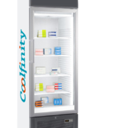
Andrea is a Dutch doctor who worked in maternity ward of the Mulago hospital in Kampala, Uganda with about 1500 beds. She is dedicated to improving the healthcare in the neonatal field and helps deliver healthy babies. She told us that she was frustrated, that oxytocin was not readily available to stop post-partum bleeding. The drug was present in the hospital, but it could not be used as it had lost its potency due to lack of reliable cooling. In Mulago, power outages were so severe that refrigerators do not work anymore. Due to the lack of cooling of oxytocin, several mothers died unnecessarily on a weekly basis. Working under these conditions is not why she had become a tropical doctor in the first place. Lack of reliable cooling in the medical field leads to huge losses of insulin, red blood cells, vaccines and lab-reagents. Coolfinity believes that long power outages and weak power grids should not limit life and opportunities in tropical countries. Cooling is the basic need that is really hampered by ineffective power supply in more than 70 countries in the world. Reliable and sustainable cooling is the catalyst for people in upcoming markets to give their quality of life a boost. Therefore, Coolfinity has developed a refrigerator, the IceVolt 300, that only needs 6 hours of power to stay cool for 24 hours to preserve the medication in the cold chain. The refrigerator is specifically designed to maintain a stable 5 degrees Celsius cooling, in line with the WHO requirements. The refrigerator works very effectively under harsh tropical conditions (35 degrees Celsius). And if the cooler is unplugged, it will even run for 2 days without power. Because of Coolfinity’s technology, the need for a back-up power generator is even eliminated. With this unique and innovative refrigerator Coolfinity will save costly medication and precious lives worldwide, where it is needed most.
https://interhospi.com/wp-content/uploads/sites/3/2020/08/Coolfinity_ijskast.jpg10708003wmediahttps://interhospi.com/wp-content/uploads/sites/3/2020/06/Component-6-–-1.png3wmedia2020-08-26 14:31:322021-01-08 12:10:23Saving lives, a refrigerator at a time
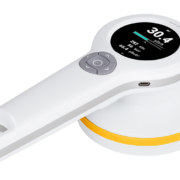
Radiation measurement often requires different devices for varying applications adding to the cost and complexity of data collection. The new RaySafe 452 Survey Meter is a versatile, powerful tool that can be used for multiple situations, reducing the number of devices technicians need to carry, learn, and calibrate. The 452 Survey Meter is the ideal tool to measure radiation in a wide variety of applications, including wall leakage, scatted radiation in a room, tube leakage, nuclear medicine, and control of treated patients. The meter helps technicians make data collection accurately and easily with fast response times and detect variations from background radiation levels due to its high sensitivity. There is no need for post processing of the data due to a flat, wide energy response range. With the automatic saving of dose rate values every second data is easily saved to be analysed later. The RaySafe 452 features an optimized modern design that provides a big, clear, easy-to-read display with all the parameters in a single backlit display. Its ergonomic and lightweight design makes it easy to hold, carry, and use even over extended periods of time. And it has an IP 64 rating so it can easily be cleaned with water without damaging the device. The survey meter comes with RaySafe View software that makes it easy to transfer data to a PC and log data quickly, easily, and efficiently. All data is time stamped and dose rate waveforms can be further analysed later. Read more
https://interhospi.com/wp-content/uploads/sites/3/2020/08/IH274_Fluke_Biomed_RaySafe_452.jpg5338003wmediahttps://interhospi.com/wp-content/uploads/sites/3/2020/06/Component-6-–-1.png3wmedia2020-08-26 14:31:322021-01-08 12:10:25Radiation meter for variety of applications
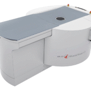
According to the German Cancer Research Center in Heidelberg, some 69,000 women in Germany are diagnosed with breast cancer every year, with almost 18,000 dying of the disease. Conventional diagnostic methods, while well established, are not always reliable. 3D imaging with high isotropic resolution, on the other hand, offers clear advantages. Nu:view, the world’s first breast CT scanner to use spiral CT technology, is the brainchild of Erlangen-based company AB-CT. With the CE marking for nu:view in place, the new scanner is already in use on patients at the University Hospital Zurich (USZ). What sets nu:view apart is the very high image resolution coupled with low radiation dose and short scan times. To obtain the best possible image quality and maximize radiation dose efficiency at the same time, the detector uses state-of-the-art single photon counting technology. Unlike conventional scintillation, nu:view uses detectors made of cadmium telluride (CdTe), transforming every X-ray photon directly into an electric pulse. In a single rotation around the female breast, 2,000 projection images are created, with a full scan taking as little as seven to 12 seconds – at very low radiation levels without breast compression, ensuring excellent patient comfort. For the first time, a CT scanner makes it possible to acquire an image of the entire female breast in a single scan in true 3D, imaging both the soft tissue and the calcifications. Compared with mammography, the three dimensional, non-superimposed images make it far easier to detect micro-calcifications. This non-compressive method also means less discomfort for the patient and reduces the number of additional ultrasound image.
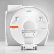
The Magnetom Sola Cardiovascular Edition is a 1.5 Tesla magnetic resonance imaging (MRI) scanner designed specifically for cardiovascular examinations. Magnetom Sola Cardiovascular Edition incorporates the latest technologies with the specific aim of providing the maximum diagnostic information in cardiovascular examinations. This leads to faster, more reliable, and definitive diagnoses for a larger number of patients with underlying ischemic, structural, and arrhythmogenic conditions. The MRI scanner’s innovative applications can cut examination times down to 20 minutes and clarify a broad range of clinical questions in cardiology. Guidelines issued by the European Society of Cardiology (ESC) have long described magnetic resonance imaging as the gold standard for assessing the morphology and function of the heart. Over 25 ESC Guidelines now contain specific recommendations describing instances where MRI should be used in cardiac diagnosis. This is primarily due to the accuracy of MRI when quantifying cardiac volume, mass, and wall motion, as well as its ability to diagnose ischemia and myocardial viability. There are also diseases such as myocarditis (inflammation of the heart muscle), for which cardiovascular MRI is the only non-invasive diagnostic option with the pre-requisite sensitivity. By receiving the right diagnosis at the right time, this group of patients can avoid unnecessary invasive procedures. The Compressed Sensing Cardiac Cine application accelerates MRI scanning sequences so that cardiac function can be measured while the patient is breathing freely. Previous scanners required patients to hold their breath for up to 20 seconds several times during the process. This means that the benefits of the gold standard are now available to new groups of patients, including individuals with arrhythmia or dyspnea who were previously not eligible for CMR. The MyoMaps application provides quantitative information on the tissue composition of the cardiac muscle. This can be used to diagnose diseases of the heart muscle, scar tissue and edema at a very early stage of the condition, allowing the right treatment option to be chosen for the patient as soon as possible. Clinical conditions such as myocarditis can even be detected without using contrast agents. Cardiac Dot Engine is a unique software package which provides step-by-step guidance for standardized diagnostic cardiac MRI exams. It is based on artificial intelligence algorithms, allows users to navigate confidently through the cardiovascular MRI scanning process. Also, BioMatrix Technology automatically adjusts to the biovariability of different patients to prevent undesirable variations in the scanning process. As a result, BioMatrix Technology reduces examination times, cuts down on the number of repeat scans, and leads to consistent, high quality scanning results. Read more
The OmniTom mobile 16-slice computed tomography (CT) scanner features an array of improvements from Samsung’s award-winning CereTom CT scanner. It is the world’s first mobile imaging device with omni-directional wheels, maximizing mobility and allowing easier and quieter movement in small spaces. Its 16-slice (0.625 mm per slice) advanced data acquisition system with effective dose optimization delivers superior image quality. The scanner maintains a small footprint ideal for mobile use while increasing the gantry opening to 40 cm for improved coverage of adult head and neck, and full body pediatric scanning. In addition, OmniTom features an internal drive system, making portability less strenuous, while also offering smart-sensing collision avoidance software to maximize control and patient safety. OmniTom is ideal for cranial procedures and is designed to deliver the highest quality non-contrast CT, CT angiography and CT perfusion scans. The combination of its rapid scan time, ultra-small footprint and immediate image viewing makes OmniTom an indispensable tool for collecting real-time data on critically ill patients. The latest product also demonstrates Samsung’s “AccE” (Access, Accuracy and Efficiency) strategy that aims to change the industry by incorporating innovations in physical access to provide care wherever it is needed. It also aims to improve accuracy in diagnosis with advanced algorithms and efficiency with advanced display, user experience and information management technology developed by Samsung Electronics. Read more
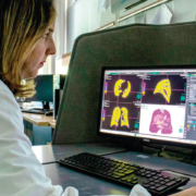
Pisa University Hospital has received support from Fujifilm Italia to improve and speed up the analysis of lung CT images which are important for the diagnosis of COVID-19. This has been possible thanks to an update by Fujifilm of the configuration of the Synapse 3D software used by emergency room radiologists.
The hospital says this new technological development has been useful to improving management of patients affected by COVID-19, during the emergency.
Synapse3D is a 3D medical image analysis system that uses Fujifilm’s image recognition technology to construct and analyze highprecision 3D images, compiled from tomographic images from CT and MRI. It delivers 3D visualization of medical images and has applications in image-based diagnosis and surgery simulation.
Synapse 3D delivers clinical value through fast, accurate, efficient and robust image processing for radiology, cardiology and surgical preoperation simulation.
It was developed in Japan (where it is named Synapse VINCENT) and it consists of more than 50 different processing modules.
The Synapse3D Lung Analysis/Airway module enables the analysis of density ranges in the lung in a quick, easy, objective and reproducible way.
At Pisa University Hospital the configuration of Synapse 3D was completed quickly, with the study of density ranges and a feasibility analysis carried out in a few days, meaning that this updated integrated workflow has been possible to use in the hospital since April 1.
The Pisa University Hospital had already set up three levels of assessing Coronavirus patients, and with the support of Synapse 3D software, doctors have had the ability to quickly and easily identify the stage of pneumonia, and therefore hospitalise patients accordingly:
normal hospitalisation for patients with mild pneumonia not requiring respiratory support
assessment by a pulmonologist or intensive care doctor for patients with moderate pneumonia to plan suitable respiratory support
intensive care assessment for patients with severe pneumonia with a view to transferring them to the intensive care unit
The density analysis provided by Synapse 3D has made it is possible to analyse the lung according to the different pixel densities of the CT images. Three groups were therefore defined based on different density ranges that allow the radiologist to evaluate the percentage of lung with lower density (emphysema), higher density (interstitial effort) and normal lung.
Dr. Chiara Romei, MD, PhD, Radiologist at the Pisa University Hospital, explained: “In a time of emergency such as this, it is crucial for us radiologists to detect accurate data as quickly as possible. Synapse 3D has enabled a quantitative analysis that is much faster and more objective than the visual analysis of the radiologist; in a couple of minutes it is possible to obtain data relating to the percentage of lung with greater and lesser density and to have a precise and objective, reproducible and shareable value.”
It is important to note that the data obtained by Synapse 3D does not replace the molecular diagnoses made through the nasopharyngeal swab (RT-PCR) and does not replace the analysis and diagnostic work by the radiologist, but instead it supports the reporting of daily exams to monitor and study the evolution of the disease, thus optimising workflow.
For medical professionals – download the take-away: https://synapse.fujifilm.eu/fujifilm-takeaway/ Read more
https://interhospi.com/wp-content/uploads/sites/3/2020/08/PRODUCT_FUJIFILM_1.jpg42010003wmediahttps://interhospi.com/wp-content/uploads/sites/3/2020/06/Component-6-–-1.png3wmedia2020-08-26 14:31:322021-01-08 12:10:17Fujifilm’s Synapse 3D provides clinical decision support, speeds up workflow during COVID-19 emergency at Pisa University Hospital
GE Healthcare recently launched the Edison Developer Program to accelerate the adoption and impact of intelligent applications and developer services across health systems. The program is based on Edison, GE Healthcare’s secure intelligence platform, and helps healthcare providers gain easier access to market ready algorithms and applications by directly integrating these technologies into existing workflows.
For developers, the Edison Developer Program offers a rich set of healthcare services to help accelerate their ability to build innovations that improve operational and clinical outcomes – and also gives them access to scale and the ability to deploy applications across GE Healthcare’s massive customer base. This program expands the existing Edison ecosystem of leading researchers, technology providers and academic institutions who develop, manage, secure and distribute advanced applications, services and AI algorithms to drive real healthcare outcomes.
GE notes that investment in healthcare startups is exploding – but implementing new innovations effectively can be cumbersome and complex, slowing adoption. For instance, deploying a one-off clinical AI application is currently a manual and disjointed process. The company points out that clinicians are looking for a single solution that can span multiple modalities and seamlessly integrate applications or AI algorithms directly into their existing workflows to harness the power of these technologies.
The Edison Developer Program addresses these needs directly by bringing market-ready AI applications to the Edison platform, integrating them into existing GE Healthcare offerings – on medical devices, in the cloud or at the edge of the network. This deep integration makes it easier for AI and analytics innovators to build, deploy and distribute their offerings. It breaks down barriers to adoption for clinicians, ultimately reducing costs and improving the value of new solutions.
“The opportunities for healthcare with a truly intelligent connected digital enterprise are significant, but no one organization can get there alone,” said Amit Phadnis, Chief Digital Officer, GE Healthcare. “The Edison Developer Program is unique in its deep technology integration and scaling through the workflow, opening the door to faster adoption by health systems. Bringing together leading technology providers, developers and academic institutions under a single, connected ecosystem will help our customers simplify and optimize data aggregation and orchestration of clinical and operational applications in ways that have the potential to create real impact from the bottom line to better patient outcomes.” Read more
https://interhospi.com/wp-content/uploads/sites/3/2020/06/logo-footer.png442003wmediahttps://interhospi.com/wp-content/uploads/sites/3/2020/06/Component-6-–-1.png3wmedia2020-08-26 14:31:322021-01-08 12:10:21GE Healthcare launches Edison Developer Program to simplify AI adoption
T2 Biosystems’ T2Bacteria® and T2Candida® Panels are the first and only FDA-cleared and CE-marked tests that identify the most serious bacterial and fungal pathogens directly from blood sample in just three to five hours, without waiting for a positive blood culture —which can take one to six or more days. These capabilities allow for faster species identification, enabling the potential for faster targeted treatment, de-escalation of empiric therapy and improved patient outcomes. All T2Direct DiagnosticsTM panels are run on the T2Dx® Instrument using a patient’s blood sample with validated clinical sensitivity of 91 to 96% and specificity of 98 to 99%. The direct from blood capability is enabled by the proprietary T2MR-powered T2Dx® Instrument which can detect organisms at concentrations as low as 1 CFU/mL. This represents a thousandfold increase in sensitivity compared to products that detect species from positive blood culture bottles where the number of cells is typically in the range of 10,000 to 10,000,000 CFUs/mL. T2 Biosystems recently received FDA Breakthrough Designation for the T2ResistanceTM Panel, a diagnostic panel that can detect 13 resistance genes from both gram-positive and gram-negative pathogens from a single patient blood sample in 3 to 5 hours. The T2Resistance Panel is also run on the T2Dx instrument and is expected to be CE-marked and available in Europe by the end of 2019, and offered as a Research Use Only product in the United States before yearend.
T2 Biosystems will showcase its latest innovations at ECCMID at Booth #1.22.
https://interhospi.com/wp-content/uploads/sites/3/2020/08/T2_Biosystems_Candida-Panel.jpg80012003wmediahttps://interhospi.com/wp-content/uploads/sites/3/2020/06/Component-6-–-1.png3wmedia2020-08-26 14:31:322021-01-08 12:10:25Rapid diagnostics from whole blood for bacterial and fungal pathogens
We may ask you to place cookies on your device. We use cookies to let us know when you visit our websites, how you interact with us, to enrich your user experience and to customise your relationship with our website.
Click on the different sections for more information. You can also change some of your preferences. Please note that blocking some types of cookies may affect your experience on our websites and the services we can provide.
Essential Website Cookies
These cookies are strictly necessary to provide you with services available through our website and to use some of its features.
Because these cookies are strictly necessary to provide the website, refusing them will affect the functioning of our site. You can always block or delete cookies by changing your browser settings and block all cookies on this website forcibly. But this will always ask you to accept/refuse cookies when you visit our site again.
We fully respect if you want to refuse cookies, but to avoid asking you each time again to kindly allow us to store a cookie for that purpose. You are always free to unsubscribe or other cookies to get a better experience. If you refuse cookies, we will delete all cookies set in our domain.
We provide you with a list of cookies stored on your computer in our domain, so that you can check what we have stored. For security reasons, we cannot display or modify cookies from other domains. You can check these in your browser's security settings.
.
Google Analytics Cookies
These cookies collect information that is used in aggregate form to help us understand how our website is used or how effective our marketing campaigns are, or to help us customise our website and application for you to improve your experience.
If you do not want us to track your visit to our site, you can disable this in your browser here:
.
Other external services
We also use various external services such as Google Webfonts, Google Maps and external video providers. Since these providers may collect personal data such as your IP address, you can block them here. Please note that this may significantly reduce the functionality and appearance of our site. Changes will only be effective once you reload the page
Google Webfont Settings:
Google Maps Settings:
Google reCaptcha settings:
Vimeo and Youtube videos embedding:
.
Privacy Beleid
U kunt meer lezen over onze cookies en privacy-instellingen op onze Privacybeleid-pagina.