

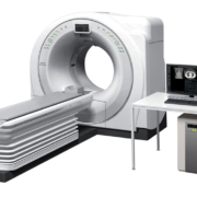
In the modern healthcare environment the demand towards CT goes beyond simple high throughput and accurate diagnosis. Efficient operator workflow, improved patient experience and easy installation into existing facilities are also main considerations. Speedia HD enables high-speed whole-body scanning with sub-millimetre slices, which is difficult to achieve on 16 slice CT systems. A single breath hold (approx. 14sec.), can produce high-resolution images in the range of 1100mm or more. Thereby allowing wide range, high resolution MPR images to be acquired as routine.
Speedia HD with its 40mm width detector and unique 3D reconstruction algorithm-CORE method, achieves the high-speed scan even when using a pitch of 1.58. Therefore, it enables a chest area of 320mm to be scanned in only 4.5sec and a thoraco-abdominal area of 570mm in just 7.5sec. This reduces the burden on the patients who have difficulty maintaining a still position or holding a breath for a long-time.
For more information please click here
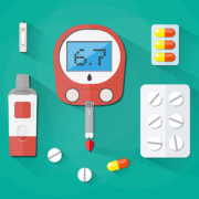
Point-of-care (POC) testing consists of diagnostic tests performed in the physical proximity of a patient, with results obtained on site. One useful definition is from the German Society of Clinical Chemistry and Laboratory Medicine, which explains it as “diagnostic testing at or near the site of patient care, with an easy-to-use instrument, under the immediate health care (e.g. emergency room, operating room, intensive care unit) and not by laboratory personnel.”
Turnaround time, regulations and guidelines
POC tests are a major contrast to their traditional counterparts, which involve taking patient specimens, transporting them to a laboratory and then returning the results to a physician. By eliminating both transport and laboratory, POC tests provide quicker turnaround time. In turn, this enables clinicians to focus on patient care rather than spending critical time on waiting for test results from the lab. This leads to better patient flow to hospitals and clinics, since patients can be diagnosed, triaged and treated earlier. Regulations and guidelines have also encouraged adoption of POC testing. For example, in Germany, certification of a chest pain clinic requires that it be able to perform blood gas analysis within 15 minutes. According to German Society of Cardiology (DGK) guidelines, “the time from blood collection to result documentation may not exceed 45-60 minutes. If this is not possible, a point-of-care test unit on site to determine cardiac markers is mandatory.”
Concerns remain
In spite of such drivers, concerns about reliability and benefits have impeded POC tests from achieving full potential. There are no universally accepted standards on their use or effectiveness. In addition, testing is often performed by personnel without training in clinical laboratory science, or occasionally (in emergency situations) by volunteers. Several POC tests are also conducted by patients themselves, in what is known as self-testing.
One meta-study in 2016, published in the peer-reviewed journal ‘Critical Reviews in Clinical Laboratory Sciences’ echoed such observations. The most prevalent barriers to growth of POC testing, it noted, were associated with the economics of adoption and regulatory issues (such as accreditation), alongside poorly trained staff. Another problem was competition with clinicians who favoured traditional centralized tests. The authors also highlighted the greater cost per POC test when compared to centralized testing, although they acknowledged the existence of difficulties in gauging its cost-effectiveness, given the complexities of making comparisons.
Integrating POC tests into health management strategy
In spite of these limitations, POC tests have yielded measurable improvements in workflow efficiency and patient care, according to numerous studies, some of which go back several years.
Two decades ago, a randomized, controlled trial at a British teaching hospital assessed the impact of POC tests on health management decisions. The results, published in the ‘British Medical Journal’ in 1998, found that physicians using POC tests reached patient management decisions an average of 1 hour and 14 minutes faster than patients evaluated through traditional means.
Indeed, the rapid turnaround time provided by POC tests allows for accelerated identification and classification of patients into high-risk and low-risk groups, leading directly to improvements in quality of care and an increase in clinical throughput.
More recently, studies have shown that POC tests can reduce revenue losses due to workflow delays of test-dependent medical procedures – such as disruptions in magnetic resonance imaging (MRI) or computer tomography (CT) queues.
Overall, POC tests make the maximum impact when they are implemented as part of an overall health management strategy to increase the efficiency of clinical decision-making. The quick availability of test results can provide clinical pathways which directly impact on outcomes.
Decentralization of healthcare
One of the key drivers of growth in POC testing consists of the progressive decentralization of healthcare and patient-centric care. In early 2018, the influential Joint Commission International highlighted these two factors as key to the growth in use of POC testing.
Interest in POC testing and its potential contribution to decentralized healthcare models, however, date back several years. In the late 2000s, ongoing efforts to optimize information and communications technologies and enhance the efficacy of healthcare began to emphasize the impact of diagnostic services at the point of care.
Such processes have accelerated more recently, after the closure of several laboratories and the emergence of new structures such as micro hospitals, community para-medicine systems etc. The pace of such developments has been accelerating in recent years.
Lab closures
In the US, the federal Centers for Medicare and Medicaid Services imposed major reductions in clinical laboratory test fees in 2018, in order to make savings, with further cuts planned in 2019-2022. The impact is expected to be profound on small community lab companies, particularly those servicing nursing homes and those in small, rural hospitals. Some labs, such as Peace Health Labs in Oregon, estimated a cutback in revenues of 20% due to the decision by Centers for Medicare and Medicaid Services, and put themselves up for sale.
Peace Health Labs was bought in late 2017 by Quest Diagnostics. Soon after, Quest began to close some of Peace Health Lab’s facilities and patient service centres in many smaller communities in Oregon and Washington. Other small labs have rationalized, for example by selling business units. Many have simply begun closing.
Such a phenomenon extends to laboratories elsewhere, too. In Britain, labs are being downsized by government programs to consolidate medical diagnostic testing at larger facilities – especially at the regional level, and target lowering costs through economies of scale. Such a process is especially pronounced at hospitals in communities where the scale of local demand is inadequate to support full-service clinical and pathology testing.
In 2006, a British government report known as the Carter Review recommended that clinical pathology labs be run as managed pathology networks. The report noted that the standard District General Hospital delivering a full range of services will become increasingly unsustainable, and impact adversely on both service quality and cost-effectiveness. The impact of the Carter Review can be seen in the recent downsizing of the pathology laboratory at Queen Elizabeth Hospital (QEH), a 480-bed facility located in Norfolk County, about 150 kilometres from London, and an accompanying effort to create a regional pathology network known as the Eastern Pathology Alliance.
Remote settings and POC testing
In spite of its vast and sparsely populated interiors, Australia too has witnessed major cutbacks and consolidation of its pathology laboratory network. Casualties include the pathology laboratories at Maryborough Base Hospital in Queensland, about 250 kilometres from Brisbane, and at Gold Coast Hospital, about 80 kilometres southeast of the same city. The economics of a high-volume laboratory functioning at a large scale ensure that officials will seek to further consolidate clinical laboratory testing in smaller hospitals, so as to make savings via increased economies of scale.
Due to issues of accessibility and distance, remote settings would be considered to be natural demonstrators of the need for POC testing. In March 2015, for example, an article on European POC testing perspectives published by researchers from Sweden, the Netherlands and Britain in ‘The Upsala Journal of Medical Sciences, observed that outside a hospital setting, POC testing “provides laboratory quality services to underserviced areas and general practitioners.”
Hybrid models
One suggested approach to replace the existing community hospital model for rural area is called a hybrid model. It is based on freestanding emergency departments which have links to primary care providers. Such a care model, however, challenges the ability of large, regional clinical laboratories to provide necessary medical laboratory testing to rural freestanding EDs, and requires the presence of small rural labs.
However, such advantages are hardly straightforward.
In Australia, a Government-funded study between 2005 and 2007 investigated POC tests covering a total of 4,968 patients in urban, rural and remote locations. This multi-centre, cluster randomized controlled trial, led by a team from Flinders University Rural Clinical School, Adelaide, sought to determine the safety, clinical effectiveness, cost-effectiveness and satisfaction of the tests.
One of the key findings of the study was that rural and remote practices showed a greater need for training compared to their urban counterparts.
POC testing in EDs and chronic care
Most such macro-processes focus on consolidation at acute care hospitals, with attention traditionally given to emergency departments. Indeed, EDs have become the gateway to unscheduled hospital care – by some estimates accounting for over three of four such admissions in the US.
However, the benefits of POC testing are also evident with individuals suffering from chronic disease, who require regular check-ups to monitor disease progression. In such patients, decisions to change or modify treatment are often directly dependent on clinical testing. POC tests provides healthcare professionals the means to perform and act upon test results during the same office visit.
Such observations have profound resonance in the context of increasingly decentralised medical care.
Compliance and adherence: the contribution of POC testing
Meanwhile, there is increasing evidence about the impact of POC testing beyond convenience to compliance. In November 2009, ‘The Medical Journal of Australia’ found similar or greater levels of self-reported medical adherence in patients undergoing long-term treatment for diabetes or coagulation disorders when POC testing was performed at regular office visits.
Having access to immediate test results through POC is associated with the same or better medication adherence compared with having test results provided by a pathology laboratory. POC tests can provide general practitioners and patients with timely and complete clinical information, facilitating important self-management behaviours such as medication adherence.
In March 2010, ‘The British Journal of General Practice’ published results of a study on POC testing in a general practice setting. The authors sought to determine if patients were more satisfied with point-of-care testing than with pathology laboratory testing for three chronic conditions, namely diabetes, hyperlipidemia and anticoagulant therapy. Their findings conclusively established that patients had significantly higher levels of confidence in physician and more motivation to look after their condition when POC testing was used. Such subjective factors, according to the authors, can translate into quantifiable improvements in disease management.
Lab staff hold key to good training
In a decentralized healthcare setting, the wider acceptance of POC testing will depend on the training of staff in key processes such as sample collection, the calibration, maintenance and use of instruments, documentation and reporting of critical findings. These vary across healthcare settings, and across countries, but this on its own may not be an impediment.
In August 2013, the ‘Journal of Clinical Nursing’ published a study on six different POC test training initiatives for nurses on three continents. One of their most interesting findings was that a key factor for success consisted of the involvement of laboratory staff.


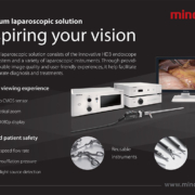
One of the most exciting recent developments in imaging consists of all-optical ultrasound. Unlike traditional ultrasound, which is achieved using piezoelectric transducers, optical systems perform ultrasonic generation via pulsed light. This is followed by optical reception of ultrasonic reflections from the tissue which is being imaged.
Reducing the need for trade-off
Though ultrasound is one of the most common medical imaging tools, conventional devices tend to be bulky and cannot typically be used at the same time as some other imaging technologies. This is why a hybrid combination of optics and ultrasound, coupled to inexpensive fibre-based probes for intravascular imaging, promises to open up new possibilities for medical imaging.
In terms of imaging, optical techniques ensure satisfactory contrast levels, while ultrasound provides high resolution. Optical technologies can also be manipulated to generate low frequency ultrasound which yields greater penetration into tissue, or high frequency ultrasound to obtain higher resolution images, albeit at a shallower depth. In practical terms, such a combination also gives flexibility to physicians in how they use imaging technology to diagnose and treat medical problems. For example, to provide intravascular imaging and detection of conditions like plaque, ultrasound can provide details of morphology while the optical imaging highlights its composition.
Broadband imaging
In technical terms, traditional ultrasound image formation covers a narrow frequency band (usually half the central frequency), while signal generation in optical ultrasound is broadband (covering sub-MHz to several hundred MHz frequencies). In addition, the tomographic principles used by optical ultrasound generally entail data collection over wide angles. This improves image quality and resolution, while minimizing image artifacts.
Efforts to develop broadband all-optical ultrasound transducers date to over a decade. One prototype was developed and tested for high-resolution ultrasound imaging in 2007 at the University of Michigan, Ann Arbor. It consisted of a two-dimensional gold nanostructure on a glass substrate, followed by polydimethylsiloxane plus gold layers. The system achieved a signal-to-noise ratio of a pulse-echo signal of over 10dB in the far field of the transducer, where the centre frequency was 40MHz with −6dB bandwidth of 57MHz. In a paper published in the August 2008 issue of ‘IEEE Transactions on Ultrasonics, Ferroelectrics, and Frequency Control’, the developers of the system concluded that preliminary imaging results “strongly suggest that all-optical ultrasound transducers can be used to build high-frequency arrays for real-time high-resolution ultrasound imaging.”
Innovation not enough to offset drawbacks
As mentioned, the major driver of research into hybrid optical alternatives has consisted of limitations in conventional ultrasound systems. Such drawbacks persist in spite of developments in processing speed, a reduction in noise-to-signal ratios and enhancement in the quality and timing of image capture.
Although innovations such as matrix transducers have enabled the emergence of volumetric ultrasound and 3-D/4-D, elastography has offered physicians the ability to view both stiffer and softer areas inside of tissue.
Elastography uses b-mode ultrasound to measure the mechanical characteristics of tissues, which are then overlaid on the ultrasound image. However, its use in clinical practice remains complicated due to a wide range of techniques used by different manufacturers, alongside differences in parameters used to characterize tissues.
Other areas of innovation include micro-ultrasound, which harnesses ultrasound at microscopic levels and provides 3- to 4-fold improvements in resolution compared to conventional ultrasound. One of the first applications of micro-ultrasound is to allow better targeting of biopsies – for example, in the treatment of prostate cancer by urologists.
Ultrasound-modulated optical tomography
In the late 2000s, ultrasound-modulated optical tomography (UOT) showed considerable promise in imaging of biological soft tissues, with promising application in several areas, including cancer detection. UOT detects ultrasonically modulated light to localize and image subjects. The key limitation of UOT, however, is weak modulated signal strength.
Photoacoustic tomography
Considerable attention has also been given to photoacoustic tomography, which converts absorbed light energy into an acoustic signal. The technique provides compositional information on body tissue in real time without requiring any contrast agents. It also allows much higher depth penetration than conventional optical techniques. Photoacoustic tomography has been used for mapping the deposition of lipids within arterial walls.
Photoacoustic tomography begins by sending pulsed light into tissue, typically from a Q-switched Nd:YAG laser. This creates a slight hike in temperature which causes the tissue to expand, creating an acoustic response which is detected by an ultrasound transducer. The data is then used to visualize the tissue.
However, photoacoustic tomography systems have proved difficult to translate into clinical applications due to their high cost, as well as a relatively large footprint which requires a dedicated optical table to house the laser. On a technical level, moreover, a low pulse repetition rate (in the dozens of hertz) prevents photoacoustic tomography from being used in high frame rate imaging, which are required for clinical applications such as cardiac related problems, where the rate of blood flow is high, or in other similarly fast-moving settings.
There are nevertheless several efforts to cope with the challenges facing photoacoustic tomography. The first is enhancing signal-to-noise ratio and the depth of penetration of optical absorbers. Researchers at Purdue University in the US, who are at the forefront of investigations into the technique, believe that new optical manipulation techniques to maximize photon density might provide a way forward. They have recently announced development of a motorized photoacoustic holder, which allows manoeuvring the aim of the device and fine-tuning the depth to where light is focused. This, they believe, could significantly improve light penetration as well as the signal-to-noise ratio.
Other efforts seek to cope with fast-moving and dynamic settings. In Singapore’s Nanyang Technological University, for example, researchers have demonstrated up to 7,000 Hz photoacoustic imaging in B-mode, using a pulsed laser diode as an excitation source and a clinical ultrasound imaging system to capture and display the photoacoustic images.
All-optical ultrasound
All-optical ultrasound, which has recently catalysed maximum interest, involves using pulsed laser light to generate ultrasound. Scanning mirrors control where the waves are transmitted into tissue. After this, a fibre optic sensor receives the reflected waves, recombines them and creates a visualization of the area being imaged.
Bandwidth, acquisition time and electromagnetic interference
Such a modality exhibits wide bandwidth and satisfactorily addresses one of the major shortcomings of previous efforts at clinical application – namely, prolonged acquisition times (ranging from minutes to hours). Unlike conventional ultrasound imagers which use electronic transducer arrays to transmit sound waves into tissue and receive the reflections for reconstruction as images by a computer, all-optical ultrasound imagers are also immune to electromagnetic interference. As a result, an all-optical ultrasound system can be safely used alongside a magnetic resonance imaging (MRI) scanner, allowing physicians to obtain a more comprehensive picture of tissues around an area of interest, such as a tumour or blood vessel. Immunity from electromagnetic interference and MRI compatibility also means that all-optical ultrasound can be used during brain or fetal surgery, or for guiding epidural needles.
Miniaturization
The absence of electronic components gives yet another advantage, too. Components of conventional ultrasound devices are difficult to miniaturize for internal use. This is due to two factors: a drop in sensitivity after a reduction in the area of the active piezoelectric transducer, and the impact on size of the transducer due to the casing of the piezoelectric element and electrical insulation.
Miniaturization is particularly important in minimally invasive measurements such as medical endoscopy or for inspection of the lumen in non-destructive testing. Small-area detectors are also preferred in tomographic applications, given that detector size inversely correlates to spatial resolution.
Due to difficulties in miniaturization, most ultrasound devices use large, handheld probes placed against the skin. Although some high-resolution probes have been developed, they are considered too expensive for routine clinical use.
Unlike the above limitations for conventional ultrasound devices, the miniaturization of optical detectors (e.g. via interferometric resonators) does not impact on active detection area. In other words, there is no loss of sensitivity. Finally, optical components are not only easily miniaturized but also significantly less expensive to manufacture, compared to compacting electronic ultrasound systems.
First video-rate all-optical ultrasound system
The world’s first all-optical ultrasound system capable of video-rate, real-time imaging of biological tissue has been demonstrated by a research team from University College London (UCL) and Queen Mary University of London (QMUL). It was used to capture the dynamics of a pulsating ex-vivo carotid artery within the beating heart of a pig, and revealed key anatomical structures required to safely perform a transseptal crossing, namely left and right atrial walls, the right atrial appendage and the limbus fossae ovalis.
The researchers believe the new technology will allow ultrasound to be integrated into a wide range of minimally invasive devices in different clinical contexts, and provide ultrasound imaging of new and previously-inaccessible regions of the body. Above all, its real-time imaging capabilities allows differentiation between tissues at significant depths, helping to guide surgeons in some of the highest risk moments of procedures. This will reduce the chances of complications occurring in cases such as cardiac ablation.
Designed for clinical advantage
The new system from UCL and QMUL uses light guided by miniature optical fibres, encased within a customized clinical needle, which generate ultrasonic pulses. Reflections of these ultrasonic pulses from tissue are detected by a sensor on a second optical fibre, in order to provide the real-time imaging.
The developers based their design on a nano-composite optical ultrasound generator coupled to a fibre-optic acoustic receiver with extremely high sensitivity. In turn, harnessing eccentric illumination provided an acoustic source with optimal directivity. This was then scanned with a fast galvo mirror which provided video-rate image acquisition (compared to a time-frame of several hours in previous experiments). It also increased image quality in both 2D and 3D, and made it possible to acquire the images in different modes.
The scanning mirrors in the new system are flexible. They allow for seamless toggling between 2D and 3D imaging as well as a dynamically adjustable trade-off between image resolution and penetration depth. Unlike conventional ultrasound systems, these are achieved without requiring a swap of imaging probe. In a minimally invasive interventional setting, in particular, such probe swapping extends procedure times and introduces risks to the patient.
The technology has been designed upfront by the researchers for use in a clinical setting, with sufficient sensitivity to image moving tissue inside the body at centimetre-scale depth and fit into existing workflow. The researchers are currently working on developing a flexible imaging probe for free-hand operation, as well as miniaturized versions for endoscopic applications.
Prins Hendrikstraat 1
5611HH Eindhoven
The Netherlands
info@interhospi.com
PanGlobal Media IS not responsible for any error or omission that might occur in the electronic display of product or company data.
