
As the threat of a COVID-19 pandemic stares us in the face, it may be opportune to consider some scenarios ahead, especially in light of lessons from other, similar outbreaks in recent decades.
The first problem is sporadic bursts of public concern, alternating with periods of denial. Managing both requires measured doses of reliable information from authoritative sources. Such measurement is a delicate art at the best of times. During a crisis, media hype can mutate it easily into mass misinformation.
Similarly, the threat of terrorism has close parallels with each of the above aspects.
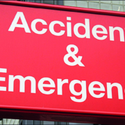
The ICU (intensive care unit) is easily one of a hospital’s highest value resources. A scarcity of intensive care beds means patients require prioritization when demand exceeds supply. As a result, there are frequent delays in admission to an ICU. Though it is accepted that such delays adversely impact patient outcomes, there has been little data on the relationship between bed availability in an ICU and processes of care for patients who develop sudden clinical deterioration – especially in the context of an emergency department (ED). Recently, studies seeking to address this gap have provided renewed momentum to such discussions. They have also dovetailed with other efforts, such as specialist training in critical care for emergency medicine students and residents. However, the area generating maximum interest is a dedicated ICU within an emergency department.
Balancing needs, finding beds
Critically ill patients are commonplace in emergency medicine. They require aggressive and timely care, but emergency medicine clinicians have to balance their needs with those of other patients in their facility. In addition, due to constraints in beds in the ICU, increasing numbers of critically ill patients require to be boarded for prolonged periods of time in the ED. Adding to this problem is a shortage of beds in EDs too.
One of the most vexed questions is whether ED physicians consider bed availability in an ICU as part of their triage decisions, thereby impacting, in a potentially profound manner, on patient outcomes and resource utilization in both the ED and ICU. In effect, does a high availability of ICU beds lead to a bias in admission of patients who are either too well or too ill to benefit ? On the other side, does a low availability then lead to denying admission to ED patients, who would otherwise have been accepted to the ICU?
ED-ICU interface demands attention
In 2013, a study by the George Washington University School of Public Health and Health Sciences in Washington, DC, found that the volume of ICU admissions from EDs in the US had increased sharply, by almost 50 percent, in the period 2001-2009.1 During this period, another study found that the number of ICU beds across the country had increased only 15%, from 67,579 to 77,809.2
In other words, it is clear that ICU admissions from EDs have been increasing at a faster rate than ED visits. The George Washington University study found that though lengths of mean ED and hospital stays had not changed significantly, the mean ICU admission spends over 5 hours in the ED prior to transfer to an ICU bed. As a result, its authors concluded, there was a need for more emphasis on the ED-ICU interface and for critical care delivered in the ED.
Training emergency physicians in the ICU
The roots of this complex combination of challenges go back several decades. One good example is a time-based study, published in 1993 in the peer-reviewed journal ‘Critical Care Medicine’.3 The authors, from Houston, Texas-based Methodist Hospital’s Department of Emergency Services, noted that not only did critically ill patients “constitute an important proportion of emergency department practice”, but also needed treatment in the ED “for significant periods of time.” One of the solutions they proposed was for emergency medicine practitioners to “receive training in the continuing management of critically ill patients.”
The above approach was also witnessed in Europe. In Belgium, for example, an official paper from 1995, titled ‘How to become an intensivist’, proposes that a candidate with an “agreement in Emergency medicine has to make another year of ICU formation.”4
Pathways remained unclear
In subsequent years, there was significant growth in emergency medicine residents pursuing critical care fellowship training, and a reconsideration of the role played by the ED in caring for the critically ill. Nevertheless, there still was a lack of clarity in ways to acquire advanced training in critical care for emergency medicine residents.
In December 2002, an article in ‘Current Opinion in Critical Care’ complained that although ED care for critically ill patients was shown to significantly impact mortality, “formal critical care training for emergency physicians” was still “limited.”5
Less than three years later, another peer-reviewed journal, ‘Annals of Emergency Medicine’, noted that in spite of growing demand for critical care services, most critical care medicine fellowships did not accept emergency medicine residents, “and those who do successfully complete a fellowship do not have access to a US certification examination in critical care medicine.”6 The authors proposed “expansion of the J-1 visa waiver program for foreign medical graduates,” but said the only sensible long-term approach was to strengthen the relationship between emergency medicine and critical care medicine.
Critical care medicine as emergency medicine sub-specialty
In the US, the Accreditation Council for Graduate Medical Education (ACGME) approved critical care medicine as a sub-specialty for emergency medicine physicians in 2011. The following year, the surgical critical care fellowship pathway was approved for emergency physicians interested in becoming board-eligible intensivists.
Currently, the most common training pathways are via combinations of critical care medicine with internal medicine and anaesthesiology, and alongside surgical critical care and neurocritical care. Career pathways for physicians trained in emergency and critical care medicine are also evolving, with options in both community and academic settings.
The role of professional societies
Leading professional societies in emergency medicine and critical care have set up focused sections on the interface between the two areas to stimulate interest as well as provide support to medical students and residents.
Examples from the US include the Emergency Medicine Residents’ Association (EMRA), whose Critical Care Division maintains a comprehensive database of training opportunities across the country,7 and regularly publishes alerts on key developments in critical care. Another interesting initiative is the Coalition for Critical Care Medicine in the Emergency Department (C3MED), which was set up in 2003 and hosts an active email discussion forum.8
Similar efforts have been undertaken by the American College of Emergency Physicians (ACEP),9 the Society of Critical Care Medicine (SCCM),10 the American Association of Emergency Medicine11 and the Society for Academic Emergency Medicine (SAEM).12
In Europe, one of the best-known initiatives to harmonize convergence of the ED and the ICU is ISICEM, the International Symposium on Intensive Care and Emergency Medicine. This non-profit organization, headquartered in Brussels, was set up in 1980. It currently runs a series of eight annual events, covering different aspects of intensive care and emergency medicine. Over the years, participation has grown from about 200 to over 6,000 from more than 100 countries.
Impact of ED on ICU: US and European studies
There have also been concerted efforts to assess the impact of emergency department volume and boarding times on ICU admission and patient outcomes. Two recent studies have catalysed considerable new attention in the topic.
The first is a retrospective cohort study on critically-ill ED patients for whom a consult for medical ICU admission had been requested over a 21-month period. It was published in ‘Critical Care Medicine’ last year by a US-based team from the Icahn School of Medicine at Mount Sinai, New York, and titled ‘Effect of Emergency Department and ICU Occupancy on Admission Decisions and Outcomes for Critically Ill Patients’.
The authors conclude that ICU admission decisions for critically ill ED patients were affected by ICU bed availability. However, higher ED volume and other ICU occupancy did not play a role. They also found that prolonged ED boarding times were associated with worse patient outcomes, suggesting a need for improved throughput and targeted care for patients awaiting ICU admission.
In August 2019, ‘Critical Care Medicine’ published findings online from another study on this topic, this one by a Dutch team from six University Medical Centres at Amsterdam, Groningen, Leiden, Nijmegen, Rotterdam and Utrecht, along with the country’s National Intensive Care Evaluation (NICE) foundation.13 The retrospective observational cohort study conducted a registry analysis of 14,788 patients from the six hospitals, and found an association between emergency department to ICU time greater than 2.4 hours and increased hospital mortality after ICU admission
Ad-hoc and hybrid models
At present, there are two approaches to the challenge of intensive care in the ED. The more common is to have an emergency physician intensivist working standard ED shifts, and lending expertise on an ad-hoc basis to critically ill patients. A recent development is a ‘hybrid’ model. This earmarks a dedicated area of the emergency department for ramping up care to critically ill patients, with a dedicated physician providing intensive care only to such patients, typically for periods longer than an hour.
Supporters of the hybrid model state that it is easier and less expensive to establish with extra costs involving only the dedicated ED-ICU physician.
The ED-ICU
One of the most watched developments in recent years in care for critically ill patients in an ED is the development of ED-ICUs (emergency department intensive care units).
Two such facilities in the US, Stony Brook Resuscitation and Acute Critical Care Unit (RACC) in New York and Emergency Critical Care Center (EC3) in Michigan are considered as being both ED-ICU pioneers and best-of-class references for the concept.
EC3 is considered to be among the world’s most advanced emergency critical care centres. It was opened in February 2015 and has five resuscitation trauma bays and nine patient rooms, located adjacent to the main adult emergency department.
Due to this reputation, the case for ED-ICUs was strengthened after a recent study by EC3 found convincing improvements in survival as well as reduced inpatient ICU admissions.14 In effect, an ED-ICU can improve care and survival rates for the entire emergency department population.15
The EC3 study covered 350,000 ED patient encounters, and found that implementation of an ED-based ICU was associated with significant reductions in risk-adjusted 30-day mortality among patients, from 2.13 to 1.83 percent. The median time to ICU-level care for critically ill patients decreased from 5.3 hours to 3.4 hours, while the hospital ICU admission rate from the ED dropped from 3.2 percent to 2.8 percent.
References
1.https://www.sciencedaily.com/releases/2013/05/130514212946.htm2.https://www.ncbi.nlm.nih.gov/pmc/articles/PMC4351597/3.https://www.ncbi.nlm.nih.gov/pubmed/8319477/4.http://www.siz.be/education/training-in-critical-care/5.https://www.ncbi.nlm.nih.gov/pubmed/124545496.https://pdfs.semanticscholar.org/4f1b/5cea333174e599784e6a2d80c9b55b868b2e.pdf7.https://www.emra.org/fellowships/critical-care-fellowships/8.c3med@yahoogroups.com9.https://www.acep.org/criticalcaresection/10.http://www.sccm.org/Member-Center/Sections/Pages/Emergency-Medicine.aspx11.http://www.aaem.org/membership/critical-care-section12.https://community.saem.org/communities/community-home?CommunityKey=5dc206d8-d248-4f71-aecd-e0490cdc3ba913.https://insights.ovid.com/pubmed?pmid=3139332114.https://jamanetwork.com/journals/jamanetworkopen/fullarticle/273862515.https://medicalxpress.com/news/2019-07-department-based-intensive-patient-survival.html
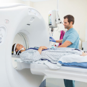
Since its introduction in 1973, X-ray computed tomography (CT) has become a leading modality for diagnostic imaging. The advantages of CT are manifold. Above all, they include rapid scanning and small spatial resolution, which allows for relatively quick and accurate diagnosis of injuries and disease. CT has also been an imaging tool of choice for the staging and treatment follow-up of cancer.
Growth in use, but variations between countries
Overall, CT use has grown rapidly. The total number of scans in the US is estimated to be in the region of 80 million a year. In England, the National Health Service (NHS) reported 4.8 million CT scans in 2016/17, which is 40 percent more than the 3.4 million MRI scans done during that year. CT usage has also been growing rapidly – in England at about 8% annually, compared to just 1.5% for X-rays and 5% for ultrasound.
Nevertheless, there are significant variations between countries in the intensity of CT use. According to data from the Paris-based Organization for Economic Cooperation and Development (OECD), the annual rate of CT scans per 1,000 inhabitants ranges from a high of 225-230 in the US and Japan, to a low of 37 in Finland. The rate is about 80 in Italy, 90 in the Netherlands, 110 in Spain, 140 in Germany and 200 in Belgium and France.
Differences in radiation dosing practice
Though large, such divergences are considered to be less significant than differences in radiation dose to which patients are exposed, for the same condition. In December 2007, a study published in ‘European Radiology Supplement’ had found dosage could have been halved in many cases without impacting on image quality. Another study two years later revealed a 13-fold difference between the lowest and highest radiation doses used for identical CT procedures by four clinical sites in the neighbourhood of San Francisco.
Concerns about such issues have been dramatically highlighted after a major new international study, which attributes differences in dosage to the person doing the scanning rather than to patients or equipment. The study, published in ‘The British Medical Journal’ (BMJ) in January 2019, found that patient characteristics, make and model of scanner, and type of hospital where the CT scan was done had little effect on the amount of radiation used.
Analysis of 2 million CT scans in 151 institutions
The BMJ study was based on a massive effort by a research team led by Dr. Rebecca Smith-Bindman, a professor in the Department of Radiology and Biomedical Imaging at the University of California San Francisco (UCSF). The researchers analysed dose data for over 2 million CT scans of the abdomen, chest and head, at 151 institutions in seven countries.
Their findings are likely to resonate strongly, given the association of radiation with cancer. Although CT scans account for a minority of diagnostic radiologic procedures, they use large amounts of radiation per image. Some estimates suggest that CT contributes nearly half the US population’s radiation dose from all medical examinations. The figure in England is higher, at 68 percent, although plain radiography is used five times more often than CT in the country (22.9 million procedures in 2016/17 versus 4.8 million).
Cancer risks of CT
The association with cancer has been controversial, especially when predictions of the impact of CT scanning have been based on a linear-no-threshold dose-response model. Some have argued that CT radiation doses are too low to produce any health effect.
There is also uncertainty about how to calculate risk accurately. This is because of a host of factors. Firstly, radiologists are not necessarily familiar with CT radiation exposure descriptors (volume CT dose index and dose length product). Secondly, there have been a series of revisions about the relative sensitivity of organs to radiation. Finally, radiation dose in units such as millisieverts (mSv) are used to estimate population risks based on generic models, not individual patient calculated dose. Indeed, the radiation dose in a typical CT scan (1–14 mSv depending on the exam) is similar to the annual dose received from natural sources, such as radon and cosmic radiation – which typically varies from 1 to 10 mSv, depending on where a person lives.
Even small risks justify search for solutions
Nevertheless, the current consensus is that, even if the risk of cancer from CT imaging is small, the economic burden of treatment of the proportionately reduced number may well be significant, given the high prices of cancer treatment.
Neither does anyone question the logic of attacking even a small cancer risk. In December 2009, a report in ‘The Archives of Internal Medicine’ made a detailed assessment of projected cancer risks due to CT scans in the US. The study was conducted by a team from the Radiation Epidemiology Branch of the National Cancer Institute (NCI), and argued that changes in practice might help to avoid the possibility of reaching an attributable risk of 29,000 cancer cases based on CT scans in the year 2007. The authors also observed that the impact would be largest in abdomen, pelvis and chest CT scans in adults aged 35 to 54 years.
Unnecessary scans
One of the most vexatious issues concerns CT scans which are not medically necessary, especially when it concerns repeat imaging of a particular patient – and the ensuing enhancement of cancer risk. According to one estimate, unnecessary scans could account for as much as 30 percent of CTs in the US. In Europe, such a figure is also likely to be high in countries such as Belgium and France where per capita CT scan levels are close to those of the US.
Though the US state of California has passed a law requiring documentation in a patient’s medical record of radiation dose used for every CT scan, compliance has been inconsistent. Perspectives in Europe are problematic too. For example, the European Union collects dose levels in Europe, but there are major differences in definitions and data collection techniques.
Progress in pediatric dosing
Until the NCI study at the end of 2009, the emphasis on reducing CT cancer risks had largely been on pediatric scans. The authors of that paper noted there was evidence of pediatric doses being reduced as a result of social marketing campaigns such as Image Gently. The latter was launched in 2008 by the Alliance for Radiation Safety in Pediatric Imaging.
Lessons from the pediatric dose control campaign
One of the key recommendations of Image Gently was to promote standardization of pediatric dose measurements and display across vendor equipment.
This is precisely what the recent BMJ study proposes to do for all patients. The authors of the study assessed mean effective doses and proportions of high dose examinations (defined as CT scans with doses above the 75th percentile defined during a baseline period) for abdomen, chest, combined chest and abdomen, and head CT. These were classified by patient characteristics (sex, age, and size), type of institution (trauma centre, 24×7 care provision, academic and private hospital), practice volumes, machine manufacturer and model, country etc. The figures were adjusted for patient characteristics, using hierarchical linear and logistic regression.
For example, after taking into account patient factors, a fourfold range in radiation doses still existed in abdominal scans. Similar variations were found for chest and combined chest-and-abdomen scans.
Huge variations in dose
The BMJ study found that variations in radiation dose across institutions and countries were huge. For abdomen CT examinations, the mean effective radiation dose differed by a factor of four, with a 17-fold range in the share of high dose examinations (4 to 69%). Variations in mean effective dose for chest scans and combined chest plus abdomen scans were also close to four times, while the share of high dose exams varied from 1 to 26%, and 2 to 78%, respectively. For head CT, the differences were less spectacular (with the range of mean effective doses less than 1.5 times and the share of high dose exams ranging from 8 to 27%.
Achievable and universal standards
However, when the UCSF group adjusted for technical parameters, that is, in terms of the way CT scanners were used by medical staff, the variations in doses nearly disappeared.
The researchers conclude that it is possible to optimize doses to a “single set of achievable quality standards” and apply this “to all hospitals and imaging facilities.” They also noted that the choice of “appropriate CT protocol parameters might be less complex than widely believed.” The key to protocol optimization lies in updating physician awareness and recalibrating expectations about what constitutes a diagnostic CT scan. The latter will be based on a better alignment of CT protocol parameter choices with diagnostic image quality requirements.
One interesting finding was that institutions with lower average doses shared scanning approaches. These institutions tended to limit the number of protocols, with each relying on the minimum dose required to answer the clinical question. They used multiple CT scanning infrequently, had lower settings for tube current and tube potential, and used higher pitch for most, if not all, imaging indications.
The way ahead
The road to CT dose reduction and standardization will vary by type of institution and country. This is due to differences in the make and model of CT scanners as well as medical cultures, in terms of radiologist preferences and personnel support. There are case studies of protocol overhauls taking a year or more, and needing to be kept up-to-date with new CT software and scanner upgrades. Examinations with higher radiation exposure generally give more acceptable images than those where exposure is lower. The challenge is to optimize a ‘correct’ minimum dose for different patient sizes, ages and conditions. Continuing improvements in scanning technology will undoubtedly also be part of the process of optimizing protocols. On their side, some companies have been experimenting with artificial intelligence algorithms to position patients correctly in a CT scanner. Off-centre CT scans can expose patients to much higher levels of radiation
than necessary.




Prins Hendrikstraat 1
5611HH Eindhoven
The Netherlands
info@interhospi.com
PanGlobal Media IS not responsible for any error or omission that might occur in the electronic display of product or company data.