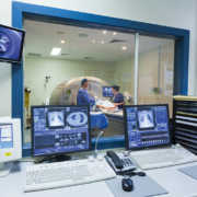
Functional MRI – opening new frontiers in the brain
Functional magnetic resonance imaging (fMRI) is by far the principal method used to investigate the brain’s cortical areas and subcortical structures. fMRI has dramatically transformed perceptions of the human brain, allowing precise delineation of regions associated with a vast range of external stimuli and moods – ranging from depression and anger to laughter and play.
Researchers are now exploring further expansion in the scope of fMRI. These range from the development of more precise sensors and probes with quicker response times to the use of fMRI in new applications such as artificial intelligence. Some have even sought to extract images seen by viewers directly out of their brains.
From dog language to crocodile music
Some have also sought to see if fMRI can work in other species of living things.
In 2016, scientists in Hungary concluded that dogs can understand the meaning and tone of human speech, and that they process language in the same way humans do. To reach this conclusion, they managed to get 13 pet dogs to lie completely motionless in an fMRI scanner for eight minutes while wearing earphones and a radio-frequency coil on their heads.
Earlier this year, a team at Germany’s Ruhr-University in Bochum went further than canines by using fMRI to study the brain of a Nile crocodile as it heard complex sounds, including classical music by Bach.
The eye sees, the brain predicts vision
Given the increasing number of ultra-high field systems available worldwide, experts expect a dramatic impact on our understanding of the brain due to sustained enhancements in resolution (both spatial and temporal), as well as in sensitivity and specificity.
Early this year, researchers from the University of Glasgow published results of a fMRI-based experiment to confirm the capability of the visual cortex to make predictions about what a viewer would see next. The study sought some answers to a seemingly perplexing question. Human beings move their eyes approximately four times per second, requiring their brains to process new visual data every 250 milliseconds. In spite of such rapid and constant variation in perspective and image, how is it that the world remains stable ?
The functional MRI used by the Glasgow researchers showed that the brain rapidly adjusts its predictions, with the visual cortex feeding back updates to a new predicted coordinate every time the eyes move.
The Glasgow study established the importance of fMRI in new frontiers of neuroscience research. fMRI is now seen as a means to contribute to research into mental illness as well as help the development of artificial intelligence. Indeed, a better understanding of the predictive mechanism in the human brain may directly lead to breakthroughs in brain-inspired artificial intelligence in the future – especially in terms of visual predictive capabilities.
The role of calcium ions in brain activity
Beyond such frontiers, MRI technology is also undergoing other forms of evolution. Some of these, which involve new sensors and pathways to monitor neural activity deep within the brain, are not just path-breaking but also offer the possibility of profound new insights into understanding how human beings think.
One of the most exciting developments in such a context involves the tracking of calcium ions, which are closely correlated to neuronal firing and brain signalling. MRI typically detects changes in blood flow, and its utility derives from the fact that when a region of the brain is in use and neuronal activation ensues, blood flow to that region also increases. However, such a process provides only indirect clues; the signals are difficult to attribute to a specific underlying cause. By contrast, sensing based on calcium ions may allow linkage of neuron activity patterns to specific brain functions, and thereby enable researchers to understand how different parts of the brain intercommunicate during particular tasks.
Indeed, it has been several years since neuroscientists know that calcium ions rush into a cell after a neuron fires an electrical impulse, and have used fluorescent molecules to label calcium and then image it via traditional microscopy. Though the technique has allowed for precisely tracking neuron activity, its practical use has been limited to small regions of the brain.
MIT designs calcium detecting molecular probe
At the Massachusetts Institute of Technology (MIT), researchers have sought a way to image calcium using MRI, in order to allow for the analysis of much larger volumes of brain tissue than was possible by fluorescent labelling. To do this, the MIT researchers designed a new molecular probe whose architecture can detect subtle changes in calcium concentrations outside of cells and respond in a way that can be tracked with MRI. Such a process allows for direct correlation to neural activity deep within the part of the brain known as the striatum.
Tests in rats enabled the MIT researchers to establish that calcium sensors accurately detect changes in neural activity from electrical or chemical stimulation. The levels of extracellular calcium correlate with low neuron activity. In other words, when calcium concentrations drop, neurons in the area are firing electrical impulses.
The goal of the researchers is to greatly enhance precision in mapping neural activity patterns. By measuring activity in different regions of the brain, they hope to find how different types of sensory stimuli are encoded by the spatial pattern of neural activity which is induced.
The MIT probe essentially consists of a sensor made up of two kinds of particles which bind in the presence of calcium. The first is synaptotagmin, a naturally occurring calcium-binding protein, and the other a lipid-coated magnetic iron oxide nanoparticle which binds to synaptotagmin, but does this only if calcium is present. Calcium binding leads to the particles clumping together, and appearing darker in the MRI image.
The researchers are now attempting to increase the speed of response by the sensor, which currently requires a few seconds after the stimulation. A more important goal is to modify the sensor such that it can pass through the blood-brain barrier. This would enable the delivery of the particles without the need to inject them directly in the test site, as is required at present.
Research into new sensors and neurochemical pathways, as being done at MIT, will no doubt open new vistas in fMRI. However, other efforts too are expected to greatly enhance the range and spectrum of its applications.
Powering up fMRI machines
In May 2013, the European Journal of Radiology published results of a study comparing fMRI at 7T compared to 3T in imaging of the amygdala, a ventral brain region of specific importance to psychiatry and psychology. Traditionally, MRI imaging of such areas is prone to signal losses along susceptibility borders – alongside signal fluctuations due to physiological artifacts from respiration and cardiac action. The increase from 3T to 7T showed a significant gain in percental signal change and demonstrated the potential benefits of ultra-high field fMRI in ventral brain areas.
UC Berkeley targets massive resolution boost in fMRI
More recent efforts are also aimed at enhancing resolution. Today’s top-of-the line scanners, incorporating 10T magnets, can typically localize activity within a region comprising 100,000 neurons or more, about the size of a grain of rice. To be able to concentrate more finely, on smaller groups of neurons, requires a bottom-up re-design of almost the entire gamut of scanner components and sub-systems.
The University of California at Berkeley is currently targeting a 20-fold boost in fMRI resolution in order to provide the most detailed images of the brain ever seen. The project is funded by a BRAIN Initiative grant from the National Institutes of Health.
New approach to fMRI design and architecture
The leap in resolution will be directly due to innovations in hardware design, scanner control and image computation. Currently, spatial resolution of fMRI recordings is based on variations in the magnetic field as well as, indirectly, on the size of detector. The latter consist of coils of wire, which are arrayed around the head of a subject and pick up signals. The Berkeley system uses a far larger number of smaller coils than clinical MRIs, which use smaller numbers of large coils. The result is straightforward – a much higher resolution of the brain’s outer surface, which is needed to identify key layers of the cortex.
Reducing dimensions in such ultra-high resolution MRI holds the key to image the brain in functional regions, where neurons are all essentially involved in the same type of processing. The target which researchers hope to reach is in the range of 0.4 millimetres This is because the cerebral cortex, the brain’s outer layer, consists of columns of neurons which correspond to a specific sensory feature (such as the vertical rather than horizontal edge of an object) and such columns are 0.4 millimetres on the side and 2 millimetres long. The Berkeley researchers are reported to be confident of their ability to build machines which can scan down to the 0.4 millimetre target by 2019.
Peering into the brain’s depths
If successful, the new fMRIs would allow researchers to study cortical microcircuits and glimpse the deepest recesses of human brain function so far. The developers of the system are ambitious. They aim to provide “the most advanced view yet of how properties of the mind, such as perception, memory and consciousness, emerge from brain operations.” This will open ways to observe disturbances in brain structures and functions, and it is hoped, radically enhance the diagnosis and understanding of neurological diseases.
Extracting images out of the brain
One of the most far-reaching possibilities of fMRI was recently announced by a team from the Japan’s Kyoto University, who used machine-learning and artificial intelligence to translate brain activity into images in test subjects.
These ranged from pictures being looked at by the subjects, to things they remembered seeing. The images included a lion, a fly, a DVD player, a postbox, alphabets and geometric shapes, and were recreated pixel by pixel, based on a deep neural network (DNN).
The images were projected on to a screen in an fMRI scanner, with the heads of subjects secured in place via a bar on which they had to bite down. The subjects, who participated in multiple scanning sessions for a period of more than 10 months, stared at each image for several seconds before taking a rest. After this, they had to recall one of the images seen previously and picture it in their mind.
The DNN was then used to decode the signals recorded by the fMRI scanner and produce a computer-generated reconstructed image of what the participants saw.


