Beukenlaan 137
5616 VD Eindhoven
The Netherlands
+31 85064 55 82
info@interhospi.com
PanGlobal Media IS not responsible for any error or omission that might occur in the electronic display of product or company data.
Sepsis is a potentially fatal condition after the immune system over-reacts to an infection, leading to shock and organ failure. It is most frequently provoked by commonplace bacteria. Globally, over 30 million people develop sepsis each year. Some 6 million die as a result. Although there has been progress in controlling deaths from sepsis in recent decades, the challenge is still a major one. Indeed, in industrialized countries, the incidence of sepsis is higher than that of new cases of cancer.
Unpredictable and terrifying
Sepsis is frequently encountered in a hospital setting. It is also a leading cause for hospital readmission. In the US, studies estimate that one of 3 people who die in hospital have sepsis.
One of the biggest challenges for clinicians is that sepsis occurs unpredictably and progresses at terrifying speed. This makes timely diagnosis a tough call.
Definitions of sepsis have also tended to vary. In 2018, a working group of 19 specialists, convened by the Society of Critical Care Medicine in the US and the European Society of Intensive Care Medicine (ESICM), updated the clinical definitions and criteria for sepsis and septic shock. The taskforce recommended defining sepsis as “life- threatening organ dysfunction caused by an inappropriate host response to infection.” It also concluded that the term ‘ severe sepsis ‘ was redundant.
Rory’s Regulations
Nevertheless, there has been some progress in recent years in understanding sepsis and standardizing approaches to diagnose, manage and treat the condition.
In 2012, Rory Staunton, a healthy 12-year-old from New York, died due to the fact that his sepsis was not diagnosed. In the wake of this, the government of New York State mandated all hospitals to comply with protocols to improve the early diagnosis and treatment of sepsis and septic shock, and made it compulsory for reporting all sepsis cases to the Department of Health.
The New York Sepsis Initiative, which the media called Rory’s Regulations after the young victim, essentially consist of two treatment bundles.
The first is a 3-hour bundle, and is indicated for patients with severe sepsis and needs to be activated within three hours of a patient’s arrival at hospital. It includes blood culturing to determine choice of antibiotics, starting antibiotic treatment and assessing blood lactate levels – an important marker for sepsis.
The second, 6-hour bundle, is earmarked for patients with septic shock and needs to be carried out within six hours of their arrival at hospital. It includes administration of intravenous fluid, vasopressors to contract blood vessels and a follow-up check on lactate levels.
Assessing the New York Sepsis Initiative
The New York Sepsis Initiative was assessed earlier this year by a team from Warren Alpert Medical School at Brown University. They studied data from 91,357 patients, treated over a period of 27 months at 183 hospitals.
The findings were encouraging. The two sepsis bundles were used in 81.3 percent of patients. After implementation of the protocols, compliance steadily increased across hospitals in the State. The study’s most important finding, however, was that patients administered the bundles saw a reduction in mortality risk over 4 percentage points, at 24.4 percent. The mortality risk in those who did not receive the bundles was 28.8 percent. In addition, hospitals complying with the protocols saw a significant reduction in average length of stay.
Limits to fighting sepsis
While the New York State initiative provides strong evidence of the potential for standardizing sepsis-fighting measures, another study this year shows there may be limits to its scope. The study, by researchers from Brigham and Women’s Hospital in Massachusetts, was published in March by ‘JAMA Network Open’. It sought to investigate the precise role of sepsis in hospital deaths and estimate how many were preventable.
The researchers studied records of 568 people from six acute care hospitals for the years 2014 and 2015, who had died in the hospital or after discharge to hospice care. Using a 6-point Likert scale, ranging from “definitely preventable” to “definitely not preventable,” they concluded that some 90 percent of deaths were not preventable in a hospital setting. On the other side, 1 in 8 sepsis-related deaths were deemed “potentially preventable with better hospital-based care.”
The key reason for such a prognosis was that most sepsis fatalities occur in medically complex, older patients with severe co-morbidities, including chronic conditions such as cancer, heart and lung disease. In the few cases of death due to sub-optimal care, the most common causes included late antibiotic administration.
The lead author of the study, Dr. Chanu Rhee, called for more “innovation in the prevention of underlying conditions” to reduce sepsis mortality by a significant margin.
Long term decline in sepsis death rates
Although the challenge of sepsis remains serious, there has been significant progress over recent decades. In October 2018, the annual meeting of ESICM (the European Society of Intensive Care Medicine) was presented with an analysis of 30-year trends in sepsis deaths. Using World Health Organization figures, researchers from Harvard Medical School and Imperial College London (ICL) found that the average death rate from sepsis in Europe, North America and Australasia fell from 36.2 per 100,000 men in 1985 to 27.1 in 2015, and for women, from 23.2 per 100,000 women to 19.6.
Countries which managed to reduce death rates most significantly were Finland, Iceland and Ireland, while increased rates were noted in both Denmark and Lithuania.
Prospects for managing and treating sepsis in future years is likely to improve due to several new weapons, ranging from targeted drug development to artificial intelligence. Growing interest in this field is indicated by more than 200 sepsis biomarkers approved by the US Food and Drug Administration (FDA), among them interleukins, C-reactive protein and procalcitonin.
MIT’s IL-6 sensor system
It is known that interleukin-6 (IL-6), a protein produced in response to inflammation, begins to increase a few hours prior to other sepsis symptoms. IL-6 levels have not been strong enough to be detected by traditional tests. However, new sensor technologies appear to offer promise.
Researchers at the Massachussets Institute of Technology (MIT) have developed a small microfluidic sensor which can reportedly detect sepsis in a small blood sample (such as that obtained from a finger prick) within 25 minutes. The system uses antibody-laced magnetic microbeads in one fluid channel, which mixes with the blood sample and identifies the IL-6 biomarker. Meanwhile, another channel attaches the biomarked beads to an electrode. When a current is run through the electrode, a signal is produced each time an IL-6 bead passes through.
The magnetic detection system is far less expensive than the high-end optics required by conventional assays, and requires far less blood. The MIT researchers state that they will eventually be able to detect minute increases in IL-6 during the test itself. They are now continuing work on researching other proteins which act as early markers for sepsis detection and would reinforce diagnostic accuracy.
Early warning sepsis indicator
A new hematological biomarker, introduced in 2018 by Beckman Coulter as the Early Sepsis Indicator, is reported as part of a routine complete blood count (CBC) and measures morphological changes in monocytes, cells which play a role in the dysregulated immune response to sepsis. A positive result alerts clinicians to a higher probability of sepsis at an early stage
Thermography tools
Another novel diagnostic technique is based on the fact that abnormal body temperature patterns accompany the earliest stages of sepsis. University of Missouri researchers have proposed using infrared thermography to measure the difference between body extremities and a patient’s core temperature. The team have developed an automatic real-time system which calculates this, based on a frontal and lateral infrared thermogram of the face. Writing in a recent edition of the ‘International Journal of Data Mining and Bioinformatics’, they state the system works successfully, irrespective of the angle of the head relative to the imager and differences in backgrounds.
Targeting enzymes
Other efforts involve new drugs. One priority consists of signalling pathways which control immune cell behaviour during sepsis. So far, most research on inflammation has focused on kinases, the enzymes which transfer phosphate groups to specific substrates.
In August 2019, researchers from the University of California San Diego (UCSD) School of Medicine discovered a wholly new target area – the enzymes which remove them. In particular, they focused on PHLPP1, an enzyme which impacts upon inflammation by removing phosphates from the transcription factor known as STAT1, which controls inflammatory genes.
Using a mouse model, the researchers administered live E. coli bacteria and lipopolysaccharide (LPS), to both PHLPP1-deficient and normal mice. They found that the former fared far better, with half surviving infection-induced sepsis after 5 days – compared to zero for normal mice. The UCSD researchers believe that inhibiting PHLPP1 might form the basis for new sepsis treatments in humans, offering the means to control the dangerous inflammation of sepsis while maintaining the critical bactericidal properties of white blood cells.
Non-antibiotic drugs against sepsis
Researchers at the Royal College of Surgeons in Ireland (RCSI) have tested a compound called cilengitide (brandname InnovoSep) in a preclinical trial. A key feature of InnovoSep is that it is not an antibiotic, and does not face the limitations associated with the latter – namely, the need for rapid identification of causative bacteria and growing resistance to antibiotics.
Cilengitide is an antagonist of alpha-v beta-3, the key endothelial cell integrin which mediates the adhesion of cells to the extracellular matrix. In everyday terms, the drug prevents bacteria “from getting into the bloodstream from the site of infection by stabilizing the blood vessels so that they cannot leak bacteria and infect the major organs,” according to Steven Kerrigan of the RCSI.
Artificial intelligence
To some, however, artificial intelligence (AI) is seen as potentially the most exciting frontier in the fight against sepsis. In 2018, the journal ‘Nature Medicine’ featured an AI system developed by scientists at Imperial College London, which proved to be more reliable predicting the best treatment for sepsis, as compared to human doctors. This was after it had ‘learned’ from an analysis of 100,000 patient records and clinical decisions in intensive care units about sepsis over a 15-year period.
Another promising AI system against sepsis has been developed by Sentara Healthcare in the US. Sentara’s sepsis prediction tool is based on identifying at-risk patients by using an algorithm to spot patterns from some 4,500 pieces of data in an electronic record. These focus on metrics such as body temperature, heart rate, blood tests, gender, medical history, etc.. Sentara had previously developed a ‘sepsis sniffer’ which detected when a patient had just begun to have sepsis. The current system goes further, and does not wait until a patient has already developed the disease.
While everyone in the healthcare industry agrees that early detection of breast cancer saves lives, much less consensus can be found across the broader conversation of breast cancer screening in general. This inconsistency is especially apparent as it pertains to breast density, an issue that carries significant weight for both clinicians and patients. It is necessary for radiologists to not only acknowledge and understand how breast density impacts screening in general, but also to recognize the discrepancies in today’s breast density protocols, best practices for handling them and how this can affect clinicians and patients.
by Tracy Accardi
To start, consider the way a patient’s breast density is currently assessed. Most commonly, radiologists complete a visual assessment, which involves looking at digital images of the patient’s breasts and determining which of the categories her tissue fits into best according to a classification system known as the Breast Imaging Reporting and Data System (BI-RADS). There are four classifications to establish breast density type, which include – from least to most dense – fatty, scattered fibroglandular, heterogeneously dense, and extremely dense. Although the four categories help establish what radiologists should be looking for visually to determine if a woman has dense breasts, each radiologist’s individual perceptions are open to interpretation, potentially leading to inconsistencies in classification. As a result, some women may be misinformed about what their breast density is, which can be problematic considering breast density has long been recognized as a risk factor for cancer. In fact, women with very dense breasts are four to five times more likely to develop breast cancer than women with less dense breasts [1,2].
Screening protocol for dense breast patients
Once a woman’s breast density is classified, there is a good deal of debate regarding next steps for breast cancer screening. In fact, in a 2017 Kadence study, only 32 percent of the surveyed radiologists in Europe indicated they have a formal screening protocol in place for patients with dense breastS [3]. There are a number of modalities radiologists can choose to utilize when screening women for breast cancer, however, very dense breasts are challenging to read, particularly when using traditional 2D mammography. This is because suspicious calcifications appear white on a mammogram, blending in with dense breast tissue that is similar in colouring that is also known as a “masking effect.” Therefore, the imaging modality used to screen patients, especially those with dense breasts, truly matters. In the U.S., for example, Hologic’s 3D Mammography Exam is the only mammogram that is FDA-approved as superior to standard 2D mammography for routine breast cancer screening of all women, including those with dense breasts [4]. Despite this, there are no official guidelines that radiologists are encouraged to follow when screening their patients with dense breasts. As a result, patients may be missing the opportunity to receive a breast cancer diagnosis earlier on so they can start treatment right away because they weren’t screened with the most appropriate technology.
Clearly, there are many ways that clinicians across the world are currently approaching breast density protocols, especially as they pertain to assessment and screening. These inconsistencies are creating confusion among clinicians and patients alike. Fortunately, there are a number of solutions for this issue. When assessing density, radiologists should consider technology available to them to help remove subjectivity from their evaluations. In fact, clinicians can combine their patient-specific knowledge with artificial intelligence (AI), which—thanks to machine learning-based algorithms—can be used to classify breast tissue within the BI-RADS category, allowing for objective, accurate assessments. As a result, women can and should be better informed about what their breast density truly is, which may help those who didn’t realize they were at risk for cancer to be more compliant with screenings. Additionally, radiologists and their facilities should offer their patients the best possible technology that exists for screening dense breasts, pending they have no extenuating limitations based on their individual patient profiles.
Healthcare professionals owe it to their patients to find solutions that provide the best possible outcomes. By making breast density and the inconsistencies surrounding it a priority for reconciliation, radiologists can best deliver care to their patients.
References
1. Boyd NF, Guo H, Martin LJ, et al. Mammographic density and the risk and detection of breast cancer. N Engl J Med. 356(3):227-36, 2007.
2. Yaghjyan L, Colditz GA, Collins LC, et al. Mammographic breast density and subsequent risk of breast cancer in postmenopausal women according to tumor characteristics. J Natl Cancer Inst. 103(15):1179-89, 2011.
3. Kadence study conducted in partnership with Hologic in 2017. Data on file.
4. FDA submissions P080003, P080003/S001, P080003/S004, P080003/S005.
The author
Tracy Accardi, Global Vice President of Research & Development for Breast & Skeletal Solutions at Hologic, Inc.
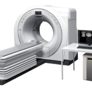
In the modern healthcare environment the demand towards CT goes beyond simple high throughput and accurate diagnosis. Efficient operator workflow, improved patient experience and easy installation into existing facilities are also main considerations. Speedia HD enables high-speed whole-body scanning with sub-millimetre slices, which is difficult to achieve on 16 slice CT systems. A single breath hold (approx. 14sec.), can produce high-resolution images in the range of 1100mm or more. Thereby allowing wide range, high resolution MPR images to be acquired as routine.
Speedia HD with its 40mm width detector and unique 3D reconstruction algorithm-CORE method, achieves the high-speed scan even when using a pitch of 1.58. Therefore, it enables a chest area of 320mm to be scanned in only 4.5sec and a thoraco-abdominal area of 570mm in just 7.5sec. This reduces the burden on the patients who have difficulty maintaining a still position or holding a breath for a long-time.
For more information please click here
The term “artificial intelligence”, defined in the Oxford Dictionary as “the capacity of a machine to simulate or surpass intelligent human behaviour”, was first used in the 1950s, but the idea that the human brain, or at least parts of it, could somehow be ‘copied’ or ‘imitated’ using a series of mechanical networks has been part of myth and literature for centuries, as far back as, for example, the automatons of Hephaestus and Daedalus.
by Prof Jean-Louis Vincent
Artificial intelligence is an umbrella term that covers multiple tools and technologies. In recent years, huge advances in machine learning algorithms, neural networks, deep learning, facial recognition, computing power and statistical techniques have increased the potential applications of artificial intelligence across multiple aspects of everyday life. In terms of medicine, artificial intelligence is already beginning to find a place in disease prediction and diagnosis, clinical decision support and therapeutic guidance, prognostication, and remote follow-up and monitoring.
So, what do these new developments mean for the medical profession – is this ever advancing technology going to decrease the need for doctors? To some extent, the answer is of course yes, perhaps particularly in the field of diagnostics. Using machine learning, computerized systems are able to study and develop pattern recognition from millions of test results over a very short period of time, whereas a doctor can only see and process a limited number of events throughout a whole professional career. The vast amounts of data becoming available as increasing numbers of patients have electronic health records and are managed with electronic monitoring systems are providing the input needed for artificial intelligence to develop, while also reducing the administrative load on doctors and other healthcare staff, a common source of complaint. Machine-based interpretation of visual data is now more accurate, effective, and rapid than the human eye and associated with less risk of error. Interpretation of X-rays and other medical images will become increasingly automated, reducing the need for radiologists to perform this role. Similarly, electrocardiograms are already read automatically; many of the reports are still validated by a cardiologist, but is this really necessary? In ophthalmology, examination of the fundus for many diagnoses, including diabetic retinopathy and macular degeneration, can be performed more accurately by computerized algorithms than by most experienced ophthalmologists. Application of deep learning to a database of eye CT-scans at a London hospital has enabled more than 50 ocular diseases to be recognized by the system. Non-image-based diagnosis is also possible. A pilot system developed in California that applied machine learning to electronic health record data has been used to diagnose common pediatric problems and shown to perform better than junior doctors.
Although artificial intelligence may therefore indeed reduce the need for doctors in some areas, this is not necessarily a bad thing; it should not be seen as the enemy, but rather as a means of improving medical practice and patient care. More rapid, accurate diagnosis can only be positive. Another important example of how artificial intelligence can be beneficial is in predictive medicine. Natural language processing of data from electronic medical records was able to more accurately predict postoperative complications in patients undergoing major surgery than were patient safety indicators. Machine learning-based sepsis prediction has been associated with reduced mortality rates. Indeed, early identification of patients with an increased risk of sepsis raises awareness and facilitates early investigation and management by sepsis-trained physicians in a condition where rapid diagnosis and treatment are crucial to maximize chances of survival. More broadly, identification of patients at risk of deterioration on the general floor or after surgery will enable appropriate monitoring systems to be offered to a selection of patients in an environment where it is not feasible, financially or practically, to monitor every hospitalized patient.
Another example is the field of therapeutics. Machine-learning can identity the most effective therapeutic algorithms for individual patients, with the most appropriate evaluation and follow-up. These algorithms are being incorporated into closed-loop systems enabling appropriate treatments to be administered and doses adjusted in real-time for individual patients according to continual personal data provision. Just some of many examples include implantable cardioversion defibrillators, diabetes management with implantable glucose monitors and insulin pumps, and use of vasopressors to prevent hypotension during surgery.
Artificial intelligence can thus be used to provide continually adjusted, up-to-date, patient-relevant information on the best therapeutic options for a specific patient much more rapidly and accurately than doctors can. Doctors can currently assimilate only a small portion of the information available on which to base such decisions. By configuring the huge amounts of patient data now available into predictive models, machine learning can provide physicians with recommendations regarding the optimum treatment schedule for a specific patient with a specific condition(s). Doctors are still needed to interpret the recommendation, explain them to the patient and their family, and initiate the chosen treatment course. Using artificial intelligence, doctors will be more enabled to select the optimum treatment regimens for individual patients, rather than the one-treatment-fits-all packages that are still used for many conditions today. Moreover, doctors will have greater access to accurate statistics to inform discussions with patients and their families, both about the likely outcomes from different treatment options, but also about likely prognosis if treatment is refused or withdrawn. The role of doctors in this situation will increasingly be one of advisor as patients become ever more involved in their own healthcare decisions.
Artificial intelligence is going to continue to invade every aspect of daily life, including medicine, and needs to be carefully developed and validated. Although currently very much associated with rich economies of the developed world, artificial intelligence could play a large role in improving healthcare in countries where qualified doctors are less widely available. These new technologies could therefore help to decrease inequalities in the health sector. Even in low income countries, the combination of artificial intelligence with telemedicine can help increase access to high quality medicine. Wherever it is employed, it will not entirely replace doctors but rather make them more efficient, provide more time for one-to-one patient contact and help provide care best adapted to individual patients, with reduced errors. People often raise the question of associated costs. But costs may not be very high, especially as the technology becomes more widely used. Indeed, artificial intelligence may even help reduce global healthcare costs by increasing efficiency.
Doctors should not fear this technology, but learn to accept and embrace it, determining and directing how it can best be incorporated into medical practice to improve patient care.
The author
Jean-Louis Vincent, MD, PhD
Dept of Intensive Care, Erasme Hospital, Université libre de Bruxelles, Brussels, Belgium
Suggested reading:
Darcy AM, Louie AK, Roberts LW. Machine learning and the profession of medicine. JAMA 2016;315:551-2.
De Fauw J, Ledsam JR, Romera-Paredes B, et al. Clinically applicable deep learning for diagnosis and referral in retinal disease. Nat Med 2018;24:1342-1350.
Joosten A, Alexander B, Duranteau J, et al. Feasibility of closed-loop titration of norepinephrine infusion in patients undergoing moderate- and high-risk surgery. Br J Anaesth 2019 Jun 26. doi: 10.1016/j.bja.2019.04.064129
Kilbride MK, Joffe S. The new age of patient autonomy implications for the patient-physician relationship. JAMA 2018;320:1973-1974
Liang H, Tsui BY, Ni H, et al. Evaluation and accurate diagnoses of pediatric diseases using artificial intelligence. Nat Med 2019;25:433-438.
McCoy A, Das R. Reducing patient mortality, length of stay and readmissions through machine learning-based sepsis prediction in the emergency department, intensive care unit and hospital floor units. BMJ Open Qual 2017;6:e000158
Murff HJ, FitzHenry F, Matheny ME, et al. Automated identification of postoperative complications within an electronic medical record using natural language processing. JAMA 2011;306:848–855.
Topol EJ. High-performance medicine: the convergence of human and artificial intelligence. Nat Med 2019;25:44-56
Vincent JL, Creteur J. Big data are here to stay! Anaesth Crit Care Pain Med 2019;38:339-340.
Vincent JL, Einav S, Pearse R, et al. Improving detection of patient deterioration in the general hospital ward environment. Eur J Anaesthesiol 2018;35:325–333.
April 2024
The medical devices information portal connecting healthcare professionals to global vendors
Beukenlaan 137
5616 VD Eindhoven
The Netherlands
+31 85064 55 82
info@interhospi.com
PanGlobal Media IS not responsible for any error or omission that might occur in the electronic display of product or company data.
This site uses cookies. By continuing to browse the site, you are agreeing to our use of cookies.
Accept settingsHide notification onlyCookie settingsWe may ask you to place cookies on your device. We use cookies to let us know when you visit our websites, how you interact with us, to enrich your user experience and to customise your relationship with our website.
Click on the different sections for more information. You can also change some of your preferences. Please note that blocking some types of cookies may affect your experience on our websites and the services we can provide.
These cookies are strictly necessary to provide you with services available through our website and to use some of its features.
Because these cookies are strictly necessary to provide the website, refusing them will affect the functioning of our site. You can always block or delete cookies by changing your browser settings and block all cookies on this website forcibly. But this will always ask you to accept/refuse cookies when you visit our site again.
We fully respect if you want to refuse cookies, but to avoid asking you each time again to kindly allow us to store a cookie for that purpose. You are always free to unsubscribe or other cookies to get a better experience. If you refuse cookies, we will delete all cookies set in our domain.
We provide you with a list of cookies stored on your computer in our domain, so that you can check what we have stored. For security reasons, we cannot display or modify cookies from other domains. You can check these in your browser's security settings.
.These cookies collect information that is used in aggregate form to help us understand how our website is used or how effective our marketing campaigns are, or to help us customise our website and application for you to improve your experience.
If you do not want us to track your visit to our site, you can disable this in your browser here:
.
We also use various external services such as Google Webfonts, Google Maps and external video providers. Since these providers may collect personal data such as your IP address, you can block them here. Please note that this may significantly reduce the functionality and appearance of our site. Changes will only be effective once you reload the page
Google Webfont Settings:
Google Maps Settings:
Google reCaptcha settings:
Vimeo and Youtube videos embedding:
.U kunt meer lezen over onze cookies en privacy-instellingen op onze Privacybeleid-pagina.
Privacy policy