• News
    • Featured Articles
    • Product News
    • E-News
  • Magazine
    • About us
    • Digital edition
    • Archived issues
    • Media kit
    • Submit Press Release
  • White Papers
  • Events
  • Suppliers
  • E-Alert
  • Contact us
  • FREE newsletter subscription
  • Search
  • Menu Menu
International Hospital
  • AI
  • Cardiology
  • Oncology
  • Neurology
  • Genetics
  • Orthopaedics
  • Research
  • Surgery
  • Innovation
  • Medical Imaging
  • MedTech
  • Obs-Gyn
  • Paediatrics

Archive for category: Featured Articles

Featured Articles

Mindray: a world class company grown in China

, 26 August 2020/in Featured Articles /by 3wmedia

Founded in 1991, Mindray is one of the leading global providers of medical devices, committed to innovation in the fields of patient monitoring & life support, in-vitro diagnostics, and medical imaging. International Hospital’s editor in chief met David Yin, Group Vice President and General Manager of International Sales and Marketing on the Mindray stand and reviewed their latest products on display at CMEF.
Headquartered in Shenzhen, China, Mindray possesses a global marketing and service network with subsidiaries and branch offices in 32 countries in North and Latin America, Europe, Africa and Asia-Pacific, as well as 31 branch offices in China. To date, Mindray has 7,600 employees. Particularly strong is its R&D department which employs 1,700 engineers and accounts for a spend of almost 10% of annual revenue. The company is dedicated to adopting advanced technologies and transforming them into accessible innovation, improving the quality of care, while helping to reduce its cost and make it more accessible to a larger part of humanity. Today, Mindray’s products and services can be found in healthcare facilities in over 190 countries besides China.
Mindray is the perfect example of a company built on growth from the domestic to the international market. Key milestones in its development include the New York Stock Exchange listing in 2006, the Datascope acquisisition in 2008 and the Zonare takeover of 2014.
Among the many products on show at CMEF was the cutting edge design BeneVision patient monitor with its rotatable landscape and portrait layout as well as its innovative clinical decision support tools like HemoSight. On the ultrasound imaging side, the Resona 6 premium system was developed with Zonare and is powered by the innovative ZONE Sonography Technology. At the other end, the M6 hand-carried ultrasound system offers a wide range of tools that maximize diagnostic capabilities at the bedside. Another highlight at CMEF was the WATO EX65 Pro anesthesia workstation which is newly launched in the Chinese market.

https://interhospi.com/wp-content/uploads/sites/3/2020/08/Mindray_pic_crop.jpg 589 588 3wmedia https://interhospi.com/wp-content/uploads/sites/3/2020/06/Component-6-–-1.png 3wmedia2020-08-26 14:17:292021-01-08 12:30:29Mindray: a world class company grown in China

Smartphones – new apps fight cardiovascular disease, drive mHealth

, 26 August 2020/in Featured Articles /by 3wmedia

Smartphones are turning out to be an exciting weapon against the scourge of cardiovascular disease, which is considered by the World Health Organization (WHO) to be the most common cause of death worldwide.
Reasons for such a development are varied. Smartphones bristle with sensors like cameras and accelerometers which can be used for making, storing and transmitting diagnostic measurements. Smartphones are also small, mobile and capable of being paired with wearable devices such as  wristbands, watches, skin patches etc.

Real-time and continuous measurement
The concept of always-on continuity, enabled by smartphones, makes sense in several cardiovascular health-related contexts. To date, most authoritative studies in this field are based on questionnaires, and focused on variables like diet, exercise, sleep etc. They have also relied almost wholly on participant recall.
By contrast, the sensors in mobile smartphones allow for real-time and continuous measurement of a range of factors. This can make a major difference.
For instance, high blood pressure (hypertension) is known to be a leading cause of strokes and heart attacks. However, blood pressure is very difficult to measure precisely. It can vary widely over just one day, and increase if one simply dangles one’s feet off a table, or for that matter becomes stressed by the exam itself. In this case, a wearable which monitors blood pressure through the day and night, and provides an average over time to compare with those from previous days or weeks, has clear advantages over the spot metric offered by blood pressure measurement at a physician’s clinic.

Applications in atrial fibrillation
Elsewhere, smartphone apps are now targeting the diagnosis of irregular heart rhythms, which can indicate atrial fibrillation (AF), another major cardiovascular risk. Such irregularities need not be symptomatic, but can be all the more dangerous because of that.
In 2014, the US Food and Drug Administration (FDA) approved the AliveCor Heart Monitor, which consists of a smartphone app plus a phone case fitted on its back with special sensors. Touching the sensors allows visualization of cardiac electrical activity on the phone screen.  
Currently, a host of other smartphone apps alert users about potential AF without requiring any special sensor-equipped case. Though yet to be cleared by the FDA, reports suggest they might be similar in accuracy to AliveCor.
Migration from fitness to the medical
Another approach to arrhythmia has been taken by the Media Lab at the Massachusetts Institute of Technology (MIT). The latter’s Cardiio spin-off also illustrates the potential for migrating smartphone apps from fitness monitoring to the medical. In 2012, Cardiio launched an eponymous fitness app to measure heart rate based on facial light reflection, given that a beating heart pumps and increases blood volume in the vessels. On its part, blood hemoglobin absorbs light, and this decreases the amount of light reflected by the skin. Though such tiny changes in reflection are invisible to the human eye, they can be sensed by smartphone cameras and interpreted by apps.
Cardiio recently deployed its light-reflection system in another app, which detects the irregular heartbeat patterns of atrial fibrillation.
An electrocardiogram (ECG), which involves the attachment of electrodes to the chest to measure electrical impulses in a heartbeat and detect irregularities, remains the standard for AF-detection. However, as in the case of the blood pressure variations mentioned above, clinicians are aware that an ECG might not pick up an AF, should the heart rhythm irregularity be sporadic. Indeed, in some cases, irregularities are discovered only after patients suffer a stroke. One of the most common of these is known as paroxysmal AF, which causes spontaneous irregular heartbeats that are not straightforward to diagnose.

Supplementing traditions, new frontiers

In general, no one believes that smartphones will replace diagnosis by traditional medical devices. However, they have begun to supplement the latter, and are expected to continue doing so. Such a process is taking smartphones into ever-newer frontiers.
For example, engineers at the California Institute of Technology (Caltech) have demonstrated a smartphone app which measures ‘left ventricular ejection fraction’ (LVEF). LVEF is the volume of blood pumped by the heart per beat as the arteries expand and contract, and is one of the principal measures of heart health. It is typically assessed by ultrasound, which can take hours and be performed only by technicians.
The Caltech app requires patients to hold a smartphone camera against the carotid artery in their neck. This feeds directly into the heart and provides especially accurate information. The procedure, which lasts under two minutes, involves the camera measuring the expansion and contraction of the carotid artery’s walls. An algorithm in the app uses this data to calculate blood flow from the heart. According to some reports, the app provides LVEF data which is as accurate as an ultrasound.
Many industry experts foresee that next generation wearables will have ECG and pulse oximetry capabilities, with some going as far as predicting that wearables, supported by sensors embedded discretely in clothing, could be used for continuous blood glucose and blood pressure monitoring.

Part of wider mHealth drive
As smartphones begin to be seen as a strategy to fight cardiovascular disease,  many manufacturers and app developers have sought to commercially capitalize on the wider mobile personal health (mHealth) movement. Indeed, it is now becoming accepted that mobile devices and apps can provide data to make meaningful and informed clinical decisions.
For some cardiovascular conditions, mHealth is also seen as enhancing the ability for pre-emptive intervention by giving patients more accessible diagnostic tools and information. 
Indeed, CVD prevention represents an ideal zone for propagating and popularizing mHealth. Cost‐efficient and scalable approaches can yield large scale insights into behaviours shaping/adversely impacting on cardiovascular health. Such solutions, in turn, can provide the raison d’etre for interventions which seek to change risky behaviour.
To make this work in the long term, however, providers, payers and professional societies concerned with cardiovascular health need to closely partner with mHealth developers. So too should regulators, especially given the emergence of a growing body of evidence about the benefits of smartphone-driven mHealth – not least in areas such as cardiovascular health.

Universities join industry in research
Until now, the gap in conclusive evidence has largely been on the clinical side, and there have been calls for more research to see how viable – and valuable – such solutions really are.
In January 2017, the authoritative ‘Journal of the American Medical Association’ published the results of a smartphone-centric cardiovascular study in the US, with physical activity patterns tracked and identified through cluster analysis and correlated with self-reported disease. The study found that a pattern of lower overall activity with more frequent transitions between active and inactive states was associated with the prevalence of equivalent self-reported cardiovascular disease as a function of higher overall activity with fewer transitions.
The JAMA study also drew several other conclusions. The first was confirmation of the existence of a sufficiently large smartphone-using population, who could be engaged to demonstrate cardiovascular health status using smartphones. Secondly, it showed that large-scale, real-time data could be gathered from mobile devices, stored, transferred and shared securely. The authors also noted that more data than any previous collected about the standard six-minute walk test could be generated in weeks.
Industry is enthusiastically upporting these efforts. The Apple Watch app Cardiogram, for example, has presented results saying that it could diagnose atrial fibrillation with 97 percent accuracy. Cardiogram had collaborated with the University of California San Francisco as part of Heart eHealth, the largest study to date on mHealth and heart disease. Apple recently announced it would be partnering with Stanford University researchers to run a study targeted at investigating AF.

Europe launches apps aimed at patients and professionals

In Europe, the European Society of Cardiology (ESC) has funded the creation of two AF apps targeted at patients, and at healthcare professionals.
The patient app provides education on AF, including sections on pathology, symptoms, prognosis, associated co-morbidities, management strategies and practical self-care tips. It is also designed to present information on individual stroke risk and provide a personal health record and symptom diary. On their part, patients fill in sections about their relevant health histories, which can facilitate consultation when shared with their healthcare professionals.
The professional app is designed as an interactive management tool incorporating new ESC Practice Guidelines on AF, and allows both conventional viewing of guideline text and recommendations, as well as interactive treatment algorithms. It is also aimed at improving consultation efficiency, via the provision of a patient register, pre-filled with data supplied by the patient app mentioned above.

Need for caution remains
In spite of all the buzz around smartphones and cardiovascular health, several experts have also been urging caution.
Major issues include a lack of representativeness. The bulk of smartphone users, not least those willing to experiment with new apps, are young, while cardiovascular disease risks rise in older age groups. In the US, for example, only 12% of adults aged over 65 years are estimated to own a smartphone.
Some studies in Europe have found such trends to be reversing. For example, a survey by consultants Deloitte in the UK last year indicates that 71% of 55-to-75 year olds now own an app-capable handset, and this age group has seen a faster adoption rate than any other over the past five years. However, members of the demographic tended to use their smartphones less than younger people.
Finally, younger users can also be fickle, with a steep drop-off in engagement over time. One survey found that only 80% of consumers continued to use their wearables regularly after three months.  Such factors can produce major risks for data integrity in a study.

The limits of mass consumer technology
Another problem is technology. In spite of dramatic progress in recent years, an Apple Watch is easy to cheat.
Accuracy is another drawback. One smartphone app to measure blood pressure required users to place a smartphone against their chest and a finger over the camera. However, it was discovered to have missed high blood pressure in as many as eight of 10 patients. This was also the problem with a phone case which sought to measure blood pressure at the fingertip, but studies were inconclusive about whether the case was good enough for use in a home environment. 
Healthcare professionals point to some more serious limitations. Devices checking heart rates infer rhythms from the pulse, and it is possible to have a normal pulse with an abnormal heart rhythm.

https://interhospi.com/wp-content/uploads/sites/3/2020/08/IH173_SmartphonesandCVhealth_Tosh_thematic.jpg 450 800 3wmedia https://interhospi.com/wp-content/uploads/sites/3/2020/06/Component-6-–-1.png 3wmedia2020-08-26 14:17:292021-01-08 12:30:11Smartphones – new apps fight cardiovascular disease, drive mHealth

Geriatric emergency medicine – growing patient numbers drive demand

, 26 August 2020/in Featured Articles /by 3wmedia

Just like pediatric emergency units were developed to serve children, healthcare experts are recognizing that older adults require specialized forms of emergency care, which differ from the general population. Indeed, emergency rooms can be unforgiving for the elderly, many of who are often traumatized by the experience.
New geriatric emergency departments have recently begun to emerge, led by the US. They not only provide more appropriate care for older people, but can bring cost savings to a hospital, too.

A major and growing challenge
In the US, up to 25% of ED patients are aged 65 years or older. Indeed, geriatric ED patients represent 43 percent of all admissions, including 48 percent admitted to the intensive care unit (ICU). Geriatric patients in the ED also have an average length of stay that is 20 percent longer than younger populations.
There are no consolidated figures for Europe. However, there are both similarities and differences vis-a-vis the US. In the UK, a Nuffield Trust report in 2009 found nearly 40 percent of all ED admissions being for the over-65s and 10 percent for people aged 85 and above. However, it also observed that “at most, 40 percent of the increased number of emergency admissions” over a four-year period could be explained by the effects of population ageing.

The numbers of elderly are not insignificant.
In the US, the 2010 Census found 13 percent of the population, corresponding to over 40 million people, were over 65 years in age. Their numbers too showed a sharper increase than other population groups, with people in the 85+ age group growing at almost three times the rate of the general population. 
The situation in Europe is even more demanding, with 19.2 percent of the population in the 65+ age group in 2016, up from 16.8 percent a decade previously.

Benefits for both elderly and hospitals

There are several benefits which the elderly can derive from a geriatric ED. The most important is optimization of care. This is achieved by focusing resources, attention and capability to their most common risks and needs; the latter differ in several respects from other age groups.
Conversely, a geriatric ED can also provide benefits to a hospital. Improved standards of care for a large patient population are a useful marketing or public relations tool. In the US, hospitals have been marketing the geriatric ED to attract older patients who utilize higher reimbursing programmes. Finally, the case for special geriatric attention has become compelling due to the Affordable Care Act. This reduces reimbursement, should a patient return to the hospital due to iatrogenic complications such as infections and wounds.

Paradigm change for both emergency and geriatric care

Traditionally, ED teams were not provided with training for the care of older people. The ED environment was instead organized according to single organ management. For elderly ED admissions, a more holistic approach was considered as best practice, especially in terms of frailty and geriatric syndromes. Several such attitudes continue to this day.

In parallel, geriatric medicine (GM) has historically avoided paying attention to emergency care contexts, and competencies specifically associated with the elderly (e.g. management of falls, confusion, dementia, delirium, the risk of adverse drug-drug or drug-food interactions); these are as important in an acute care setting as in a geriatric ward. Indeed, various studies have pointed out that underlying vulnerabilities which led to an ER visit may go undetected and unaddressed by emergency room staff.

Compelling evidence

However, it has also become clear that dedicated geriatric EDs can make a major difference in delivering quality care to the elderly. One study used Medicare data from 2012 and 2013 to study falls by the elderly, a significant cause of morbidity – leading to hip fractures and nursing home admissions. The researchers found that less than 4 percent received a physical therapy (PT) consult. On the other hand, they also discovered that readmission rates for another fall within 60 and 180 days dropped significantly in patients who had a PT consult.

A brief history of the geriatric ED

The concept of a geriatric ED took root in the US in 2008. Since then, such facilities have become increasingly common in the country. Figures from the non-profit ECRI institute state there were 50 geriatric EDs in operation in the US in early 2014, with another 150 in development.
The first American hospital to develop a geriatric ED model was Holy Cross Hospital in Silver Spring, Maryland, part of the St. Joseph Mercy Health Systems. The geriatric practice was inspired by the fact that nearly one of five of its ED patients was 65 or older. Moreover, its CEO made a more prosaic observation – that the hospital’s ED was not well suited to take care of his mother.
The Holy Cross Hospital was used to pilot the concept of a geriatric ED. Since then, other St. Joseph Mercy’s hospitals have developed geriatric EDs, as have other hospital groups.
In 2012, the Icahn School of Medicine at Mount Sinai received an award from the US government’s Department of Health and Human Services to implement a geriatric ED model at three major urban hospitals, namely Mount Sinai Medical Center in New York City, Northwestern Memorial Hospital in Chicago and St. Joseph’s Regional Medical Center at Paterson, New Jersey.

Common sense innovations
The practices prescribed by Holy Cross for its pioneering geriatric ED involved simple environmental standards such as natural glare-free lighting, soothing colours, beds rather than gurneys equipped with better mattresses and non-skid flooring.  Posters and scales were equipped with larger print, and reading glasses made available. The designers also ensured that rooms/units were large enough to accommodate family members, whose role in care delivery of the elderly is now widely acknowledged.

Staff training
However, the most important developments at the Holy Cross ED concerned staff training and responsibilities.  ED staff were given special training in geriatrics, while pharmacists were charged with reviewing medications of every elderly patient, to monitor and analyse them as causative  factors for a medical emergency.  Lessons from Holy Cross, including the maxim that geriatrics care is the ‘ultimate team environment’, have been transferred to other US healthcare facilities and to hospitals in Europe and elsewhere too.

The expertise a well-trained ED team bring to interactions with a geriatric patient directly impact the latter’s condition. Studies have shown that trained ED staff also lead to the use of relatively less expensive outpatient treatments.
The advantage of training nurses for an ED role was highlighted by the ‘Journal of the American Geriatrics Society’ in January 2018. The article, which studied 57,287 patients over 65, reported that an ED-based transitional care nurse (TCN) programme focused on geriatric care was able to reduce the number of unnecessary hospitalizations by 33 percent. Its co-author, Scott Dresden, MD, an Assistant Professor of Emergency Medicine at Northwestern University wrote that the programme “created an otherwise non-existent safety net for this vulnerable population.”
Holy Cross’ first ED also ushered in a full-time, trained geriatric social worker, dedicated to emergency rooms. According to some estimates, geriatric ED patients are 400% more likely to require social services than the general population. Indeed, social workers play a key role in advising and assisting elderly patients to get post-ED care, after discharge. They also seek to know the patients and discover underlying reasons for their coming to the ED.

Reducing re-admissions and penalties
Overall, US hospitals are being compelled by the Affordable Care Act to reduce iatrogenic  complications in the elderly. One study showed that 40 percent of emergency room patients older than 65, who had been denied admission, returned to EDs with conditions which had worsened.  An article in ‘Modern Physician’ found that 27 percent of elderly patients either returned to the ED for admission or died, in the first three months after a hospital visit. 
The ‘Modern Physician’ article, however, observed that 30-day readmission rates for the elderly at Holy Cross Hospital halved after it set up a geriatric ED, from 10.9 percent to 5.2 percent.  Results at another geriatric ED, at St. Joseph Regional Medical Center in Paterson, New Jersey, were even more dramatic: returns of elderly ED patients dropped from 20 percent to just over 1 percent.

Guidelines
Geriatric ED practices are the target of new guidelines in the US, developed by The American College of Emergency Physicians (ACEP), the American Geriatrics Society (AGS) and the Society for Academic Emergency Medicine (SAEM). These call for education and training of medical staff, making specific risk-assessments of senior patients and screening those considered to be vulnerable for co-morbidities such as cognitive problems, falls, etc., performing a comprehensive review of medication, and providing a comprehensive discharge plan.
As part of their geriatric risk management, some hospitals are emphasizing the screening and triaging of elderly patients beyond their primary complaint. One popular tool here is the Identification of Seniors at Risk (ISAR), a simple patient checklist to be completed at the point of entry.
Another innovation is the use of telemedicine as part of ED discharge plans, with a typical 72 hours of coverage at home via video monitoring, and then transitioning care to a primary care physician.

Accreditation
On its part, ACEP has recently launched an accreditation programme for emergency rooms, with three levels of accreditation — basic, intermediate and advanced.
All ACEP accredited facilities must provide elderly patients with walkers, canes and reading glasses. Intermediate accreditation requires provision of suitable lighting and non-slip floors, along with hearing aids, thicker mattresses and warm blankets. Advanced accreditation targets physician-supervised improvement initiatives, such as limiting the use of urinary catheters in older patients.

Europe launches GEM curriculum
In Europe, too, efforts are being made by professional societies to develop a validated curriculum on geriatric emergency medicine (GEM). The curriculum is thorough and covers a full spectrum of activity: pre-hospital care, primary clinical assessment and stabilization, secondary clinical assessment, medication, pain management, palliative care and transitional care, along with  continuous attention to typical co-morbidities in the elderly and to differences in care paradigms and challenges vis-a-vis younger age groups.

Geriatric friendly – a new standard?
In the long run, we may well witness some major re-thinking about the impact of geriatric ED.  Mark Rosenberg, who heads geriatric emergency medicine at St. Joseph’s – one of the three hospitals that received US government funding in 2012 for implementing a geriatric emergency practice – suggests that if an ED is designed for the most vulnerable patients, it will work for the strongest patients as well. In other words, he argues that all EDs should be designed to be geriatric-friendly, as a baseline standard.


https://interhospi.com/wp-content/uploads/sites/3/2020/08/IH168_Geriatrics-and-Eb_Tosh_thematic.jpg 496 800 3wmedia https://interhospi.com/wp-content/uploads/sites/3/2020/06/Component-6-–-1.png 3wmedia2020-08-26 14:17:292021-01-08 12:30:15Geriatric emergency medicine – growing patient numbers drive demand

DBT makes major stride with Hologic’s launch of innovative mammography system

, 26 August 2020/in Featured Articles /by 3wmedia

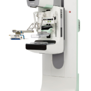
At the European Society for Breast Imaging (EUSOBI) meeting last September in Berlin, Hologic officially launched the 3Dimensions™ mammography system which offers a variety of groundbreaking features designed to provide higher quality 3D™ images for radiologists, enhanced workflow for technologists, and a more comfortable mammography experience, with low-dose options, for patients (see featured item).
On this occasion, International Hospital talked to Lori Fontaine, Vice President of Clinical Affairs for Hologic.

Is the launch at EUSOBI only for Europe or is it global?
The 3Dimensions™ mammography system received CE Mark in July 2017 making it commercially available in EMEA, followed shortly thereafter by the U.S. launch in August 2017.

Can you give some details and figures on dose reduction for the new system?
We know that dose is a common concern across Europe, and the 3Dimensions system helps address this by providing low-dose options for patients, among many other benefits. The 3Dimensions system results in a 45 percent dose reduction with a generated 2D image compared to 2D FFDM alone. 

Is the improvement in image clarity regardless of breast density likely to reduce the need for a secondary ultrasound in the screening of high density breasts?
We already know the 3Dimensions system’s Clarity HD high-resolution 3D™ imaging reduces recalls by up to 40 percent compared to 2D alone, and given Clarity HD works to deliver exceptional 3D™ images, regardless of breast size or density, it makes sense that the 3Dimensions system would be an ideal option for women with dense breasts. This is especially true since the 3Dimensions system operates in tandem with Hologic’s 3D Mammography™ exam, the only mammogram approved by the U.S. Food and Drug Administration as superior for women with dense breasts compared to 2D alone, which further demonstrates that tomosynthesis should be the standard of care for women across the globe when it comes to breast cancer screening.

Do you have any information and figures on the adoption rate of DBT by radiologists in the various European countries, are there significant country variations (or regional between US, Europe and Asia)?
Digital Breast Tomosynthesis (DBT) adoption rates vary by country. While DBT has been approved in EMEA since 2009, the majority of EMEA countries limit the use of DBT to diagnostic imaging as they have concerns regarding dose and reading time. Hologic remains at the forefront of technology innovation and is working to overcome these barriers, so that all women can be screening with DBT.

Hologic was the first company to receive FDA approval for DBT use in both the screening and diagnostic setting in the U.S. in 2011. Today, DBT is used in approximately 40 percent of all U.S. screening mammography exams and is covered by the majority of insurance companies. The evidence of the benefit of Hologic’s 3D Mammography exam as a better mammogram continues to expand and resulted in the addition of DBT to the National Comprehensive Cancer Network (NCCN) Guidelines in 2016. NCCN is recognized globally as an alliance of 27 U.S. cancer centers that develop recommendations designed to help healthcare professionals diagnose, treat and manage cancer care.

https://interhospi.com/wp-content/uploads/sites/3/2020/08/IH159_Hologic-_1__01.jpg 800 521 3wmedia https://interhospi.com/wp-content/uploads/sites/3/2020/06/Component-6-–-1.png 3wmedia2020-08-26 14:17:292021-01-08 12:30:24DBT makes major stride with Hologic’s launch of innovative mammography system

Scientific literature review – pathology

, 26 August 2020/in Featured Articles /by 3wmedia

Improvements in cell block processing: The Cell-Gel method

La Fortune KA, Randolph ML, Wu HH, Cramer HM. Cancer 2017; 125(4): 267–276

BACKGROUND: The ability to produce adequate cell blocks profoundly impacts the diagnostic usefulness of cytology specimens. Cell blocks are routinely processed from fine-needle aspiration specimens or concentrated fluid samples. Obtaining directed passes for the sole purpose of producing a cell block is common practice, particularly when the cytopathologist anticipates the need for ancillary immunocytochemical stains and/or molecular studies.
METHODS: The authors developed an effective and inexpensive process for producing cell blocks that consistently yields abundant cellular material, which they have termed the Cell-Gel method. This method can be simplified into 3 main steps: (1) preparing the sample; (2) constructing the cell block; and (3) processing the cell block. Highlights of the protocol include using a hemolytic fixative for sample preparation and disposable base moulds for cell block construction.
RESULTS: The cell block failure rate in the current study decreased from 18% with the HistoGel Tube method (January 2014 – December 2014) to 6% with the Cell-Gel method (January 2015 – December 2016). The authors evaluated 110 cell blocks processed with the HistoGel Tube method and 110 cell blocks processed with the Cell-Gel method, for a total evaluation of 220 cell blocks.
CONCLUSIONS: The authors have developed an effective and inexpensive protocol for producing cell blocks that consistently yields abundant cellular material. The Cell-Gel method uses a hemolytic fixative and disposable base moulds to produce adequate cell blocks. When the method was implemented, the cell block failure rate of the study laboratory decreased by approximately 67%.

Lung carcinoma predictive biomarker testing by immunoperoxidase stains in cytology and small biopsy specimens: advantages and limitations

Zhou F, Moreira AL. Arch Pathol Lab Med 2016; 140(12): 1331–1337

CONTEXT: In the burgeoning era of molecular genomics, immunoperoxidase (IPOX) testing grows increasingly relevant as an efficient and effective molecular screening tool. Patients with lung carcinoma may especially benefit from the use of IPOX because most lung carcinomas are inoperable at diagnosis and only diagnosed by small tissue biopsy or fine-needle sampling. When such small specimens are at times inadequate for molecular testing, positive IPOX results still provide actionable information.
OBJECTIVE: To describe the benefits and pitfalls of IPOX in the detection of biomarkers in lung carcinoma cytology specimens and small biopsies by summarizing the currently available commercial antibodies, pre-analytic variables, and analytic considerations.
DATA SOURCES: PubMed.
CONCLUSIONS: Commercial antibodies exist for IPOX detection of aberrant protein expression due to EGFR L858R mutation, EGFR E746_A750 deletion, ALK rearrangement, ROS1 rearrangement, and BRAF V600E mutation, as well as PD-L1 expression in tumour cells. Automated IPOX protocols for ALK and PD-L1 detection were recently approved by the Food and Drug Administration as companion diagnostics for targeted therapies, but consistent interpretive criteria remain to be elucidated, and such protocols do not yet exist for other biomarkers. The inclusion of cytology specimens in clinical trials would expand patients’ access to testing and treatment, yet there is a scarcity of clinical trial data regarding the application of IPOX to cytology, which can be attributed to trial designers’ lack of familiarity with the advantages and limitations of cytology. The content of this review may be used to inform clinical trial design and advance IPOX validation studies.

DUX4 immunohistochemistry is a highly sensitive and specific marker for CIC-DUX4 fusion-positive round cell tumor

Siegele B, Roberts J, Black JO, Rudzinski E et al. Am J Surg Pathol 2017; 41(3): 423–429

The histologic differential diagnosis of pediatric and adult round cell tumours is vast and includes the recently recognized entity CIC-DUX4 fusion-positive round cell tumour. The diagnosis of CIC-DUX4 tumour can be suggested by light microscopic and immunohistochemical features, but currently, definitive diagnosis requires ancillary genetic testing such as conventional karyotyping, fluorescence in situ hybridization, or molecular methods. We sought to determine whether DUX4 expression would serve as a fusion-specific immunohistochemical marker distinguishing CIC-DUX4 tumour from potential histologic mimics. A cohort of CIC-DUX4 fusion-positive round cell tumours harbouring t(4;19)(q35;q13) and t(10;19)(q26;q13) translocations was designed, with additional inclusion of a case with a translocation confirmed to involve the CIC gene without delineation of the partner. Round cell tumours with potentially overlapping histologic features were also collected. Staining with a monoclonal antibody raised against the C-terminus of the DUX4 protein was applied to all cases. DUX4 immunohistochemistry exhibited diffuse, crisp, strong nuclear staining in all CIC-DUX4 fusion-positive round cell tumours (5/5, 100% sensitivity), and exhibited negative staining in nuclei of all of the other tested round cell tumours, including 20 Ewing sarcomas, 1 Ewing-like sarcoma, 11 alveolar rhabdomyosarcomas, 9 embryonal rhabdomyosarcomas, 12 synovial sarcomas, 7 desmoplastic small round cell tumours, 3 malignant rhabdoid tumours, 9 neuroblastomas, and 4 clear cell sarcomas (0/76, 100% specificity). Thus, in our experience, DUX4 immunostaining distinguishes CIC-DUX4 tumours from other round cell mimics. We recommend its use when CIC-DUX4 fusion-positive round cell tumour enters the histologic differential diagnosis.


Role of quantitative p16INK4A mRNA assay and digital reading of p16INK4A immunostained sections in diagnosis of cervical intraepithelial neoplasia

Vasiljević N, Carter PD, Reuter C et al. Int J Cancer 2017; 141(4): 829–836

Visual interpretation of cervical biopsies is subjective and variable, generally showing fair to moderate inter-reader agreement in distinguishing high from low grade cervical intraepithelial neoplasia (CIN). We investigated the performance of two objective p16 quantitative tests in comparison with visual assessment: (i) p16-mRNA assay and (ii) digital analysis of sections stained for p16 protein. The primary analysis considered 232 high-risk human papilloma virus positive (HPV+) samples from diagnostic cervical specimens. A p16 RT-qPCR (p16-mRNA assay) was run on mRNA extracted from formalin-fixed paraffin-embedded sections. Two p16 immunohistochemistry (IHC) readings, a visual read by a histopathologist (Visual IHC) and a digital read of a high-resolution scan (Digital IHC), were done on adjacent sections. The worst reviewed CIN grade (agreed by at least two histopathologists) from up to two biopsies and a loop excision was taken, with CIN2/3 as the primary endpoint. Visual IHC attained a specificity of 70% (95%CI 61–77) for 85% (95%CI 77–91%) sensitivity. The four-point Visual IHC staining area under the curve (AUC) was 0.77 (95%CI 0.71–0.82), compared with 0.71 (95%CI 0.64–0.77) for p16-mRNA and 0.67 (95%CI 0.60–0.74) for Digital IHC. Spearman rank-order correlations were: visual to p16-mRNA 0.41, visual to digital 0.49 and p16-mRNA to digital: 0.22. The addition of p16-mRNA assay to visual reading of p16 IHC improved the AUC from 0.77 to 0.84 (P=0.0049). p16-mRNA testing may be complementary to visual IHC p16 staining for a more accurate diagnosis of CIN, or perhaps a substitute in locations with a lack of skilled pathologists.

Biomarkers for pathology diagnosis of uterine cervix malignant glandular lesions

Lee S, Rose MS, Sahasrabuddhe VV et al. Int J Gynecol Pathol 2017;36(4): 310–322

Immunohistochemistry is widely used to support a pathology diagnosis of cervical adenocarcinoma despite the absence of a systematic review and meta-analysis of the published data. This systematic review and meta-analysis was performed to investigate the sensitivity and specificity of immunohistochemistry biomarkers in the tissue-based diagnosis of cervical adenocarcinoma histotypes compared with normal endocervix and benign glandular lesions. The systematic review and meta-analysis used a PICOT framework and QUADAS-2 to evaluate the quality of included studies. The literature search spanned 40 years and ended June 30, 2015. Abstracts of identified records were independently screened by two of the authors who then conducted a full-text review of selected articles. Sensitivity and specificity of immunohistochemistry expression in malignant glandular lesions of the cervix classified per WHO 2003 compared with 5 benign comparators (normal/benign endocervix, and benign endocervical, endometrioid, gastric, and mesonephric lesions) were calculated. Of 902 abstracts screened, 154 articles were selected for full review. Twenty-five articles with results for 36 biomarkers were included. The only biomarker with enough studies for a meta-analysis was p16 and the definition of positive p16 staining among them was variable. Nevertheless, any positive p16 expression was sensitive, ranging from 0.94 to 0.98 with narrow confidence intervals (CIs), for adenocarcinoma in situ (AIS) and mucinous adenocarcinomas in comparison with normal/benign endocervix and benign endocervical and endometrioid lesions. Specificity for AIS and mucinous adenocarcinomas was also high with narrow CIs compared with benign endocervical lesions. The specificity was high for AIS, 0.99 (0.24, 1.0), and mucinous adenocarcinoma, 0.95 (0.52, 1.0), compared with normal/benign endocervix but with wider CIs, and low with very wide CIs compared with benign endometrioid lesions: 0.31 (0.00, 0.99) and 0.34 (0.00, 0.99), respectively. Results from single studies showed that p16, p16/Ki67 dual stain, ProExC, CEA, ESA, HIK1083, Claudin 18, and ER loss in perilesional stromal cells were useful with high (≥0.75) sensitivity and specificity estimates in ≥1 malignant versus benign comparisons. None of the biomarkers had highly useful sensitivity and specificity estimates for AIS, mucinous adenocarcinomas, or minimal deviation adenocarcinoma/gastric adenocarcinoma compared with benign gastric or mesonephric lesions or for mesonephric carcinoma compared with normal/benign endocervix, benign endocervical, endometrial, or mesonephric lesions. Any expression of p16 supports a diagnosis of AIS and mucinous adenocarcinomas in comparison with normal/benign endocervix and benign endocervical lesions. The majority of studies did not separate mosaic/focal p16 staining from diffuse staining as a distinct pattern of p16 overexpression and this may have contributed to the poor performance of p16 in distinguishing AIS and mucinous adenocarcinomas from benign endometrioid lesions. Single studies support further investigation of 8 additional biomarkers that have highly useful sensitivity and specificity estimates for ≥1 malignant glandular lesions compared with ≥1 of the 5 benign comparators.

GATA3 expression in triple-negative breast cancers

Byrne DJ, Deb S, Takano EA, Fox SB. Histopathology 2017; 71(1): 63–71

AIMS: GATA-binding protein 3 (GATA3) is a well-studied transcription factor found to be essential in the development of luminal breast epithelium and has been identified in a variety of tumour types, including breast and urothelial carcinomas, making it a useful immunohistochemistry marker in the diagnosis of both primary and metastatic disease.
METHODS AND RESULTS: We investigated GATA3 protein expression in a 106 primary triple-negative breast carcinomas (100 basal-like, six non-basal-like) using Cell Marque mouse monoclonal anti-GATA3 (L50-823). Reverse transcription-quantitative polymerase chain reaction (RT-qPCR) was used to quantify mRNA expression in 22 triple-negative breast cancers (TNBCs) (20 primary and two cell lines), four luminal (three primary and one cell line) and five human epidermal growth factor receptor 2 (HER2) (four primary and one cell line) amplified tumours. In 98 TNBCs where IHC was assessable, 47 (48%) had a 1+ or greater staining with 20 (21%) having high GATA3 expression when using a weighted scoring.
CONCLUSION: Our study has demonstrated that GATA3 expression is common in primary triple-negative breast carcinomas. It also suggests that although GATA3 is an estrogen receptor (ER) regulated gene, it still proves useful in differentiating between primary and metastatic tumours in patients with a history of breast cancer regardless of its molecular subtype.

Accuracy of fine needle cytology in histological prediction of papillary thyroid carcinoma variants: a prospective study

Cipolletta Campanile A, Malzone MG, Losito NS et al. 2017; 28(3): 187–197

Fine needle cytology (FNC) is a crucial procedure in the preoperative diagnosis of thyroid tumours. Papillary thyroid carcinoma (PTC), in its classic variant (cPTC), is the most common malignant neoplasm of the thyroid. Several histological variants of PTC have been described, each one with its own characteristics and prognosis. The ability of FNC to identify the variants represents a challenge even for a skilled pathologist. The aim of this study was to evaluate the diagnostic cytological accuracy of FNC in PTC and to look for specific features that could predict the different variants. This was a single centre prospective study on 128 patients who received a diagnosis of PTC on FNC. The smears were blindly reviewed by two cytopathologists to create a frequency score (0, 1, 2, 3) of the features for each variant. The cytological parameters were divided into three groups: architectural, nucleo-cytoplasmic, and background features. Univariate analysis was performed by chi-square test with Yates correction and Fisher exact test as appropriate. Multiple regression analysis was performed among the variables correlated at the linear correlation. The correlation study between cytology and histology showed an accuracy of FNC in classic, follicular, and oncocytic PTC variants of 63.5, 87.5, and 87%, respectively. Familiarity with cytological features may allow an early diagnosis of a given PTC variant on FNC samples. This is fundamental in a preoperative evaluation for the best surgical approach and subsequent treatment.

https://interhospi.com/wp-content/uploads/sites/3/2020/08/Scientific-Lit-picture.jpg 533 800 3wmedia https://interhospi.com/wp-content/uploads/sites/3/2020/06/Component-6-–-1.png 3wmedia2020-08-26 14:17:292021-01-08 12:30:29Scientific literature review – pathology

Remote monitoring – a new frontier in the fight against cardiovascular disease

, 26 August 2020/in Featured Articles /by 3wmedia

The growth in the use of modern, implantable cardiovascular devices has been accompanied by efforts to have them monitored by professionals at a distance. The principal driver for this has been convenience. However, over recent years, remote monitoring (RM) of cardiovascular devices is emerging not only as an alternative to the clinic, but in some cases as a source for enhancements to quality of care. Several professional societies have issued authoritative guidelines recommending RM for all eligible patients.

Device complexity and data transfer
Formally known as cardiovascular implantable electronic devices (CIEDs), equipment such as pacemakers, cardioverter defibrillators, loop recorders and hemodynamic monitors are technologically complex and equipped with an array of microelectronics, high computational capability and onboard firmware.
In turn, this allows for assessment, storage and remote transfer of a range of data via a transmitter placed in proximity to the patient. Examples of such data include device function, diagnostics and fault codes, to therapy delivery and intra-cardiac hemodynamics, as well as reports on patient clinical status and alerts on cardiovascular events.

Developments in remote monitoring technology

On their part, remote monitoring techniques too have undergone their own evolution  – from the original telephonic check-up of pacemaker battery levels and wand-based systems with patient-driven downloads, to current generation products which transmit data through stationary or mobile transmitters by either analogue/digital wired or wireless communication. Once transmitted, medical staff can check the information via a secure Website. Both the type and volume of transmitted data is similar to that obtained from direct interrogation.
Technically, it is important to differentiate between ‘remote interrogation’ and ‘remote monitoring’. The former involves periodic device interrogation performed manually at home by the patient or automatically at predefined points by the monitoring system. RM involves continuous device monitoring, one of whose key features is to trigger transmissions in case of alerts.

Convenience and workflow bottlenecks
Remote monitoring eliminates the need for routine, periodic visits to a clinic after CIED implantation. Most international guidelines specify that patients fitted with CIEDs should be followed up routinely, with the frequency depending on the device type and model – for instance, at Months 1 and 3  for implantable cardioverter defibrillators (ICDs). Key checks include those on battery, lead impedance, sensing amplitude, pacing threshold and arrhythmic events.
One of the most perceptible advantages of RM is of course convenience. Before it became available, patients with CIEDs had to visit clinics for periodic checks. This was a problem for several categories of patients – above all, those living in rural areas and those needing to be escorted by families due to frailty. These factors assume additional significance since the number of CIED patients has not only been increasing due to maturing technology and expanded indications, but also because an ageing society means that more people are in need of the devices. As a result, it is becoming ever-tougher to make appointments for CIED checks, and many patients who do not have RM can spend several hours waiting at a hospital for their turn.
Remote monitoring eliminates such bottlenecks and choke points. Analysis of RM data before a patient visit can shorten the time required for direct interrogation and intervention, especially should a need arise to determine the cause and management of a problem. If such a problem is only detected during a clinic visit, a patient would have to wait for the results to be verified, while the problem is detected, analysed and resolved.
According to some estimates, time required by physician to review RM data is approximately 10 minutes compared to a half-hour to complete CIED follow-up visits in a clinic.
Apart from routine transmission, special real-time protocols exist in RM for alerts, such as data anomalies, inappropriate therapy or other abnormalities. In such cases, the transmitter is usually linked to a central secure server to back up or distribute the results to a larger number of experts for further analysis and opinion.

RM data essentials
Typical data reported by RM include arrhythmic events (real-time intra-cardiac electrocardiogram, to determine if the event is supra-ventricular or ventricular), premature ventricular contractions (based on PVC frequency recording), atrial fibrillation (especially promising for patients with no prior history of AF, to allow rapid anticoagulant drug administration and prevent stroke), non-sustained VT (although this is mainly considered for ICD or CRT-D patients, rather than those with pacemakers) and VT/VF (to enable a therapy decision and whether it can be managed at home).

The Finnish ICD study
Meanwhile, rigorous observational and randomized studies have demonstrated a variety of clinical benefits, along with a high degree of patient satisfaction as well as cost effectiveness.
One of the first major studies on remote monitoring of ICDs was carried out in 2005-2006 at the Oulu University Hospital, Finland. The system consisted of a portable patient monitor, a secure database and website, at which clinicians could view and analyse data.
The study’s goal was to provide comprehensive information on the safety, ease of use, satisfaction and data acceptance by both clinicians and patients, and the cost-effectiveness of remote monitoring in a location characterized by long travel distances to the clinic.
The outcomes were satisfactory.
There were, first of all, no device-related adverse events.
80% of the remote-monitoring sessions were performed by the patients without any assistance. Indeed, ease of use and satisfaction by both patients and clinicians made an especially strong case. Most patients found the instructions ‘clear’ or ‘very clear’, with monitor set up ‘easy’ or ‘very easy’. What was equally significant was the lack of any major difference in patient feedback from the first test, at 3 and 6 months, and even during unscheduled visits.
On their side, clinicians too drew similar conclusions on ease of use and satisfaction, with the majority finding data comparable to traditional device interrogation. Just two of 137 physicians felt an in-office visit would have provided more detailed information on device function, as it was not possible to measure the pacing threshold remotely.

Early detection of clinical events
Since then, other studies have reconfirmed the immense promise of RM.
In 2010 ‘Circulation’ published results of a trial on automated remote monitoring of implantable cardioverter-defibrillator called TRUST (Lumos-T Safely Reduces Routine Office Device Follow-up).
This study, on 1,339 patients, confirmed that the burden of visiting a clinic was greatly reduced by using RM, and that it saved valuable time and resources. The study found that in-hospital evaluation numbers dropped by 45% without affecting morbidity.
The TRUST trial also established that RM facilitated early detection of clinical events, in some cases dramatically. For example, the median period from onset to physician evaluation of combined first atrial fibrillation (AF), ventricular tachycardia (VT), and ventricular fibrillation (VF) events with RM was 1 day. By comparison, conventional care reported a median period of 35.5 days. System-related problems (such as lead out-of-range impedance) occurred over four times less frequently with the RM group, although the incidence in either setting was far too low to make meaningful comparisons.

Wireless RM and cardiac hospitalization stays
The utility of wireless remote monitoring with automatic clinician alerts was the subject of another trial called CONNECT (Clinical Evaluation of Remote Notification to Reduce Time to Clinical Decision).  This multicentre, prospective, randomized study of almost 2,000 patients with high-energy CIEDs lasted for 15 months. Its results were published in ‘The Journal of the American College of Cardiology’ in 2011, and reported a decrease in mean length of stay per cardiovascular hospitalization visit from 4 days in an in-office setting to 3.3 days with RM. The CONNECT study also found a dramatic reduction in the median time to a clinical decision in response to events, from 22 days at a clinic to 4.6 days using RM.

Other benefits of RM
RM has also established some other dramatic benefits. In 2013, ‘The European Heart Journal’ reported on ECOST, a randomized study on remote follow-up of ICDs. ECOST found that patients with RM had a 52% reduction in inappropriate shocks, fewer hospital admissions after such events and 76% fewer capacitor charges, leading to longer battery life.
In December 2014, a report in ‘The Journal of Arrhythmia’ noted that in prophylactic ICD recipients, the recommended 3-month in-office follow-up interval could be extended to 12 months with automatic daily RM, and that this reduced the ICD follow-up burden over a 27-month period after implantation. The 12-month interval resulted in more than halving the total number of in-clinic ICD follow-ups. In addition, no significant difference was found between the two groups (3-month in-clinic follow-up versus 12-month RM) in mortality, hospitalization rate, or hospitalization length over the observation period.

Mortality reduction with RM
Indeed, some experts propose that RM may reduce mortality in patients with CIEDs.
One study called ALTITUDE assessed long-term outcomes after ICD and cardiac resynchronization therapy (CRT) implantation and the impact of RM on almost 70,000 ICD and CRT-plus-defibrillator (CRT-D) patients. It found that one- and 5-year survival rates were 50% higher in comparison to about 115,000 patients who received CIED follow-up in office visits.

The future: patients generally satisfied with RM
As technology continues to evolve, both new possibilities and questions are emerging.  In the years to come, remote monitoring holds forth considerable promise for future research, given that massive amounts of data have already been collected from patients. 
In spite of some typical first-mover tech concerns, RM has proven to be easy to use and well accepted, even by the elderly people and patients with low education levels. There are some patients, however, who do not accept RM. This is mainly due to suspicions about technology and the risk of losing human contact with nurses and physicians. In such cases, patient education is critical.
The other challenge involves keeping track of a flood of data and alerts from a fast-growing pool of patients. As described previously, RM detects cardiovascular events much earlier than conventional follow-up. As a result, it is becoming essential to assess whether this translates into clinical benefits for patients, or whether earlier detection of events due to RM excessively increases clinic visits; the latter might well reduce clinical benefits.
On their part, patients continue to be satisfied with RM in terms of ease of use. One Italian study at San Filippo Neri Hospital in Rome has reported a more favourable change in quality of life over a 16-month period in RM patients, compared to those lacking access to RM. Benefits which have been specifically highlighted include the patients’ peace of mind, psychological well-being, and safety.

https://interhospi.com/wp-content/uploads/sites/3/2020/08/IH174_CV-Remote-Moniitoring_Tosh_thematic.jpg 533 800 3wmedia https://interhospi.com/wp-content/uploads/sites/3/2020/06/Component-6-–-1.png 3wmedia2020-08-26 14:17:292021-01-08 12:30:11Remote monitoring – a new frontier in the fight against cardiovascular disease

Big data and imaging – algorithms and analytics aid clinical decision making

, 26 August 2020/in Featured Articles /by 3wmedia

A fluid, game-changing combination of mathematical tools and Big Data seems ready to disrupt the field of radiology. However, it also promises to pave the way for what may turn out to be potentially-dramatic advances in healthcare.
There is some irony here. Data was once seen as a liability, to maintain and pay for. It is now being considered a potentially major asset. The key to this turnaround in perspectives lies in increasingly sophisticated, deep learning algorithms, advanced analytics and artificial intelligence which interpret the Big Data and make it usable.

Explosion in image numbers and volume
There is no hyperbole in the use of the term Big Data, as far as radiology is concerned. In recent years, there has been a veritable explosion in the stock of medical images. Emergency room radiologists often examine up to 200 cases a day, and each patient’s imaging studies can be around 250 GB of data. At the upper end, a ‘pan scan’ CT of a trauma patient can render 4,000 images. Currently, about 450-500 petabytes of medical imaging data are generated per year, but this is accelerating. Decisions are made on the basis of small parts of imaging data, the proverbial tip of the iceberg. Much of the information in this data has still to be deciphered and used.

Medical imaging and disease
Medical imaging provides important information on anatomy and organ function as well as detecting diseases states. Its analysis covers a gamut of areas from image acquisition and compression, to  transmission, enhancement, segmentation, de-noising and reconstruction. 
Technology has enabled often-dramatic leaps in image resolution, size and availability. Sophisticated picture archiving and communications systems (PACS) have allowed for the merger of patient images from different modalities and their integration with other patient data for analysis and use in a clinical setting.

Limits to vision – from digital to analogue
So far, radiology information to identify disease or other clinical conditions is presented in the form of images. Although scanners digitize data into pixels, this is reconstructed into shapes and shades or colours for display in a form that can be understood by the human brain. 
This is where the ‘tip of the iceberg’ statement above comes into play. Medical scanners encode an image pixel in 56 bits, equivalent to 72,000 trillion ‘shades’. However, the scanner reduces the data amount to 16 bits, just 65,536 shades, for the human eye. As a result, 40 bits of information is lost, in just one pixel.
At some point in the future, it seems likely that radiologists use numbers rather than images to numerically define and detect patterns of diseases. The process may in fact have already begun.

Imaging analytics and deep learning
Such trends are being fuelled by rapid advances in imaging analytics. Smart, deep learning (DL) algorithms, which analyse pixels and other digital data bytes within an image, have the capacity to detect specific patterns associated with a pathology and provide conclusions in terms of a numerical metric.
One example of the use of numbers as a diagnostic definition concerns the use of algorithms in CT images to calculate bone density. The result is compared to a reference number, which au tomatically trigger alerts on low bone density. Avoiding the need for another dedicated examination, a physician can determine if a patient needs calcium supplements or another preventative measure.
Such algorithms also learn over time, and become better at what they do, resulting in even greater speed and more confidence in the future. Such a process has been driven by the steady acceleration, over the years, in computer processing speed. Indeed, while training an algorithm at the turn of the century took 2-3 months, the same results can now be achieved and iterated within minutes.

Neural systems and algorithms

Technically, deep learning produces a direct mapping from raw inputs to outputs such as image classes. Many DL algorithms are inspired by biologic neural systems. They are different from traditional machine learning, which requires manual feature extraction from inputs, and face limitations to use in the face of the large volumes of information associated with Big Data.
Big Data’s virtuous circle
Many DL algorithms directly seek to harness Big Data in radiology. Gigantic (and fast-growing) image libraries are being accessed for investigation to develop, test, validate and continuously refine algorithms, with the aim of covering a whole range of pathologies.
For radiologists, analytic results from an examination can be comprehensively evaluated against similar data obtained over a long period of time and evaluated to suggest appropriate diagnosis in current scenarios.

Such a virtuous cycle of algorithms and Big Data have become the focus for a host of major medical technology vendors as well as start-ups. However, the key enabling players are radiology departments, who own the data repositories and are uniquely placed to curate the data, in other words, organize it from fragments and make it available for running analytical algorithms.
The above process has, in some senses, been jump-started by previous efforts to data mine reports from radiology departments as they transitioned from PACS to enterprise imaging. The next step in this Big Data-driven opportunity will consist of linking information in radiology reports to the pixels of medical images.

The pixel goldmine
Few doubt any more that pixels are a goldmine, holding wholly new insights into a medical image and how best they could be utilized, not just by radiologists but other clinicians offering patient care. Alongside data mined from electronic medical records, quantitative pixel-based analysis algorithms are increasingly likely to be used to find patterns in images.

Big Data-based screening algorithms, for example, can be used to highlight subtle, multi-dimensional changes in a nodule or a lesion. This can be followed by applications such as curved planar or 3D multi-planar reconstructions, or dynamic contrast enhancement (DCE) texture analysis on highly targeted data subsets, instead of making the time-consuming effort of querying a complete imaging dataset. 

Specific examples of such an approach might include diagnosis of lesions in the liver and identification of disease-free liver parenchyma. Another would be volume analysis of lung tumours and solitary pulmonary nodules to decide temporal evolution of lesion. Big data based pattern analysis modules can detect areas of opacities, honeycombing, reticular densities and fibrosis, and thereby provide a list of differentials, using computer aided diagnostic tools.
For tumours, in general, radiologists can run algorithms to check contrast enhancement characteristics, and such metrics can be compared to prior results as well as other pathology data to provide a specific differential list.

Decision support systems
One decision support system based on Big Data assists physicians in providing treatment planning for patients suffering from traumatic brain injury (TBI). The algorithm couples demographic data and medical records of the patient to specific features extracted from CT scans in order to predict intracranial pressure (ICP) levels.
Google’s entry into this field seeks to address real world limitations – not just in terms of human capacities but also trained medical personnel. Its first deep learning imaging algorithm sought to recognize diabetic retinopathy, the fastest growing cause of blindness in poor countries, where a shortage of specialists meant many patients lost their sight before diagnosis.

The promise of AI
Google’s algorithm is based on artificial intelligence (AI), seen as an especially promising catalyst for advances in such areas.
AI-based algorithms, for example,  can calculate the volume of bleed on the basis of multiple brain CT slices in stroke patients, with the size of bleed volume indicating urgency as well as care pathway. Another recent algorithm assesses recent infarcts on CT, which can be missed if they are hyper-acute (less than 8-12 hours old), and is therefore relevant to all patients with sudden onset weakness. The University of California in San Francisco has been testing an algorithm to identify pneumothorax in chest radiographs of surgery patients, before they exit the OR (operating room).  The aim is to not only avoid the huge costs of a collapsed lung but also ensure that the OR is freed from being used for an otherwise-avoidable procedure.
AI is also being considered for workflow management and triaging. In the near future, it is almost certain that images are screened as data is acquired by a scanner, to distinguish between ‘normal’ and ‘abnormal’ images, prioritize cases according to the likelihood of disease and alerting radiologists to conditions that require urgent attention. The results are tangible and impressive. One algorithm has helped physicians to shrink the time for cardiac diagnoses from 30 minutes to 15 seconds.

Certain vendors are leveraging AI to correlate findings on properties like morphology, cell density or physiological characteristics to expert radiologist’s reports, while taking additional clinical data such as biopsy results into account. Others use reasoning protocols as well as visual technologies such as virtual rendering to analyse medical images. This is then combined with data from a patient’s medical record to offer radiologists and clinicians decision-making support.

AI and the radiologist
So far, algorithms and emerging metrics are expected to be largely used as a complement to decisions made by radiologists.
However, at some point in the future, it seems plausible that radiologists no longer need to look at images at all. Instead, they would simply analyse outcomes of the algorithms.
Once again, AI is at play here. Apart from deep learning algorithms, radiology can claim to be witness to the first successes with the emerging science of ‘swarm’ AI, which helps form a diagnostic consensus by turning groups of human experts into super experts.  Swarm AI is directly based on nature, which sees species accomplishing more by participating in a flock, school or colony (a ‘swarm’) than they can individually. One report, published in ‘Public Library of Science (PLOS)’, stated that swarm intelligence could improve other types of medical decision-making, ”including many areas of diagnostic imaging.”
In December 2015, a study in ‘IET Systems Biology’ reported about a swarm intelligence algorithm which assisted “in the identification of metastasis in bone scans and micro-calcifications on mammographs.” The authors, from universities in the UK and India, also reported about the use of the algorithm in assessing CT images of the aorta and in chest X-ray. They proposed a hybrid swarm intelligence approach to detect tumour regions in an abnormal MR brain image.

The future: human-machine symbiosis

AI is unlikely to become a replacement for radiologists, but a tool to help them. According to Curt Langlotz, MD, PhD, professor of radiology and biomedical informatics at Stanford, the “human-machine system always performs better than either alone.”

https://interhospi.com/wp-content/uploads/sites/3/2020/08/IH167_Big-Data_Tosh_thematic_crop.jpg 591 800 3wmedia https://interhospi.com/wp-content/uploads/sites/3/2020/06/Component-6-–-1.png 3wmedia2020-08-26 14:17:292021-01-08 12:30:15Big data and imaging – algorithms and analytics aid clinical decision making

Kimes 2018, 15-18 March, Seoul

, 26 August 2020/in Featured Articles /by 3wmedia
https://interhospi.com/wp-content/uploads/sites/3/2020/08/47217_KIMES-2018_IHE.jpg 1500 1030 3wmedia https://interhospi.com/wp-content/uploads/sites/3/2020/06/Component-6-–-1.png 3wmedia2020-08-26 14:17:292021-01-08 12:30:19Kimes 2018, 15-18 March, Seoul

Arab Health 2018, 29 Jan – 1 Feb, Dubai

, 26 August 2020/in Featured Articles /by 3wmedia
https://interhospi.com/wp-content/uploads/sites/3/2020/08/47240_AH18_ADVERT_MEDIA_PARTNERS.jpg 2116 1500 3wmedia https://interhospi.com/wp-content/uploads/sites/3/2020/06/Component-6-–-1.png 3wmedia2020-08-26 14:17:292021-01-08 12:30:25Arab Health 2018, 29 Jan – 1 Feb, Dubai

SpiroScout SP – easily add spirometry to your ECG device

, 26 August 2020/in Featured Articles /by 3wmedia
https://interhospi.com/wp-content/uploads/sites/3/2020/08/47194__schiller.jpg 1500 1052 3wmedia https://interhospi.com/wp-content/uploads/sites/3/2020/06/Component-6-–-1.png 3wmedia2020-08-26 14:17:292021-01-08 12:30:29SpiroScout SP – easily add spirometry to your ECG device
Page 90 of 103«‹8889909192›»

Latest issue of International Hospital

April 2024

2 June 2026

DeepHealth achieves multiple regulatory milestones for Neuro, Prostate and LumbarMR

7 January 2026

Gulf Aorta Summit 2026 Returns to Dubai with a Global Lineup of Aortic Experts

17 December 2025

GE HealthCare receives CE mark for 128cm total body PET/CT

Digital edition
All articles Archived issues

Free subscription

View more product news

Get our e-alert

The medical devices information portal connecting healthcare professionals to global vendors

Sign in for our newsletter
  • News
    • Featured Articles
    • Product News
    • E-News
  • Magazine
    • About us
    • Archived issues
    • Media kit
    • Submit Press Release

Prins Hendrikstraat 1
5611HH Eindhoven
The Netherlands
info@interhospi.com

PanGlobal Media IS not responsible for any error or omission that might occur in the electronic display of product or company data.

Scroll to top

This site uses cookies. By continuing to browse the site, you are agreeing to our use of cookies.

Accept settingsHide notification onlyCookie settings

Cookie and Privacy Settings


How we use cookies

We may ask you to place cookies on your device. We use cookies to let us know when you visit our websites, how you interact with us, to enrich your user experience and to customise your relationship with our website.

Click on the different sections for more information. You can also change some of your preferences. Please note that blocking some types of cookies may affect your experience on our websites and the services we can provide.

Essential Website Cookies

These cookies are strictly necessary to provide you with services available through our website and to use some of its features.

Because these cookies are strictly necessary to provide the website, refusing them will affect the functioning of our site. You can always block or delete cookies by changing your browser settings and block all cookies on this website forcibly. But this will always ask you to accept/refuse cookies when you visit our site again.

We fully respect if you want to refuse cookies, but to avoid asking you each time again to kindly allow us to store a cookie for that purpose. You are always free to unsubscribe or other cookies to get a better experience. If you refuse cookies, we will delete all cookies set in our domain.

We provide you with a list of cookies stored on your computer in our domain, so that you can check what we have stored. For security reasons, we cannot display or modify cookies from other domains. You can check these in your browser's security settings.

.

Google Analytics Cookies

These cookies collect information that is used in aggregate form to help us understand how our website is used or how effective our marketing campaigns are, or to help us customise our website and application for you to improve your experience.

If you do not want us to track your visit to our site, you can disable this in your browser here:

.

Other external services

We also use various external services such as Google Webfonts, Google Maps and external video providers. Since these providers may collect personal data such as your IP address, you can block them here. Please note that this may significantly reduce the functionality and appearance of our site. Changes will only be effective once you reload the page

Google Webfont Settings:

Google Maps Settings:

Google reCaptcha settings:

Vimeo and Youtube videos embedding:

.

Privacy Beleid

U kunt meer lezen over onze cookies en privacy-instellingen op onze Privacybeleid-pagina.

Privacy policy
Accept settingsHide notification only

Sign in for our newsletter

Free subscription