• News
    • Featured Articles
    • Product News
    • E-News
  • Magazine
    • About us
    • Digital edition
    • Archived issues
    • Media kit
    • Submit Press Release
  • White Papers
  • Events
  • Suppliers
  • E-Alert
  • Contact us
  • Subscribe newsletter
  • Search
  • Menu Menu
International Hospital
  • AI
  • Cardiology
  • Oncology
  • Neurology
  • Genetics
  • Orthopaedics
  • Research
  • Surgery
  • Innovation
  • Medical Imaging
  • MedTech
  • Obs-Gyn
  • Paediatrics

Archive for category: Featured Articles

Featured Articles

MEDICAL FAIR ASIA 2018: Future-ready products and industry-leading conferences & forums

, 26 August 2020/in Featured Articles /by 3wmedia

MEDICAL FAIR ASIA 2018, is set to continue its growth path with its 12th edition. An expected 1,000 exhibitors from 50 countries and 20 national pavilions will grace the exhibition to be held in Singapore from 29th to 31st August. Visitors will get to source from a comprehensive range of more than 5,000 products ranging from digital health technology, electromedical equipment, rehabilitation supplies to consumables.

There will be a total of 20 National Pavilions and Country Groups, this edition will see debut group participations from Belgium, Brazil, the Netherlands, Iran, Denmark, European Union Business Avenue, Russia, Spain and Qatar, adding to the internationality of the exhibition.

Inaugural Community Care Pavilion
On the show floor, visitors can also expect to see products relating to the current healthcare trends and needs of the Asian region. The debuting Community Care Pavilion, with its keen focus on geriatrics and digital health technology seeks to address the healthcare needs of both the ageing population and the region’s remote population by bringing healthcare beyond traditional healthcare institutions and into the community. Exhibitors have already arranged for product launches to take place during the 3-day period. France Bed Co Ltd will be showcasing their unique powered turning bed. It features an automatic turning support function that prevents users from bed sores. Xiaoniu Health Co Ltd will be unveiling their intelligent sleep machine that can perform both CPAP and AutoCPAP to sleep apnea patients. 

Another first on the show floor is the inaugural Start-Up Park. Providing a platform for young and exciting start-ups, the exhibits will feature products that could transform the market in the near future including the latest innovations in big data, and IoT. Australian start-up Rapid Response Revival Research will unveil a prototype of their phone case defibrillator, CellAED, the world’s smallest, lightest and first truly mobile AED (Automatic External Defibrillator) for the very first time.

Conferences and forums
Back by popular demand, the exhibition will play host to the second edition of the MEDICINE + SPORTS Conference. This benchmark event for sports medicine will discuss topics ranging from digital innovations in sports and healthcare, exercise medicine to tailored exercise programmes for patients and athletes. A stellar lineup of speakers including experts Dr. Paul Gastin, Director for the Centre for Sport Research at Deakin University, Mr. Christian Stammel, CEO of WT | Wearable Technologies and Prof. James S. Skinner, Professor Emeritus in the Department of Kinesiology, Indiana University, have been confirmed.

With Start-Ups and SMEs deepening their presence in global business, the exhibition will also host the Medtech SME Workshop. Organized by the first and only regional medical technology association, Asia Pacific Medical Technology Association (APACMed), the workshop will provide small businesses with concise knowledge on clinical trials, product validation, patent laws and many others. Through this workshop, Medtech start-ups and SMEs can learn to navigate processes to develop cost-effective solutions to meet the region’s healthcare needs.

In line with the highlight on Community Care, the exhibition will also feature the first-ever Paradigm Shifts in Healthcare seminar from 30th to 31st August. Leading speakers will discuss the evolution of the healthcare industry while attendees learn how to overcome future challenges as healthcare goes beyond hospitals to the community. 

Supported by the Robotic Surgery Society of Singapore, the Medical Innovation & Technology Forum will focus on robotic surgery and discuss how patients evolve from passive healthcare recipients to active value-seekers, encouraging healthcare providers to tap into the latest technological advances to provide more efficient treatment options.

www.medicalfair-thailand.com
https://interhospi.com/wp-content/uploads/sites/3/2020/08/NJD_0733.jpg 533 800 3wmedia https://interhospi.com/wp-content/uploads/sites/3/2020/06/Component-6-–-1.png 3wmedia2020-08-26 14:17:292021-01-08 12:30:10MEDICAL FAIR ASIA 2018: Future-ready products and industry-leading conferences & forums

IHF Journal – World Hospitals and Health Service

, 26 August 2020/in Featured Articles /by 3wmedia
https://interhospi.com/wp-content/uploads/sites/3/2020/08/IH164_IHF_page1.jpg 1500 986 3wmedia https://interhospi.com/wp-content/uploads/sites/3/2020/06/Component-6-–-1.png 3wmedia2020-08-26 14:17:292021-01-08 12:30:18IHF Journal – World Hospitals and Health Service

Kimes, March 15-18 2018, Seoul, Korea

, 26 August 2020/in Featured Articles /by 3wmedia
https://interhospi.com/wp-content/uploads/sites/3/2020/08/47216_KIMES-2018_IHE.jpg 1000 685 3wmedia https://interhospi.com/wp-content/uploads/sites/3/2020/06/Component-6-–-1.png 3wmedia2020-08-26 14:17:292021-01-08 12:30:29Kimes, March 15-18 2018, Seoul, Korea

Pre-hospital, Hospital, Homecare

, 26 August 2020/in Featured Articles /by 3wmedia
https://interhospi.com/wp-content/uploads/sites/3/2020/08/47258_691703-B-2017_17_01_Eagle_Dv2_QtrPageRT-NEW.jpg 1462 1012 3wmedia https://interhospi.com/wp-content/uploads/sites/3/2020/06/Component-6-–-1.png 3wmedia2020-08-26 14:17:292021-01-08 12:30:21Pre-hospital, Hospital, Homecare

KIMES, Seoul, 15-18 March 2018

, 26 August 2020/in Featured Articles /by 3wmedia
https://interhospi.com/wp-content/uploads/sites/3/2020/08/47218_KIMES-2018_IHE.jpg 1500 1030 3wmedia https://interhospi.com/wp-content/uploads/sites/3/2020/06/Component-6-–-1.png 3wmedia2020-08-26 14:17:292021-01-08 12:30:22KIMES, Seoul, 15-18 March 2018

Carotid artery stenting – the challenge of plaque protrusion

, 26 August 2020/in Featured Articles /by 3wmedia

Although not commonplace, carotid artery stenting (CAS) is occasionally accompanied by the protrusion of plaque into the stent lumen, and a variety of ischemic complications during intra- and post-operative periods. Among these complications, plaque protrusion (PP) into the stent and thrombus on the stent after CAS are some of the most worrying for clinicians.
The correction of PP is achieved by additional post-dilations or stent-in-stent implantation.

First noticed in mid-1990s
The first observations of PP date over two decades and provided the impetus for embolic protection devices. In a paper published in the mid-1990s, a team led by Frank Veith, M.D (from the Mayo Clinic) suggested that restoration of flow and removal of protection devices might lead to the continuing break off of protrusions and provoke some delayed strokes.
Today, apart from PP, key causes of late in-stent stenosis seen in CAS include neo-intimal proliferation due to self-expanding stents as well as restricted post-procedural stent dimensions from inefficient balloon dilation.

The covered stent
One way to prevent PP is by covering the plaque with a stent graft. Covered stents also offer advantage in usability, without the need for distal protection devices in difficult internal carotid arteries, where a protection filter may be near-impossible to place. However, covered stents are accompanied by a high rate of restenosis. In one randomized trial in the mid-2000s, researchers at Vienna Medical University reported a 38% restenosis rate in patients treated with covered stents for carotid artery stenosis, while restenosis was absent in the bare stent group. The precise causes of high restenosis rates with stent grafts require further research. One suspected factor is the buckling of a covered stent’s proximal and distal ends and the prevention of endothelization.

Both symptomatic and asymptomatic events
During stent implantation, plaque disruption and distal migration of plaque particles may cause symptomatic or asymptomatic ischemic events, in spite of protection devices. These can be viewed on diffusion-weighted (DW) magnetic resonance imaging (MRI).
In some CAS cases, physicians encounter plaque particles filling filters leading to symptomatic cerebral embolism. This is particularly true with ulcerated plaque and severe stenosis.  What is now of growing concern is that, after stent implantation, plaque protrusion into the lumen can lead to peri-procedural stent thrombosis, 30-day stroke, and late in-stent stenosis.

Stroke biggest complication
The biggest complication with carotid artery stenting is stroke. This can occur during CAS and for up to 30 days after the procedure. Although the cause of late stroke after CAS remains unknown, PP is generally suspected to be a key cause.
PP incidence is evaluated by IVUS (intra-venous ultrasound) and angiography, although as we shall see, there are variations in results based on methodology. The prognosis of PP (over a 30-day period) and the incidence of ischemic lesions (48 hours after CAS) are usually assessed by diffusion-weighted images.

The Tokai study
In recent years, one oft-cited report concerns a study by the Department of Cardiology at Tokai University School of Medicine (Isehara). The findings were published in October 2014 by the ‘Journal of Stroke and Cerebrovascular Disorders’. During their study, the Tokai researchers evaluated 77 CAS procedures, which were performed consecutively with IVUS between May 2008 and December 2012. All cases were distally protected with filter devices. The rate of PP was assessed at the end of each procedure using IVUS and angiography.
Six plaque protrusions (7.8%) through the stent struts were detected by IVUS but only two (2.6%) by angiography. One of the major predictors of PP was pre-procedural severe stenosis with flow delay.   Overall stroke rate was 2.6% (major 0%, minor 2.6%), and these occurred in the catheterization laboratory. However, no late stroke was observed at 30 days after procedure.
One of the key outcomes of the Tokai study was that IVUS seems to detect plaque protrusion better than angiography. Since the adequate management of plaque protrusion is considered as a means to reduce stroke complications, IVUS usage is worth considering.

The Yao-Nara study
In 2017, results of another, broader Japanese effort by researchers at Ishinkai Yao General Hospital (Yao) and Nara Medical University (Nara) suggested different conclusions. The study, which was published in the April 17 issue of ‘JACC: Cardiovascular Interventions’, sought to clarify the frequency and prognosis of plaque protrusions in CAS by analysing data on 328 patients treated under IVUS guidance in the period 2007-2016, using different types of stents and embolic protection devices.
At 30 days, the rate of ipsilateral ischemic stroke was 2.8% and the rate of transient ischemic attack was 2.6%. There were no patient deaths. Moreover, in most stroke cases, symptoms were observed immediately after dilatation.  New ischemic lesions were found in 35.7% of patients within 48 hours of the procedure, based on diffusion-weighted imaging (DWI).

Lack in lesion variation, but stent type matters, as does evaluation method
One of the most intriguing conclusions was the lack of difference in the incidence of new ischemic lesions, in terms of stable versus unstable plaques. Analysis by stent type, however, did indicate difference. There were more ipsilateral ischemic lesions with open-cell stents as compared to closed-cell stents.
The authors suggest the findings indicate a necessity to minimize PP “to prevent periprocedural ischemic stroke” and that the placement of open-cell stents with high radial force may disintegrate unstable plaque, causing protrusions. One strategy mentioned by the authors to manage PP is to perform IVUS to check for large-volume protrusions. The latter are then sought to be differentiated as being either ‘convex’ or ‘non-convex’. For the former, stent-in-stent placement is performed using closed-cell stents until the disappearance of the protrusion. In the case of ‘nonconvex’ protrusions, the authors recommend 5-10 minutes of observation, followed again by stent-in-stent placement should the protrusion enlarge, or clinical follow-up within 30 days after CAS in case of no enlargement.

As we observed previously, there are differences in PP incidence based on whether it is evaluated by angiography or IVUS. One of the most significant limitations of the Japanese study above was the occurrence of 27 cases of plaque protrusion on IVUS, but just nine cases on angiography. The study protocol required confirmation by both modalities.

Limitations to Yao-Nara study
In an editorial accompanying the study, William A. Gray, MD (Lankenau Heart Institute, Wynnewood, PA), cautioned that this two thirds difference “will clearly affect many of the subsequent associations and conclusions.” Gray also underlined that by treating plaque protrusion with stent-in-stent placement in approximately half of the cases, the researchers might have potentially changed the clinical and imaging outcomes. Furthermore, he cautioned, the study was not core-lab controlled, with no routine use of MRI before and after procedures, and that the assessors were not blinded. Finally, they did not mandate use of specific stents or perform independent neurological assessment of clinical outcomes.
As a result, the association between stent type and plaque protrusion is ‘likely’. However, it may not be as strong as the authors contend.

Such shortcomings are likely to be addressed when the Japanese effort is paired with emerging data showing reductions in both plaque protrusion and ischemic lesions via the use of mesh-covered stents. Gray agrees that this is strengthening the case for “improvements in stent design.” Indeed, emerging micromesh stent designs are expected to contribute greatly to prevention of plaque protrusion and may become a new standard for CAS.

SCAFFOLD trial
In the United States, the SCAFFOLD trial, led by Peter A. Schneider, MD, of Kaiser Foundation Hospital at Honolulu (Hawaii) is completing evaluation of a mesh-covered, open-cell heparin coated stent in patients at high surgical risk. The objectives are to make the first 30 days safer, with the understanding that reduced cell size equates to less plaque prolapse and fewer delayed events.
Other similar trials using different mesh technologies are also under way, and more are imminent. In Italy, for instance, University of Roma La Sapienza has begun a positive-control study to analyze and compare the rate of off-table subclinical neurological events in two groups of patients submitted to CAS with a close-cell stent, and a new mesh-covered carotid stent called C-Guard.
Overall, new parameters are coming into place, via stents with differences in pore size, flexibility etc. The drivers for such efforts range from new materials to a broad range of cardiovascular conditions. In France, for example, University Hospital Grenoble is conducting trials with Mguard, a stainless-steel closed cell stent covered with an ultra-thin polymer mesh sleeve, to prevent distal embolization during percutaneous coronary intervention in ST-segment-elevation myocardial infarction.

Case selection and stenting success

One of the observations of SCAFFOLD was that case selection for stenting was a key “to good clinical results.”
So far, patient selection criteria for CAS is largely based on surgical risk related to other co-morbidities. The morphology of the atherosclerotic plaque is given little attention, although studies have demonstrated the existence of extensive variability, which in turn confers specific risks for plaque vulnerability. Overall, the detection of unstable plaque on MR plaque imaging and the use of open cell stent are considered to be significant predictive factors of PP.

In recent years, there have been growing calls for devising best practices in peri-procedural management and follow-up, and for continuous feedback from clinicians to industry to improve stent design.
In general, achieving better outcomes of CAS is seen as the best method to solidify its place as a frontline treatment of carotid vascular disease. 

One promising approach for patient selection and identification of plaque, has been the use of virtual histology intravascular ultrasound imaging (VH IVUS). Researchers have suggested a strong correlation between VH IVUS plaque characterization and the true histological examination of plaque following endarterectomy, especially in ‘vulnerable’ plaque types.
The results of one of the earliest efforts in this area were published in October 2007 in ‘The Journal of Endovascular Therapy’. This followed a prospective, two-arm study by the Arizona Heart Hospital & Translational Research Center. The researchers enrolled 30 patients.
In the first arm of the study, 15 patients underwent VH IVUS examination of carotid plaque with a cerebral protection device. This was immediately followed by carotid endarterectomy (CEA), and the comparison of ‘virtual’ with true histology (classifying plaque type by VH IVUS and histopathology in a blinded study).
In the second arm, 15 patients undergoing CAS had a preliminary VH IVUS scan performed with cerebral protection. Debris collected from the filter following stenting was examined histologically and compared with the VH IVUS data.
The diagnostic accuracy of VH IVUS to agree with true histology in different carotid plaque types was 99.4% in thin-cap fibroatheroma, 96.1% for calcified thin-cap fibroatheroma, 85.9% in fibroatheroma, 85.5% for fibrocalcific, 83.4% in pathological intimal thickening, and 72.4% for calcified fibroatheroma.

https://interhospi.com/wp-content/uploads/sites/3/2020/08/IH145_thematic_crop.jpg 300 800 3wmedia https://interhospi.com/wp-content/uploads/sites/3/2020/06/Component-6-–-1.png 3wmedia2020-08-26 14:17:292021-01-08 12:30:34Carotid artery stenting – the challenge of plaque protrusion

Extremity Cone Beam CT imaging demonstrates value of weight-bearing scans

, 26 August 2020/in Featured Articles /by 3wmedia

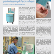
Commercially launched in early 2017, the Carestream OnSight 3D Extremity System is a Cone Beam Computed Tomography (CBCT) scanner designed for point-of-care extremity imaging in weight-bearing patient position for orthopedic clinics, imaging centres, specialty offices, hospitals and emergency departments. The system uses a high-performance amorphous-Silicon (a-Si(H)) flat-panel detector and a unique three-source X-ray tube design. This detector allows for the rapid acquisition of X-ray projections, which helps minimize the negative impact of patient motion. The three-source X-ray tube was designed to reduce the “cone beam” artifact that has traditionally impacted large volume CBCT reconstructions as reported in scientific literature.
The detector and source rotate around the patient’s anatomy, acquiring a multitude of projections from different angles, axially and rotationally.  The images are then reconstructed into a 3D volume using advanced software reconstruction techniques. This produces high resolution volumetric 3D images that have the same spatial resolution in any plane.

Cobalt Health, a leading UK medical charity, has installed the world’s first Carestream OnSight 3D Extremity Cone Beam CT system at their Imaging Centre in Cheltenham, Gloucestershire UK.

Founded in 1964, Cobalt provides a wide range of oncology services across the south-western UK counties of Herefordshire,Worcestershire and Gloucestershire. Cobalt has a history of early investment in new technologies such as MRI and PET/CT. A long-standing Carestream customer, Cobalt was the first facility in the UK and Ireland to implement the Carestream MyVue Patient Portal and currently has both Carestream Vue RIS and Vue PACS installed. Peter Sharpe, CEO of Cobalt Health said: ‘As a charity we’re very used to introducing new technology to support our patients and referring clinicians and this seemed like an ideal opportunity. The Carestream OnSight 3D Extremity CBCT scanner really fitted very nicely, particularly in supporting our orthopedic clinics. It provides something that we couldn’t offer previously, in terms of image resolution and flexibility; it seemed like a really good fit.’ ‘It provides you with true weight bearing images, high resolution and low radiation dose. I think there’s a huge opportunity to embed it in the patIent pathway in A&E and orthopaedic clinics across the UK.’ To introduce the benefits of the OnSight system to the patient pathway, Cobalt held a series of evening seminars where they showed case studies and orthopedic surgeons demonstrated how patients could benefit from the cone beam CT system. ‘It’s the best way of marketing the new technique,’ said Peter Sharpe. ‘The referrers need to come and understand how it works, what the image quality is, and what the benefits are.’

One-stop clinics

Cobalt runs regular one-stop clinics with orthopedic surgeons who refer their patients on the same day for X-rays, MRI or CT scans. Roisin Dobbin-Stacey, PET CT and CT Manager for Cobalt Health explained: ‘The Carestream On-Sight 3D CBCT doesn’t discriminate; it’s not just for sports injuries or for one-stop clinics, it will be available to all patients.’ ‘The weight-bearing feet and ankle exams that we’ve been doing, on people of all ages, have been made considerably easier; it only takes 25 seconds to get these incredible images. They step into the scanner and all they have to do is keep still for 25 seconds.’
‘In the past, when you had a patient who said they had a pain in their foot or ankle when they were walking or running, you would lie them down and do a CT scan and it wouldn’t show anything. You can now put them into the CBCT scanner and see the true condition of a patient who’s got all their weight going through that joint and you can see the difference; you can see why they’ve got that pain.’
‘The dose, of course, is something else that is talked about a lot; referrers ask why they would send their patient for a CT scan when they can have an X-ray; but actually if a patient is having a CBCT scan, the dose is only slightly higher than with an X-ray, and it’s a weight bearing exam. And it’s a lot less than with a CT, so that again is very encouraging.’

Exquisite detail
Consultant Radiologist, Prof. Iain Lyburn, has had a very positive experience with the Carestream OnSight 3D scanner. ‘It’s very high quality, very high resolution,’ he said. ‘The detail is exquisite, so you can see very small bony defects, very small osteophytes, with great clarity. It’s also much quicker than some other investigations, taking less than a minute for many body parts, so you get a cross sectional slice through  the area in a relatively quick time.’ ‘We recently examined a young man with hind foot pain and, whereas an MRI scan showed some edema, with the Carestream CBCT image you could see the bony detail wit absolutely exquisite clarity and what we hadn’t appreciated properly was an ill-defined irregularity around the os trigonum, which was the cause of the pain. It was a very small detail that you couldn’t pick up on the MRI, these small fragments of bone causing the pain. It was very helpful. We had another patient with pain below the ankle joint whose MRI showed some edema across the joint in the calcaneum, so we thought that was probably the cause of the pain. Remember the MRI would be done with the patient lying supine with their ankle on the bed, whereas with the CBCT the patient was standing in the functional  position, and what it highlighted beautifully was a protuberance in the subtalar joint.We could see the impingement far more clearly demonstrated because of the way the image was taken and realized that it was going to be the cause of the symptoms. There was possibly a suspicion of it on the MRI with the edema, but having the cone beam CT showing it in position clarified that that was the source of the symptoms. And that might change the management of the patient, because many times we would do a plain radiograph, see how the patient gets on then get them back.With the Carestream OnSight CBCT you would get the diagnosis straight away and would see most fractures earlier than you would on an X-ray. In imaging, as with many other aspects of medical technology, you’ll look back in a few year’s and see that the Carestream CBCT is irreplaceable.’

Plug and play
Installing the OnSight 3D Extremity system at Cobalt’s Imaging Centre was straightforward, as Roisin Dobbin-Stacey explained. ‘Planning and getting the room ready for delivery of the equipment was very easy; the room size had to be a minimum of 8 feet by 12 feet (Ed. 2.5m x 3.7m). The equipment arrived, it was brought up in the lift, wheeled in and plugged into a 240 volt socket. It literally is plug and play!’ ‘The Carestream engineers were fantastic, they got it all up and running within a couple of days, and the Apps training was brilliant. I think the system itself, how it’s been designed, is so user friendly. As a radiographer you want something that’s easy to use, and for me it’s fantastic, it’s such good fun to use. Cobalt CEO Peter Sharpe summed up his feelings about the Carestream OnSight CBCT system: ‘we have no regrets. It’s an excellent device, it works well and uptime has been 100 percent. It’s easy to use, patients love it and the image quality is superb so yes, it’s been a great investment.’

Carestream Healthwww.carestream.com

https://interhospi.com/wp-content/uploads/sites/3/2020/08/IH133_Carestream_Peter-Sharpe_crop.jpg 554 658 3wmedia https://interhospi.com/wp-content/uploads/sites/3/2020/06/Component-6-–-1.png 3wmedia2020-08-26 14:17:292021-01-08 12:30:16Extremity Cone Beam CT imaging demonstrates value of weight-bearing scans

Series 1140 SmartLab

, 26 August 2020/in Featured Articles /by 3wmedia
https://interhospi.com/wp-content/uploads/sites/3/2020/08/47198_hans-rudolph.jpg 1462 1012 3wmedia https://interhospi.com/wp-content/uploads/sites/3/2020/06/Component-6-–-1.png 3wmedia2020-08-26 14:17:292021-01-08 12:30:32Series 1140 SmartLab

IT and patient safety – demands for a change in culture

, 26 August 2020/in Featured Articles /by 3wmedia

Two studies on information technology and patient safety were released in the US in April 2018.
The first, by ECRI Institute and its Partnership for Health IT Patient Safety stakeholder collaborative, takes a searching look at the hidden risks of healthcare IT (information technology) systems.
The second report, by Pew Charitable Trusts, focuses on electronic health records (EHRs), and is based on its long-standing view that in spite of more than 30 billion dollars (£25 billion) of federal health IT investment over the past decade, the transition from paper to electronic records has yet to reach its potential to enhance healthcare coordination and improve patient safety.
Both studies call for a much greater degree of proactivity to anticipate problems before they run out of control. Interestingly, there also seem to be several parallels in their respective analyses of the challenges.

A broad-based look at patient safety
One of the first priorities for healthcare organizations is to tie health IT into existing patient safety initiatives. The challenges that exist whenever older systems are updated or replaced, or integrated with successor technologies, can ideally become learning opportunities. However, this requires the organization to have a collaborative management culture that makes initiatives such as IT safety and other best practices part of their daily workflow.
Both the ECRI and Pew are emphatic about the need for collaboration. ECRI lays down specific recommendations on obtaining feedback from stakeholders (patients, payers, cybersecurity experts, regulators, providers and others). The Pew report also maintains that collaboratively gathering inputs from across the healthcare stakeholder spectrum is critical to improve patient care and reduce provider burden in the context of EHR use.

Tip of the iceberg
The ECRI Safe Practice Recommendations begin with an ominous warning – that the known universe of IT-related safety problems are likely to be just the tip of the proverbial iceberg. The net risk from such issues is that a variety of bugs are lurking beneath the surface, and, in the ECRI’s view, pose permanent dangers to patients. Such risks, in turn, are compounded by the sharp growth in recent years of ransomware and other cybersecurity threats which seek to exploit loopholes in codes. For ECRI, this is a key reason why both providers and vendors need to make health IT safety an integral part of their overall patient safety program.

The ‘over’-customization quandary
In general, the roots of healthcare IT problems stretch back to the 1980s and 1990s when demands for customization led to ad-hoc rewriting of legacy programs, so as to avoid loss of functionalities. So-called patches, as part of platform updates, also sought to retain some of these functionalities, many of which had been proven – or otherwise become indispensable – over the years.  During this process, poor documentation of code changes was commonplace. In addition, since legacy systems faced many problems communicating with one another across their proverbial silos, layers of integrative middleware were added in sequence until they became cluttered and unmanageable.
Many such concerns are reflected by Pew. Over customization, auto-refresh mix-ups and unclear default settings with EHRs, as well as alert fatigue, can all result in patient harm.
An earlier Pew report in December 2017 had explained that safety problems could be caused by the very design of an EHR system (e.g. complex interfaces and guidance terminology) or by its customization during implementation and adaptation. Like the middleware in an IT system, Pew explains that an EHR “interface that is cluttered may cause confusion or an inability to locate key information, whereas an overly bare display may force the clinician to search for information in multiple places.”
Indeed, some of the reasons for risky over-customization of EHRs directly reflect those made during program rewrites to legacy IT systems in the 1980s and 1990s.
As the Pew study observes, healthcare facilities often work with vendors to customize certain aspects of their EHR system which fit their workflow, for example by displaying data which is critical for specific clinicians at a particular facility. However, it warns, such customizations “may not have undergone rigorous testing and could lead to unintended safety consequences.”

Specific risks with EHRs
The Pew report highlights a range of specific risks. One of the most commonplace may consist of mix-ups in auto-refresh of patient lists when EHRs revert to the default view. In such cases, providers can inadvertently make medical decisions based on another patient, rather than the one being treated, if they do not realize the system has just refreshed a particular list.
Default EHR settings are also seen as a specific danger for medication dosages. Healthcare providers may think they are ordering a fixed dose of a particular drug, while what they enter instead “is multiplied by the patient’s weight, potentially contributing to overdoses,” Pew notes.
Yet another problem consists of incomplete laboratory results, which can result in erroneous medical decisions since a provider lacks complete information on a particular patient. In effect, physicians may fail to realize that not all lab results are displayed on a screen, or that results may have been delayed – among other reasons, because samples might still be undergoing testing.

In its recommendation to integrate health IT safety into a broader safety program, the ECRI report also pointed explicitly to the rise in duplicate medication errors after a new EHR implementation. Contributing factors include the design of the EHR as well as the occurrence of task-related changes (such as multiple persons entering orders for the same patient at the same time).
Other EHR-related problems mentioned as cases by ECRI included data entry of a pediatric patient’s weight in pounds rather than kilograms – followed by erroneous medication dose, incorrect alert responses due to the simultaneous opening of multiple patient charts, the inability to account for problems such as a pending swallow evaluation before a dietary order, or an allergy to eggs which contraindicate propofol.
Some of the most frequent EHR problems concerned mix-ups of patients, in one instance due to two having the same name !

New safety approaches: collaboration and workflow integration
Beyond EHRs, as the hitherto-unknown problems in the health IT iceberg become more apparent with time, users are advised by the ECRI to collaborate in order to integrate and embed new safety practices into their daily workflow. Suggestions include the provision of inc entives for actively working together on safety-related efforts, and to learn and share analysis of near-misses and other hazards, as well as workaround strategies.

Pew too had stressed the importance of collaboration for improved EHR use, in its December 2017 report.  One way towards this is to bring stakeholders together to share data on patient safety incidents and do this in a “nonpunitive environment.” After this, stakeholders can be encouraged to “develop solutions for common and significant usability issues.”
Pew also suggests that safety tests on functionality and usability are conducted by entities throughout the entire EHR life-cycle – from development and after implementation. Such a process should bring together developers, IT professionals, clinicians, nurses and pharmacists – in essence, every one using the EHR system.
Pew cautions that one specific area for attention is alert fatigue (due to too many unnecessary or false alerts). This can result in genuine life-saving warnings being missed. One concrete means to avoid such conundrums is by designing EHR systems to specifically verify and red flag certain potential problems (e.g. dangerous drug interactions with different medications).

The need for encouraging a collaborative culture to enhance healthcare IT safety has also been identified in Europe. In 2015, a team led by Solvejg Kristensen at Denmark’s Aalborg University studied the association of quality management systems with teamwork and safety from seven European countries. Although they found different approaches to quality management systems and to perceptions of teamwork and safety climate, they noted the importance of organizations investing in leadership, time, capital and technical expertise to attain continuous quality improvement and enhance patient safety.
Indeed, at whichever facet of the healthcare technology spectrum one looks at, proactive, meaningfully structured collaboration may be the only way to achieve a unified vision of safe health IT and a wider culture of safety in the health enterprise.
As healthcare IT becomes increasingly pervasive, such concerns are bound to demand increasing attention.
The National Patient Safety Foundation and IHI
Many of these issues – in terms of both challenge and response – were the subject of a set of eight recommendations made in 2015 in a report from the National Patient Safety Foundation (NPSF) in the US to ensure “that technology is safe and optimized to improve patient safety”. The recommendations are as follows:

  1. Ensure that leaders establish and sustain a safety culture
  2. Create centralized and coordinated oversight of patient safety
  3. Create a common set of safety metrics that reflect meaningful outcomes
  4. Increase funding for research in patient safety and implementation science
  5. Address safety across the entire care continuum
  6. Support the healthcare workforce
  7. Partner with patients and families for the safest care
  8. Ensure that technology is safe and optimized to improve patient safety

European initiatives
In 2016, the NPSF added that it was also important  to make health IT-related patient safety an organizational priority by securing management commitment, and to develop an environment which was “conducive to  detecting, fixing and learning from system vulnerabilities.”
The NPSF was merged with the Institute for Healthcare Improvement (IHI) last year, and some of its initiatives are likely to be transferred to Europe via the IHI Health Improvement Alliance Europe (HIAE), which aims to improve work processes and create new delivery models relevant to European health systems. The HIAE has already established connections with professional societies in several countries, including Britain, Denmark and Belgium.
One good example of HIAE efforts is the The Platform for Continuous Improvement of Quality of Care and Patient Safety (PAQS), a Belgium-based initiative which aims to consolidate relationships between various stakeholders in healthcare in order to work together, in a consistent and cohesive manner.
Other facets of IHI which are expected to make a mark in Europe include the so-called Open School Online Courses. Several of these are directly concerned with IT-focused elements of patient safety and the need to build a culture of safety in a health organization.

https://interhospi.com/wp-content/uploads/sites/3/2020/08/IH172_Health-IT-Safety_Tosh_thematic.jpg 449 800 3wmedia https://interhospi.com/wp-content/uploads/sites/3/2020/06/Component-6-–-1.png 3wmedia2020-08-26 14:17:292021-01-08 12:30:11IT and patient safety – demands for a change in culture

The world’s fastest hemoglobin analyzer

, 26 August 2020/in Featured Articles /by 3wmedia
https://interhospi.com/wp-content/uploads/sites/3/2020/08/47212_IHE-92x178mm_DiaSpect-POC-Connect_Jan-18.jpg 1500 775 3wmedia https://interhospi.com/wp-content/uploads/sites/3/2020/06/Component-6-–-1.png 3wmedia2020-08-26 14:17:292021-01-08 12:30:18The world’s fastest hemoglobin analyzer
Page 88 of 102«‹8687888990›»

Latest issue of International Hospital

April 2024

27 June 2025

Scientists launch world first project to create synthetic human genome with £10mn Wellcome funding

27 June 2025

GE HealthCare advances precision imaging with MIM Encore platform

27 June 2025

GE HealthCare advances cardiac PET diagnostics with Flyrcado launch

Digital edition
All articles Archived issues

Free subscription

View more product news

Get our e-alert

The medical devices information portal connecting healthcare professionals to global vendors

Sign in for our newsletter
  • News
    • Featured Articles
    • Product News
    • E-News
  • Magazine
    • About us
    • Archived issues
    • Media kit
    • Submit Press Release

Beukenlaan 137
5616 VD Eindhoven
The Netherlands
+31 85064 55 82
info@interhospi.com

PanGlobal Media IS not responsible for any error or omission that might occur in the electronic display of product or company data.

Scroll to top

This site uses cookies. By continuing to browse the site, you are agreeing to our use of cookies.

Accept settingsHide notification onlyCookie settings

Cookie and Privacy Settings


How we use cookies

We may ask you to place cookies on your device. We use cookies to let us know when you visit our websites, how you interact with us, to enrich your user experience and to customise your relationship with our website.

Click on the different sections for more information. You can also change some of your preferences. Please note that blocking some types of cookies may affect your experience on our websites and the services we can provide.

Essential Website Cookies

These cookies are strictly necessary to provide you with services available through our website and to use some of its features.

Because these cookies are strictly necessary to provide the website, refusing them will affect the functioning of our site. You can always block or delete cookies by changing your browser settings and block all cookies on this website forcibly. But this will always ask you to accept/refuse cookies when you visit our site again.

We fully respect if you want to refuse cookies, but to avoid asking you each time again to kindly allow us to store a cookie for that purpose. You are always free to unsubscribe or other cookies to get a better experience. If you refuse cookies, we will delete all cookies set in our domain.

We provide you with a list of cookies stored on your computer in our domain, so that you can check what we have stored. For security reasons, we cannot display or modify cookies from other domains. You can check these in your browser's security settings.

.

Google Analytics Cookies

These cookies collect information that is used in aggregate form to help us understand how our website is used or how effective our marketing campaigns are, or to help us customise our website and application for you to improve your experience.

If you do not want us to track your visit to our site, you can disable this in your browser here:

.

Other external services

We also use various external services such as Google Webfonts, Google Maps and external video providers. Since these providers may collect personal data such as your IP address, you can block them here. Please note that this may significantly reduce the functionality and appearance of our site. Changes will only be effective once you reload the page

Google Webfont Settings:

Google Maps Settings:

Google reCaptcha settings:

Vimeo and Youtube videos embedding:

.

Privacy Beleid

U kunt meer lezen over onze cookies en privacy-instellingen op onze Privacybeleid-pagina.

Privacy policy
Accept settingsHide notification only

Sign in for our newsletter

Free subscription