

Globally, rising antibiotic resistance is mainly due to the increasing use of antibiotics [1]. There are several reasons for this increased use, such as ageing of the patient population, increased use of medical devices and increased possibilities for extensive surgery. However, it is also known that 50% of all medical antibiotic use is incorrect, that is in terms of indication, choice, way of administration and /or duration [2]. In this article, the focus is on the empiric choice and steps to be taken to make an optimal choice.
Dr C. van der Donk, Dr C. den Heijer and Dr E. Stobberingh
An example of wrong indication is the use of antibiotics in case of a respiratory tract infection which is caused by a viral agent. An incorrect way of administration is the topical use of an antibiotic in case of a severe wound infection. An example of an incorrect − too short − duration is a one-day therapy with amoxicillin for the treatment of an uncomplicated urinary tract infection; likewise, a 14-day therapy for the same indication indicates a too long duration. An incorrect choice would focus mainly on the empiric choice of an antimicrobial agent. An empiric antibiotic choice means to make a choice without knowing the causative agent of the infection to be treated and/or the antibiotic susceptibility pattern of this putative agent.
Before making an optimal empiric choice
Most infections are caused by the patient

Last March, the 29th edition of the Korean International Medical & Hospital Equipment Show attracted a record number of visitors, over 68,000 in total, which represents a growth of nearly 14% over last year. That
At ECR 2013 Carestream is showcasing a raft of new solutions designed to help radiology professionals improve patient care. These include a new portal that allows patients to electronically access and manage their X-ray exams; new lesion management tools that can help enhance accuracy in assessing changes in cancerous lesions and, as a works-in-progress, a smaller-format digital radiography detector that provides high-quality, dose-sensitive X-ray images for pediatric, orthopedic and general radiology exams.
Healthcare IT
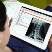
The MyVue patient portal empowers patients to electronically access and manage their X-ray exams—and then share that data with specialists and other healthcare professionals. The Web-based portal is implemented by healthcare providers to allow patients to download information to PCs, laptops, iPads or other devices. It is easy to use and reduces the time and cost of outputting medical exams onto DVD/CDs or other physical storage formats for medical records. My Vue is currently available as an option for Vue PACS and Vue Archive users and is now available for order as a Vue Cloud Service.
New lesion management tools can help enhance accuracy in assessing changes in cancerous lesions as part of diagnosis and treatment for oncology patients. A native follow-up application that can be added to Vue PACS, the new module provides semi-automatic tracking and segmentation of lesions from modalities. Each measurement generates an anatomical bookmark within the exam.
Enhancements to Carestream’s RIS including the storage and tracking of radiation dose information and other capabilities that lay the groundwork to support cumulative dose tracking, which is an important patient care initiative worldwide.
Digital Capture
In the wireless digital radiography market, a smaller-format 25 cm x 30 cm Carestream DRX 2530C detector is demonstrated as a work in progress. The new cesium iodide detector is designed to offer high efficiency for dose sensitive pediatric, orthopedic and general radiology exams. The smaller detector is designed to fit into pediatric incubator trays and offer higher DQE (detective quantum efficiency), which can lead to lower dose requirements than CR cassettes or gadolinium scintillator detectors. The new DRX 2530C detector is intended to be used with Carestream DRX-Revolution or Carestream DRX-Mobile Retrofit Kits for mobile imaging of neonatal or pediatric patients. In orthopedic and general radiology imaging, the smaller detector is designed to aid in positioning for tabletop exams such as knee, elbow, skull and other exams.
Also highlighted will be a new non-motorized option for the Carestream DRX-Evolution, a versatile DR system with modular components. The new DRX-Evolution Standard-Q offers DR capability at an affordable cost. The ergonomically designed wall stand makes standing exams easier with its extensive vertical travel range. In addition, the extra-wide Standard-Q elevating float-top table lowers easily to accommodate stretcher and wheelchair patients and offers increased patient weight capacity.
Mammography
A new module displays digital breast tomosynthesis (DBT) exams from DICOM-compliant acquisition devices on its Carestream Vue Mammo Workstation, where radiologists can also view traditional mammograms, breast ultrasound, breast MRI and general radiology exams from a single desktop. The module streamlines workflow by allowing healthcare providers to store, route, display and query/retrieve DBT exams from DICOM-compliant acquisition devices. Comparison tools enable radiologists to use personalized hanging protocols for DBT exams along with other procedures.
Digital Output
The new DryView 5950 Laser Imaging System produces 508 pixels-per-inch output for general radiology and mammography images. The new imager can support efficient printing and time-saving film cartridges and delivers an enhanced quality control system for mammography images. This innovative internal quality control system includes a built-in densitometer that will produce test prints and display data needed to support mammography quality control charting.
Dental
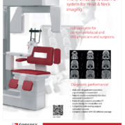
The CS 9300 System is a high-quality cone beam CT (CBCT – 3D imaging) and true panoramic imaging system for ENT and dental indications. The system can be used for a variety of ENT and dental applications – including sinus, temporal bone and maxillofacial exams; dental implantology; oral surgery; orthodontics; periodontics and endodontics. The CS 9300 is a cost-effective solution to offload ENT and dental CT, delivering up to 94 % less radiation dose than conventional CT units, and images at a much higher resolution, making it ideal for visualizing fine bony structures in the
middle ear and radicular (root) structures.
Carestream Health
www.ihe-online.com & search 46315
Bladder cancer is a significant cause of morbidity and mortality worldwide, but with the exception of countries where schistosomiasis is prevalent, such as Egypt, the disease most commonly affects older men in developed countries. In EU residents it is the fourth most frequent cancer among men, accounting for around 7% of total cancers and 6% of cancer deaths; in women it is the tenth most frequent cancer accounting for 2% of cancer deaths.
Surgery is involved in the management of most bladder cancers, with transurethral resection being the usual approach to facilitate diagnosis and staging, and to remove small superficial tumours, but for areas of the bladder that are not easily accessible, manipulation of the endoscope and other instruments may injure healthy tissues. For the approximately 30% of tumours that are muscle-invasive, open radical cystectomy (ORC) is the normal standard of care. This procedure is medically challenging for the patient, and because of the duration of the surgery together with the inevitable loss of blood and other fluids, it is contraindicated for many elderly patients.
The answer to these problems is robotic-assisted surgery. An innovative telerobotic system, which is under development at Vanderbilt and Colombia universities, USA, incorporates a segmented working arm of only 5.5 mm diameter for viewing as well as supporting micro-instruments. The inherent flexibility of the device should eventually allow micrometer-resolution images of the entire bladder wall and removal of all superficial tumour cells.
A recently published pilot trial in the US involving 47 randomized patients with invasive bladder cancer compared ORC with robotic-assisted laparoscopic radical cystectomy (RARC). While there was no reported difference in treatment outcomes, the latter group lost less blood and needed fewer transfusions, experienced a quicker return of bowel function and had shorter hospital stays. A larger scale trial involving several healthcare centres is now ongoing, which will also follow long-term outcomes.
Robotic-assisted surgery is expensive, though. Already in terms of average healthcare costs per patient, bladder cancer is the most expensive adult cancer in the West, mainly due to the high rate of recurrence. But telerobots could actually go a long way towards preventing recurrence and thus reducing costs, and while the actual procedure of RARC is currently more expensive than ORC, shorter hospital stays with fewer interventions should help balance the books.
Prins Hendrikstraat 1
5611HH Eindhoven
The Netherlands
info@interhospi.com
PanGlobal Media IS not responsible for any error or omission that might occur in the electronic display of product or company data.
