• News
    • Featured Articles
    • Product News
    • E-News
  • Magazine
    • About us
    • Digital edition
    • Archived issues
    • Media kit
    • Submit Press Release
  • White Papers
  • Events
  • Suppliers
  • E-Alert
  • Contact us
  • FREE newsletter subscription
  • Search
  • Menu Menu
International Hospital
  • AI
  • Cardiology
  • Oncology
  • Neurology
  • Genetics
  • Orthopaedics
  • Research
  • Surgery
  • Innovation
  • Medical Imaging
  • MedTech
  • Obs-Gyn
  • Paediatrics

Archive for category: Product News

Products News

Chest CT software

, 26 August 2020/in Product News /by 3wmedia

AI-Rad Companion Chest CT, an intelligent software assistant for radiology, was recently
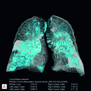
awarded the CE mark. This artificial intelligence (AI)-based software helps radiologists interpret CT (Computed Tomography) images of the thorax faster and more precisely, and to document the findings in less time with the help of automatic measurements. AI-Rad Companion Chest CT is the first AI-based application on the new AI-Rad Companion platform, and is vendor-neutral, which means the software can evaluate image data from many CT system manufacturers. Siemens Healthineers plans to expand this platform so that more and more intelligent algorithms will be available for additional organs and modalities. This will enable the company to consistently expand its portfolio of effective solutions for AI-based clinical decision support. Results drawn up by radiologists can vary by some 10-20 percent, depending on the investigator. Algorithm-based diagnostics completely obviate this variability, generating constant results. That alone is a huge advantage of AI platforms like AI-Rad Companion. Using CT images of the thorax (chest), the software can differentiate between the various structures of the chest, highlight them individually, and mark and measure potential abnormalities automatically. This applies equally to organs such as the heart and lungs, aorta, and vertebral bodies. The software automatically turns the findings into a quantitative report, which can be called up via the image viewing system used by the radiologist in the clinical routine. In certain circumstances, the intelligent assistant also alerts physicians to potential abnormalities that would otherwise have been missed because they were not the focus of the original examination, e.g. chance discoveries of pathological dilations of the aorta (aneurysms). Examination using a chest CT is a procedure often used in everyday clinical practice. For radiologists, this means more examinations in a limited amount of time and usually for low reimbursement rates. AI-Rad Companion Chest CT is a tool that can actually simultaneously increase productivity and quality in diagnostic radiology. AI-Rad Companion Chest CT is a cloud-based solution and uses certified, secure teamplay infrastructure that complies with the Health Information Portability and Accountability Act (HIPAA) in the U.S., and with the General Data Protection Regulation (GDPR) in the EU. The software conforms to Digital Imaging and Communications in Medicine (DICOM) standards. The images and all supporting information can be made automatically available in the picture archiving and communication system (PACS) in line with the radiologist’s individual requirements.
Read more

https://interhospi.com/wp-content/uploads/sites/3/2020/08/IH278_Siemens_2_image_of_the_lungs.jpg 430 800 3wmedia https://interhospi.com/wp-content/uploads/sites/3/2020/06/Component-6-–-1.png 3wmedia2020-08-26 14:31:322021-01-08 12:10:23Chest CT software

Digital mammography system

, 26 August 2020/in Product News /by 3wmedia

AMULET Innovality is Fujifilm’s flagship in digital mammography solutions. Images resulting from its innovative approach to tomosynthesis clinical application – two different acquisition modes, to better fulfil breast screening and diagnostic mammography needs – with unsurpassed 50 micron pixel size, will be shown together at the booth.

Read more

https://interhospi.com/wp-content/uploads/sites/3/2020/08/IH189_Fujifilm_Amulet-Innovality_crop.jpg 800 382 3wmedia https://interhospi.com/wp-content/uploads/sites/3/2020/06/Component-6-–-1.png 3wmedia2020-08-26 14:31:322021-01-08 12:10:27Digital mammography system

QA measurement system for specific X-ray modalities

, 26 August 2020/in Product News /by 3wmedia

RaySafe X2 Solo is a new product line that covers the measurement needs of specific X-ray modalities. It’s based on the same technology as RaySafe X2, but instead of multi-modality capability, each model meets specific needs. RaySafe X2 Solo features a large touch screen showing all parameters simultaneously, sensors ready for measurements without special settings or modes and a base unit storing all readings and showing full waveforms. It offers  true ease-of-use, which saves valuable time and minimizes the risk of making faulty measurements. RaySafe X2 Solo removes unnecessary steps in taking a measurement – like positioning the sensor, choosing a setting, or interpreting results. The R/F and DENT sensors are both orientation-independent so the only thing to do is to place the sensor in the X-ray beam and turn on the instrument. The rest is automatic – no menus or special settings needed. RaySafe X2 Solo combines state-of-the-art sensor technology with an intuitive and proven user interface, ensuring user friendliness. Each X2 Solo includes a specific sensor to cover specific needs.

Read more

https://interhospi.com/wp-content/uploads/sites/3/2020/08/IH274_Raysafe_shirley.jpg 385 700 3wmedia https://interhospi.com/wp-content/uploads/sites/3/2020/06/Component-6-–-1.png 3wmedia2020-08-26 14:31:322021-01-08 12:10:31QA measurement system for specific X-ray modalities

Premium 1.5T MRI system

, 26 August 2020/in Product News /by 3wmedia

Canon Medical Systems Corporation enters the premium 1.5T MRI market with the launch of its new Vantage Orian system. Vantage Orian is one of Canon Medical Systems’ first major product releases since officially changing their company name from Toshiba Medical on January 4, 2018, and represents a major foray into the premium wide bore 1.5T market along with new technology designed to boost productivity, enhance patient comfort and deliver diagnostic clinical confidence to users and patients alike.
With a new slim gradient that delivers a gradient performance with a maximum amplitude of 45 mT/m, combined with a slew rate of 200 T/m/s that enhances high resolution and diffusion imaging, Vantage Orian offers high performance imaging capability previously not available on 1.5T systems from Canon Medical Systems and utilizes migrated high-end 3T technology.  Canon Medical System’s PURERF and Saturn Gradient technology means that stable and consistent imaging performance is ensured, increasing signal to noise ratio (SNR) by up to 38%.
The Vantage Orian offers a range of new Rapid Scan and Easy Tech technology that reduces scan time and helps improve workflow. A redesigned gantry interface and dockable table ensure seamless patient handling. New applications like Multiband SPEEDER, that shortens acquisitions such as high angular resolution diffusion imaging (HARDI), and up to x8 accelerated k-t SPEEDER allows high frame rate cardiac cine and perfusion imaging with free breathing.
Vantage Orian also offers one of the smallest footprints in the 1.5T wide-bore class and low power consumption eco features that reduce energy use by up to 21%. The reliable system is equipped with excellent maintenance programs to satisfy the needs of hospital administrators.
A relaxed patient is key in MRI, and Vantage Orian features quiet scan sequences that reduce ambient noise by Pianissimo Zen technology. The 71cm wide bore and immersive virtual MR Theater are designed to put patients at ease. Vantage Orian also addresses challenging patients with Pediatric SPEEDER coils, contrast- free applications and ForeSee View for simplified scan planning.
Read more

https://interhospi.com/wp-content/uploads/sites/3/2020/08/IH262_Canon.jpg 448 618 3wmedia https://interhospi.com/wp-content/uploads/sites/3/2020/06/Component-6-–-1.png 3wmedia2020-08-26 14:31:322021-01-08 12:10:35Premium 1.5T MRI system

Dedicated neonatal MRI system installed in the NICU

, 26 August 2020/in Product News /by 3wmedia

Aspect Imaging’s Embrace Neonatal MRI System is the world’s first FDA-cleared and CE-approved dedicated MRI system installed inside the NICU. It can provide a high-resolution MRI scan for babies weighing from 1.0 Kg to 4.5 Kg and a head circumference up to 38 cm. Embrace has been designed specially to be placed inside the NICU to avoid the risks of transporting infants to an external MRI facility, enabling safer imaging of newborns and allowing medical staff and parents to be present during the scan. Due to its unique design, the system can also scan intubated babies without having to disconnect and reconnect any tubing. The total Prep and Scan process takes less than an hour. Embrace is quieter than traditional whole-body scanners and features a permanent magnet that has low power consumption and requires no electrical, cryogenic or water cooling or a special safety zone or RF-shielded room. This means that for the first time a dedicated neonatal MRI – FDA-cleared and CE-Approved – can be placed inside the NICU without the risks and limitations of traditional MRI systems. Additionally, Embrace features the Neonatal Capsule – an integrated, temperature-controlled, incubator-like patient bed which minimizes movement of the baby, allowing continuous control of the baby’s environmental temperature and continuous monitoring of its vital parameters. Shaare Zedek Medical Center in Jerusalem, Israel, has recently installed Aspect Imaging’s Embrace Neonatal MRI System inside its Neonatal Intensive Care Unit (NICU), where it is in regular use. 
Read more

https://interhospi.com/wp-content/uploads/sites/3/2020/08/Aspect_Imaging_Embrace_Neonatal_MRI_System_Formal_Image.png 1152 2048 3wmedia https://interhospi.com/wp-content/uploads/sites/3/2020/06/Component-6-–-1.png 3wmedia2020-08-26 14:31:322021-01-08 12:10:42Dedicated neonatal MRI system installed in the NICU

Hologic was due to showcase the following new products at ECR 2020, which has been postponed due to the COVID-19 pandemic

, 26 August 2020/in Corona News, Product News /by 3wmedia

Breast Imaging
The 3Dimensions Mammography System is the industry’s fastest and highest resolution breast tomosynthesis system on the market. The system offers a variety of features designed to provide higher quality 3D images for radiologists, and enhanced workflow for technologists, with lowdose options, for patients. For example, the system includes Intelligent 2D imaging technology, which works with the system’s Clarity HD highresolution 3D imaging to deliver unprecedented clarity, contrast and detail at a lower dose.
Another feature available with the 3Dimensions system is the Quantra 2.2 breast density assessment software, which enables standardization in patient protocols, providing reproducible and consistent breast density assessment. Furthermore, thanks to the addition of the SmartCurve breast stabilization system, the 3Dimensions system is also clinically proven to deliver a more comfortable mammogram compared to standard compression, without compromising on speed, dose or accuracy.
Interventional
The Brevera® Breast Biopsy System with CorLumina® Imaging Technology is a real-time breast biopsy and verification system that improves the patient experience and streamlines the biopsy process from start to finish. Before the Brevera system, radiologists performing stereotactic breast biopsy procedures to diagnose breast cancer were often required to leave the patient under compression while they moved to another room to image and verify tissue samples. With the Brevera system, radiologists are able to obtain and image tissue samples in the procedure room in just a few seconds, potentially saving up to 10 minutes per patient and cutting the procedure time by up to 25%.
The Affirm® Prone Breast Biopsy System is the only dedicated prone biopsy system offering superior 2D or tomosynthesis imaging. Thanks to this high-quality 2D or tomosynthesis imaging and a field of view that’s more than 6.5 times larger than older generations of prone biopsy systems, clinicians can visualize more tissue and pinpoint subtle lesions and faint calcifications that may not be visible on older systems. Of note, more than 95% of patients reported their Affirm prone biopsy procedure was faster, more comfortable and less painful than expected. The system is designed with patients’ physical and emotional comfort in mind. Direct view of the biopsy needle is eliminated helping to reduce patient stress, and clinicians can quickly identify, target and access lesions to help reduce time spent in compression.
The Faxitron™ Core Specimen Radiography System is designed for efficiency. Within seconds this self-contained, table top unit provides highresolution imaging for immediate core sample verification in the biopsy room.
Breast Surgery
The LOCalizer™ Wireless Radio Frequency Identification (RFID) Breast Lesion Localization System is a non-radioactive, radiofrequency localization system designed for precise marking and targeting of lesions in breast-conserving surgery. It was designed to replace the traditional wire-guided localization method, which requires placement of a wire on the day of surgery. With the tag, placement can be done weeks or months prior to surgery. For patients, this means they can arrive closer to their surgery time and experience fewer interventional procedures.
The Trident® HD Specimen Radiography System delivers enhanced image quality, improved workflow and instant sample verification during breastconserving surgeries and stereotactic breast biopsies. The Trident products are the only specimen radiographs on the market to use amorphous selenium direct capture imaging – the same detector technology used in Hologic’s 3Dimensions mammography system – to generate crisp, clear, high-resolution images.
Ultrasound
The Viera™ Portable Breast Ultrasound System is a wireless, handheld ultrasound device that delivers accurate, high-resolution diagnostic images at the point of care, enabling optimization of clinical workflow and patient pathway. The system seamlessly transmits images to smart devices and communication systems (PACS) in the office, exam room or surgical suite, and allows facilities to add interventional breast ultrasound services for a fraction of the cost of comparable cart systems. Additionally, the Supersonic Mach 30 Breast Ultrasound System provides fast, high quality imaging and is the first to offer an intuitive touchpad control and large adjustable touchscreen display. The system’s enhanced ergonomics are designed to increase usability.
Read more

https://interhospi.com/wp-content/uploads/sites/3/2020/08/hologic_3dimensions.jpg 1700 1106 3wmedia https://interhospi.com/wp-content/uploads/sites/3/2020/06/Component-6-–-1.png 3wmedia2020-08-26 14:31:322021-01-08 12:10:20Hologic was due to showcase the following new products at ECR 2020, which has been postponed due to the COVID-19 pandemic

Ultrasound quality assurance software

, 26 August 2020/in Product News /by 3wmedia

The CIRS Ultrasound Quality Assurance Portal helps users of CIRS general-purpose ultrasound phantoms manage their ultrasound testing programme with an easy-to-use, cloud-based application. Video and written instructions for all tests are provided within the app, and test results can either be entered directly to the app, or a data entry form can be downloaded for offline collection. The Ultrasound QA Portal provides convenient reporting features and charting features to track results. It comes pre-loaded with default testing protocols that are designed to meet the recommended practices of the American College of Radiology, the American Institute of Ultrasound in Medicine and the American Association of Physicists in Medicine. Users can easily modify to meet their specific requirements. The QA Portal will make starting and managing ultrasound quality assurance testing programme as easy as possible. Use of the QA Portal Software is complimentary for any CIRS customer who owns one or more of the following phantoms; 040GSE, 054GS, ATS539 or ATS570. To get started, the user has to register with the following information; phantom serial number, model number, institution name, institution contact phone number, first name, last name and email at http://qaportal.cirsinc.com.
Read more

https://interhospi.com/wp-content/uploads/sites/3/2020/08/IH277_CIRS_QA_PORTAL.jpg 488 800 3wmedia https://interhospi.com/wp-content/uploads/sites/3/2020/06/Component-6-–-1.png 3wmedia2020-08-26 14:31:322021-01-08 12:10:24Ultrasound quality assurance software

Certified app for detecting atrial fibrillation and other arrhythmias

, 26 August 2020/in Product News /by 3wmedia

FibriCheck is a medically certified app (CE-class IIa and FDA-cleared) capable of detecting atrial fibrillation and other heart rhythm disorders, thereby preventing strokes. FibriCheck is leveraging mobile technologies for a better health. FibriCheck uses the built-in photoplethysmographic (PPG) sensors commonly found in smartphones and smartwatches to measure the rhythm of the user’s heartbeat. Employing medically-certified algorithms together with an artificially intelligent deep neural network, FibriCheck detects the presence of atrial fibrillation and other potentially fatal cardiac arrhythmias. It is as good as a 1-lead ECG and 96,5% accurate compared with a 12-lead ECG. By decentralizing the diagnostic process, FibriCheck is paving the way for broader access to effective care for individuals while reducing the total cost of healthcare for governments and insurers, thus helping to make Health 2.0 a reality for communities around the globe. FibriCheck is commercially available since May 2017, prescribed by over 1,200 doctors, used by over 110,000 people and reimbursed by selected healthcare funds.
Read more

https://interhospi.com/wp-content/uploads/sites/3/2020/08/IH265_Qompium.jpg 533 800 3wmedia https://interhospi.com/wp-content/uploads/sites/3/2020/06/Component-6-–-1.png 3wmedia2020-08-26 14:31:322021-01-08 12:10:27Certified app for detecting atrial fibrillation and other arrhythmias

Dolphin™ (FIS) Fluid Immersion Simulation®

, 26 August 2020/in Product News /by 3wmedia

A unique, patented technology that re-distributes pressure to prevent tissue damage by emulating the effects of a body "floating" in a fluid medium. All Dolphin FIS surfaces provide an environment that promotes tissue perfusion and prevents injury related to compromised blood flow.

Dolphin FIS has been proven clinically effective at speeding the healing of advanced stage or multiple pressure ulcers, flaps, skin grafts, burns and other wound conditions and is available for beds and seat cushions, providing protection for high risk patients throughout their hospital stay or at home.

To find out more about how Dolphin can save your clinical team time and energy, while ensuring best practice, reduced risk and costs, visit www.dolphinfis.com. Alternatively, please contact Joerns Healthcare on +44 1905 842 211 or send an email to export@joerns.com. Distributor enquiries welcome.
Read more

https://interhospi.com/wp-content/uploads/sites/3/2020/08/Joerns_Dolphin.jpg 1639 2362 3wmedia https://interhospi.com/wp-content/uploads/sites/3/2020/06/Component-6-–-1.png 3wmedia2020-08-26 14:31:322021-01-08 12:10:35Dolphin™ (FIS) Fluid Immersion Simulation®

Premium mammography system

, 26 August 2020/in Product News /by 3wmedia

The unique 50 degree 3D HD Breast Tomosynthesis provides the highest depth resolution in tomosynthesis, and thus delivers excellent quality 3D images. Now also biopsies can be performed leveraging this wide tomosynthesis angle. The HD Breast Biopsy solution allows targeting suspicious areas with one click with a +/- 1mm accuracy. The new integrated specimen imaging tool facilitates the immediate control of the biopsy directly at the mammography system. The new Mammomat Revelation also provides automated breast density measurements at the point of examination. This allows for personalized risk stratification and necessary adjunct imaging exams can be triggered before the patient leaves. As soon as the tissue sample has been taken, it has to be X-rayed to confirm that the biopsy was successful. In current settings, samples have to be imaged to a second system or a dedicated specimen scanner, usually in a different room. For this entire time, the patient’s breast has to remain compressed – an uncomfortable or even painful situation that the Mammomat Revelation can substantially reduce. The system has an integrated specimen imaging tool called InSpect. Biopsy samples can be controlled within 20 seconds directly at the system without radiation exposure to the patient, improving the biopsy workflow and shortening the compression time for the patient. In case of high breast density, additional examinations such as tomosynthesis or breast ultrasound might be indicated. Radiologists currently have to visually estimate breast density during the image reading process, which usually takes place after the patient has left. If additional imaging procedures become necessary, the patient has to be called back. Mammomat Revelation is the first mammography system that provides automated breast density measurements at the point of examination. This enables direct, personalized risk stratification and adjunct imaging can be initiated before the patient leaves. Patients get results faster, which also minimizes uncertainty. Purely morphological information from a mammogram or tomosynthesis may sometimes be insufficient for a precise diagnosis. For example, it can be difficult to distinguish scar tissue from new tumours in a post-surgical examination. In addition to purely morphological information, there can be a need for functional information that can be obtained using contrast-enhanced imaging, currently usually performed using breast MRI. Mammomat Revelation now makes functional imaging possible directly on the mammography system. As contrast-enhanced mammography is still not a standard procedure, the modular arrangement of the new system enables it to be expanded to include this to suit customer requirements. With Personalized Soft Compression, the breast compression process is softened and the compression force is automatically and individually adjusted. Coupled with ergonomic SoftComp Paddles, Personalized Soft Compressions allows for better breast positioning, and a more consistent image quality while reducing discomfort.

Read more

https://interhospi.com/wp-content/uploads/sites/3/2020/08/IH248_Siemens_image-1.jpg 600 800 3wmedia https://interhospi.com/wp-content/uploads/sites/3/2020/06/Component-6-–-1.png 3wmedia2020-08-26 14:31:322021-01-08 12:10:41Premium mammography system
Page 80 of 86«‹7879808182›»

Latest issue of International Hospital

April 2024

2 June 2026

DeepHealth achieves multiple regulatory milestones for Neuro, Prostate and LumbarMR

7 January 2026

Gulf Aorta Summit 2026 Returns to Dubai with a Global Lineup of Aortic Experts

17 December 2025

GE HealthCare receives CE mark for 128cm total body PET/CT

Digital edition
All articles Archived issues

Free subscription

View more product news

Get our e-alert

The medical devices information portal connecting healthcare professionals to global vendors

Sign in for our newsletter
  • News
    • Featured Articles
    • Product News
    • E-News
  • Magazine
    • About us
    • Archived issues
    • Media kit
    • Submit Press Release

Prins Hendrikstraat 1
5611HH Eindhoven
The Netherlands
info@interhospi.com

PanGlobal Media IS not responsible for any error or omission that might occur in the electronic display of product or company data.

Scroll to top

This site uses cookies. By continuing to browse the site, you are agreeing to our use of cookies.

Accept settingsHide notification onlyCookie settings

Cookie and Privacy Settings


How we use cookies

We may ask you to place cookies on your device. We use cookies to let us know when you visit our websites, how you interact with us, to enrich your user experience and to customise your relationship with our website.

Click on the different sections for more information. You can also change some of your preferences. Please note that blocking some types of cookies may affect your experience on our websites and the services we can provide.

Essential Website Cookies

These cookies are strictly necessary to provide you with services available through our website and to use some of its features.

Because these cookies are strictly necessary to provide the website, refusing them will affect the functioning of our site. You can always block or delete cookies by changing your browser settings and block all cookies on this website forcibly. But this will always ask you to accept/refuse cookies when you visit our site again.

We fully respect if you want to refuse cookies, but to avoid asking you each time again to kindly allow us to store a cookie for that purpose. You are always free to unsubscribe or other cookies to get a better experience. If you refuse cookies, we will delete all cookies set in our domain.

We provide you with a list of cookies stored on your computer in our domain, so that you can check what we have stored. For security reasons, we cannot display or modify cookies from other domains. You can check these in your browser's security settings.

.

Google Analytics Cookies

These cookies collect information that is used in aggregate form to help us understand how our website is used or how effective our marketing campaigns are, or to help us customise our website and application for you to improve your experience.

If you do not want us to track your visit to our site, you can disable this in your browser here:

.

Other external services

We also use various external services such as Google Webfonts, Google Maps and external video providers. Since these providers may collect personal data such as your IP address, you can block them here. Please note that this may significantly reduce the functionality and appearance of our site. Changes will only be effective once you reload the page

Google Webfont Settings:

Google Maps Settings:

Google reCaptcha settings:

Vimeo and Youtube videos embedding:

.

Privacy Beleid

U kunt meer lezen over onze cookies en privacy-instellingen op onze Privacybeleid-pagina.

Privacy policy
Accept settingsHide notification only

Sign in for our newsletter

Free subscription