
Premium mammography system
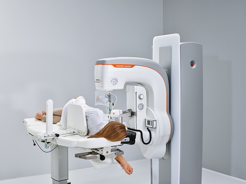
The unique 50 degree 3D HD Breast Tomosynthesis provides the highest depth resolution in tomosynthesis, and thus delivers excellent quality 3D images. Now also biopsies can be performed leveraging this wide tomosynthesis angle. The HD Breast Biopsy solution allows targeting suspicious areas with one click with a +/- 1mm accuracy. The new integrated specimen imaging tool facilitates the immediate control of the biopsy directly at the mammography system. The new Mammomat Revelation also provides automated breast density measurements at the point of examination. This allows for personalized risk stratification and necessary adjunct imaging exams can be triggered before the patient leaves. As soon as the tissue sample has been taken, it has to be X-rayed to confirm that the biopsy was successful. In current settings, samples have to be imaged to a second system or a dedicated specimen scanner, usually in a different room. For this entire time, the patient’s breast has to remain compressed – an uncomfortable or even painful situation that the Mammomat Revelation can substantially reduce. The system has an integrated specimen imaging tool called InSpect. Biopsy samples can be controlled within 20 seconds directly at the system without radiation exposure to the patient, improving the biopsy workflow and shortening the compression time for the patient. In case of high breast density, additional examinations such as tomosynthesis or breast ultrasound might be indicated. Radiologists currently have to visually estimate breast density during the image reading process, which usually takes place after the patient has left. If additional imaging procedures become necessary, the patient has to be called back. Mammomat Revelation is the first mammography system that provides automated breast density measurements at the point of examination. This enables direct, personalized risk stratification and adjunct imaging can be initiated before the patient leaves. Patients get results faster, which also minimizes uncertainty. Purely morphological information from a mammogram or tomosynthesis may sometimes be insufficient for a precise diagnosis. For example, it can be difficult to distinguish scar tissue from new tumours in a post-surgical examination. In addition to purely morphological information, there can be a need for functional information that can be obtained using contrast-enhanced imaging, currently usually performed using breast MRI. Mammomat Revelation now makes functional imaging possible directly on the mammography system. As contrast-enhanced mammography is still not a standard procedure, the modular arrangement of the new system enables it to be expanded to include this to suit customer requirements. With Personalized Soft Compression, the breast compression process is softened and the compression force is automatically and individually adjusted. Coupled with ergonomic SoftComp Paddles, Personalized Soft Compressions allows for better breast positioning, and a more consistent image quality while reducing discomfort.
