


In 2012 there were 1.9 million cardiovascular related deaths within the European Union and 4 million deaths in Europe with the higher rate of deaths to be found in the Central and Eastern European countries. During June 2013, the European Society of Cardiology (ECS) released the results of their study comparing death from a cardiac event between the 1980s and the present. At a glance, the report appears to make happy reading. On average, the rate of heart disease related deaths have halved during that period within the European Union countries. This rate applies to both sexes as well as most age groups.
Dr Melanie Nichols, a Research Associate from the British Heart foundation Health Promotional Research Groups and her colleagues in the Oxford Research Group have looked at trends in death from coronary heart disease between 1980 and 2009 in both sexes and four age groups: under 45, 45-54, 55-64 and 65 years and over [1].
Lack of comparable data in Europe
One of the problems that Dr Nichols and her group have faced in assessing the data is the differences in the way that countries record and code data. That Europe suffers from a lack of data and comparable data is evident when reading the European Cardiovascular Disease Statistics published by the European Heart Network [2]. This is especially true for incidence rates, rates of surgical procedures as well as effects of regional diets etc. To better serve assessments such as this report, the European Union has the task of developing standard methods for collecting information and procedures for calibration of locally appropriate methods and questionnaires.
Nevertheless, while working within these constraints, Dr Nichols and colleagues found that:


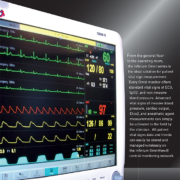
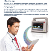
Telemedicine has been around for many years, evolving in tandem with technological change and improvements. In essence, telemedicine delivers healthcare services by means of technology, in cases where the healthcare professional cannot attend to the patient physically. Tele-anesthesia takes it a step further by doing pre-operative consultations by means of telemedicine systems. In this article, the authors report of the growing use of tele-anesthesia.
by Dr Nora Terrasini, Dr Erik Arbeid, Dr Riccardo Taddei, Dr Cedrick Zaouter, Dr Shantale Cyr and Dr Thomas M Hemmerling
What is telemedicine?
Telemedicine is defined as the delivery of healthcare and sharing of medical knowledge over a distance, using telecommunications systems [1]. The World Health Organization gives a more extensive definition of what should be considered as telemedicine and describes it as
Partial support for patients with respiratory failure can be provided with invasive mechanical ventilation or non-invasive mechanical ventilation. The role of non-invasive positive pressure ventilation (NPPV) in preventing intubation for various conditions has been well studied. Less well studied is the role for NPPV in the weaning and peri-extubation period.
by Dr Brooks A. Fallis and Dr Karen E. A. Burns
Mechanical ventilation is essential for supporting patients through episodes of respiratory failure by unloading respiratory muscles and improving gas exchange. While potentially lifesaving, invasive mechanical ventilation with an endotracheal tube or tracheostomy can be associated with important harm, in part due to complications such as ventilator associated pneumonia (VAP). VAP has been shown to increase morbidity and trends towards increasing mortality [1].
Critical care physicians strive to extubate patients as early as possible, while minimising the risk of re-intubation, which has been shown to be independently associated with increased risk of developing VAP [2]. Spontaneous breathing trials (SBT) are used to decide if a patient has been weaned. An SBT involves a focused assessment of a patient
The very first intensive care units (ICUs), introduced towards the end of the 19th century, consisted of a few beds reserved for the sickest patients and put together in one area of the general hospital ward so that the patients could be watched more closely. Patients continued to be treated by their admitting physician with consultation by other specialists as dictated by the course of their disease. Later, separate rooms were created to gather together the special monitoring and organ support equipment and specially trained nursing and medical staff considered necessary for the optimal management of critically ill patients.
Over the years, especially in larger hospitals and more commonly in the United States than in other countries, subspecialty ICUs have developed, catering for specific groups of patients, such as those with neurological, respiratory, cardiac, surgical, trauma diagnoses. This division of intensive care into subspecialties reflects a general trend across medicine towards the creation of increasingly specialized subspecialties. In the United States, one third of all ICUs are now (sub)specialty units. But is there any evidence that such units provide better care than general, multidisciplinary ICUs catering for all critically ill patients regardless of diagnosis?
by Prof. Jean-Louis Vincent
Benefits of subspecialty ICUs
Proponents of specialized ICUs suggest that patient outcomes can be improved in such units because they are managed by staff with increased expertise and training in the particular field of diagnosis. Such units are thus able to provide more focused, relevant care. However, although highly trained in their particular specialty, staff in such units may be less experienced in diagnosing and managing other systemic complications of critical illness. There is relatively little data available comparing the benefits of specialty versus general ICUs. In one study, patients with intracerebral hemorrhage had improved survival rates when admitted to a specialized neurosurgical ICU compared to a general ICU [1]. However, in another analysis, admission to a diagnosis-appropriate specialty ICU was associated with no survival benefit compared to admission to a general ICU for a selection of common diagnoses, including acute coronary syndrome, ischemic stroke, intracranial hemorrhage, abdominal surgery, and coronary-artery bypass graft surgery [2]. Interestingly, admission to a diagnosis-inappropriate specialty ICU, e.g., a renal patient admitted to a neurosurgical ICU because the renal ICU was full, was associated with increased mortality rates [2]. Performing such comparative studies is, however, fraught with difficulty, largely because there is no set definition of a
Prins Hendrikstraat 1
5611HH Eindhoven
The Netherlands
info@interhospi.com
PanGlobal Media IS not responsible for any error or omission that might occur in the electronic display of product or company data.
