• News
    • Featured Articles
    • Product News
    • E-News
  • Magazine
    • About us
    • Digital edition
    • Archived issues
    • Media kit
    • Submit Press Release
  • White Papers
  • Events
  • Suppliers
  • E-Alert
  • Contact us
  • FREE newsletter subscription
  • Search
  • Menu Menu
International Hospital
  • AI
  • Cardiology
  • Oncology
  • Neurology
  • Genetics
  • Orthopaedics
  • Research
  • Surgery
  • Innovation
  • Medical Imaging
  • MedTech
  • Obs-Gyn
  • Paediatrics

Archive for category: Featured Articles

Featured Articles

Contrast Enhancement: Expanding Frontiers of Ultrasound

, 26 August 2020/in Featured Articles /by 3wmedia

Contrast enhanced agents have been key to enhancing the diagnostic capability of computed tomography (CT), magnetic resonance imaging (MRI) and clinical radiography. Since the turn of the millennium, contrast enhancement for ultrasound (CEUS) has also emerged as an imaging tool. Along with developments in scanning hardware, new contrast agents have expanded the application envelope of ultrasound. During CEUS, tiny liquid suspensions of biodegradable gas-filled microspheres (also known as ‘microbubbles’) are injected as tracer for microscopic ultrasound imaging examinations. The microbubbles are metabolized and expelled from the body within minutes. Clinical applications for ultrasound contrast agents potentially extend to any organ or physiological system that is evaluated with conventional ultrasound, with the singular exception of the fetus. As of now, major applications are in cardiac and hepatic imaging. Other applications are being explored, including paediatric CEUS.

From imaging complement to alternative
There is growing evidence that CEUS is valuable, accurate and cost-effective. It often complements CT and MRI, and in several instances, has become an important alternative to either. This especially concerns patients with renal failure, those who wish to avoid the radiation risk of CT or cannot cope with being shut inside a scanner.
Interest in CEUS has grown sharply since 2016, after the Food and Drug Administration (FDA) approved a microbubble contrast agent for liver CEUS, paving the way for much faster growth in the US market.
The microvascular challenge
Clinically, one of the key drivers for CEUS has been limits to the performance of ultrasound imaging and Doppler techniques. While B-Mode provides anatomical information, Doppler allows for visualization of the larger vessels in the macrovascular system, based on the velocity of blood flow in the intravascular lumen. However, there are limits to both spatial resolution and Doppler sensitivity.
The utility of conventional ultrasound reduces rapidly when a clinician needs to visualise smaller vessels and capillaries, lying within deeper structures of the body’s microvascular system.
To achieve this, and more specifically, determine differences in arrival-, dwell- and wash-out time within specific regions of parenchymal tissue, there is a need for direct imaging via tracers. It is in this capacity that contrast agents play a useful role. They improve the sensitivity and specificity of ultrasound and greatly expand its scope for application.

The advantages of CEUS

CEUS has certain intrinsic advantages when compared to other imaging modalities.
It permits ultra-high temporal imaging of contrast enhancement profiles at between 20 and 50 images per second, for a duration of about 5-8 minutes. This makes it possible for continuous visualization of images in all phases – from the early arterial to the late phase – and seek to ensure no patterns are missed. CEUS also allows for both follow-up examinations at short intervals, and, given its lack of ionising radiation, for repeated examinations over a long period of time – a common requirement for chronic diseases. CEUS is also convenient. It can be used at multiple bedsite locations – from intensive care units (ICUs) and operating rooms to recovery rooms and ambulatory units.
Contrast agents for ultrasound have been found to be safe with no cardio-, hepato-, or nephro-toxic effects. Laboratory checks to assess liver, renal or thyroid function before administration are therefore not required.
Evaluating liver lesions
In the liver, CEUS has proven its utility when clinicians encounter focal lesions during cross-sectional imaging of an asymptomatic patient. Though most such collateral encounters are benign, it is necessary to pursue dedicated imaging characterization and diagnosis, in order to exclude malignancy. This is especially true when the lesions are large or otherwise atypical and when the patient is from a high-risk group.
Traditionally, the evaluation of lesions was undertaken with magnetic resonance imaging (MRI) or multiphase CT. However, the former was generally limited in availability, while multiphase CT invoked concerns about radiation. CEUS is seen to be safe, non-invasive and available.
When CEUS is used in the liver, microbubble delivery occurs via two routes, namely the hepatic artery and portal vein. Blood flow through the latter needs to first transit gastrointestinal circulation, and therefore arrives at a later time point. This permits differentiation between the two wash-in phases.
CEUS enhances the display of vascularity in liver lesions, and is both accurate and reproducible. The vascular supply for focal liver lesions is characteristic of a particular lesion type and different from normal liver tissue. While abnormal vascularity of hepatocellular carcinoma can be demonstrated early during the contrast inflow phase, metastases are characterised in the late phase. In addition, the timing and the intensity of washout can differentiate hepatocellular malignancies from non-hepatocellular ones. The former demonstrate delayed and weak washout. Non-hepatocellular tumours show strong, early washout.

The need for right dosing
Using the optimal dose is important. Too high a contrast agent dose results in artefacts, particularly in the early phases of enhancement. These include acoustic shadowing, over-enhancement of small structures and signal saturation, which is also detrimental for quantification.
On the other hand, a low dosage causes the concentration of microbubbles to be sub-diagnostic in the late phase, challenging the detection of wash out.
If the wash out is early, the dose was probably too low. Here, it can be important to evaluate the status of the liver as being healthy or diseased. In difficult cases, a second (higher) dose may be administered, with no or only limited scanning in the early phases to reduce bubble destruction. The exact dose depends on the contrast agent, ultrasound equipment (software version, transducer), type of examination, organ and target lesion, size and age of the patient.

Other challenges for CEUS in the liver
Apart from the challenge of dosing, there are other limitations too in the use of CEUS in the liver. Very small lesions may be overlooked. The smallest detectable lesions are considered to be 3-5 mm in diameter.
There are also some specific shortcomings, such as fat layers surrounding the falciform ligament. These can cause enhancement defects which might be confused with a lesion.
Given limits to penetration, deep-seated lesions may also not always be accessible. However, some clinicians suggest that bringing the liver closer to the transducer via use of left lateral decubitus positioning can overcome such a limitation.

CEUS and cardiology
CEUS has also shown remarkable utility in cardiology.  After the tracer injection, micro-bubbles follow the flow and distribution of red blood cells. opacify the cardiac chambers and enhance delineation of the left ventricular border. The microbubbles are then ejected into the arterial circulatory system, allowing for visualization of blood flow into the parenchymal organs.
An assessment of cardiac function depends on proper delineation of the endocardial border and wall motion patterns. This is where conventional ultrasound faces serious limits. Intracardiac echo reflections couple to weak signals from structures in parallel to the echo beam. The ensuing delineation of the endocardial border can therefore be unclear, resulting in an inaccurate left ventricle assessment.
What contrast agents achieve here is to completely fill the ventricular cavity, and thereby delineate it in a similar fashion to cardiac MRI.
Proper assessment of cardiac function is especially important for stress echo tests in order to demonstrate inducible ischaemia. Here, the risk of a stress examination means that inadequate image quality is unacceptable. In addition, precise delineation of the cardiac chamber is required to make an assessment of heart insufficiency and decide on whether an automatic implantable cardioverter defibrillator (AICD) is indicated. Such precision is also required with cancer chemotherapy patients, in order to assess cardiotoxicity.

New contrast agents
First-generation ultrasound contrast agents were based on air, which was sufficiently soluble in blood for use with the equipment of the time. Second-generation agents contain an inert lipophilic gas with very low solubility, thus avoiding early leakage of the gas. This provides more stability to the microbubbles.
Modern contrast agents have a shell made out of a thin and flexible phospholipid membrane. One side, which faces the surrounding blood, has hydrophilic properties. On the other, lipophilic chains make contact with the encapsulated gas.
Over recent years, technology development has focused on ultrasound contrast agents which reduce microbubble size and increase persistence within the blood in the circulatory system, to 10 or more minutes. Researchers are also seeking to develop new materials and gases to control the encapsulating shell or surface of the microbubble, in order to inhibit dissolution and diffusion.

Constraints faced by microbubbles
In spite of the above developments, there are some constraints with microbubbles.
They do not last long in circulation, due to being taken up by immune system cells, the liver or spleen. They also have low adhesion efficiency, which means only a small fraction bind to an area of interest. Microbubbles can also burst at low ultrasound frequencies and at high mechanical indices, which, in turn, can lead to local microvasculature ruptures and haemolysis.

Guidelines on CEUS
The use of CEUS varies widely from one country to another, and even between different healthcare facilities in the same country.
Guidelines were first issued for the use of CEUS for liver applications in 2004. They were updated in 2008, reflecting growth in the availability of contrast agents. CEUS has also been recommended in guidelines for several non-liver applications, under the auspices of EFSUMB.
The latest guidelines date to 2012. They are published under the auspices of the World Federation for Ultrasound in Medicine and Biology (WFUMB) and the European Federation of Societies for Ultrasound in Medicine and Biology (EFSUMB). The aim is to create standard protocols for CEUS in liver applications across the world.
According to the guidelines, CEUS is indicated for liver lesion characterization in the following clinical situations:
• Incidental findings on routine ultrasound
• Lesion(s) or suspected lesion(s) detected with US in patients with a known history of a malignancy, as an alternative to CT or MRI
• Need for a contrast study when CT and MRI contrast are contraindicated
• Inconclusive MRI/CT
• Inconclusive cytology/histology results

Paediatric applications
One new frontier for CEUS applications consist of children.
Currently, sulphur hexafluoride gas microbubbles have been approved by the FDA in the US for characterising focal liver lesions in children and vesico-ureteral reflux. In Europe, CEUS in children is indicated for vesico-ureteral reflux, although there is
significant off-label use too.

https://interhospi.com/wp-content/uploads/sites/3/2020/08/dreamstime_l_54650037_cropped.jpg 523 800 3wmedia https://interhospi.com/wp-content/uploads/sites/3/2020/06/Component-6-–-1.png 3wmedia2020-08-26 14:16:482021-01-08 12:29:46Contrast Enhancement: Expanding Frontiers of Ultrasound

IHF 2019, 6-9 November, Muscat, Oman

, 26 August 2020/in Featured Articles /by 3wmedia
https://interhospi.com/wp-content/uploads/sites/3/2020/08/47407-IHF2019_WHC19-Print-ad-190205.jpg 2000 1414 3wmedia https://interhospi.com/wp-content/uploads/sites/3/2020/06/Component-6-–-1.png 3wmedia2020-08-26 14:16:482021-01-08 12:29:54IHF 2019, 6-9 November, Muscat, Oman

KIMES 2019, 14-17 March, Coex, Seoul

, 26 August 2020/in Featured Articles /by 3wmedia
https://interhospi.com/wp-content/uploads/sites/3/2020/08/47373_KIMES-2019_140x204_IHE-Junior-page.jpg 1500 1029 3wmedia https://interhospi.com/wp-content/uploads/sites/3/2020/06/Component-6-–-1.png 3wmedia2020-08-26 14:16:482021-01-08 12:29:58KIMES 2019, 14-17 March, Coex, Seoul

Pregnancy and critical care

, 26 August 2020/in Featured Articles /by 3wmedia

Though global maternal mortality has declined by 1.3 percent a year since 1990, the rate continues to remain stubbornly high in certain regions. At present, fewer than one of 50 pregnant women require critical care. However, both maternal and fetal mortality can be high when it is required.
In industrialized countries, the rate of obstetric ICU admissions varies from 50 to over 400 per 100,000 deliveries with an overall case-fatality rate of 2%. In developing countries, fatality in obstetric ICU patients can be 3-5 times higher.

Obstetric disorders in the ICU
Obstetric ICU patients include those with obstetric disorders as well as pregnant patients with medical/surgical disorders. The bulk of patients are admitted to the ICU for obstetric disorders. In general, obstetricians are aware of all obstetric patients in hospital, whether they have a medical or obstetric problem.
There are several obstetric conditions which can require ICU admission. The most common are hemorrhage and hypertensive disorders, above all pre-eclampsia toxemia (PET) and eclampsia. In industrialiZed Western countries, these typically account for 30-35% and 20% of admissions to the ICU.

Hemorrhage leading cause of mortality, ICU admission
Obstetric hemorrhage is either antepartum or postpartum, and remains the leading cause of maternal mortality. Antepartum hemorrhage occurs in 5 percent of pregnant women, and in the bulk of cases carries no risk to the mother or fetus. Major causes involve the placenta and uterine rupture.
Postpartum hemorrhage (PPH) is the single most frequent indication for ICU admission, and involves major blood loss, regardless of the mode of birth. Though there is no universally accepted definition, it typically means more than a half litre of blood loss within 24 hours. In about two out of three cases, PPH is due to failure of uterine contraction after delivery, with most of the rest caused by placental retention. Genital trauma, due to laceration of the vagina or cervix because of instrument delivery, is also increasingly implicated in PPH.

Patient management of obstetric hemorrhage depends on identifying the cause and whether or not delivery has occurred. In PPH, management is aggressive, beginning with administration of oxytocin, emptying of the bladder and massage of the uterus. The care team also begins intravenous prostaglandin therapy, coupled with uterine tamponade via balloon compression or packing. If bleeding persists, surgical intervention is indicated: arterial ligation, suture, Cesarean hysterectomy or uterine artery embolization. Should bleeding continue, recombinant factor VIIa is administered and repeated, if there is no response.

Hypertensive disorders
Pre-eclampsia toxemia (PET) occurs in 2 to 3 percent of all pregnancies after 20 weeks of gestation, and is classified as mild, moderate or severe. Its pathogenesis results from abnormal placenta formation, and it is characterized by impaired organ perfusion due to impaired vasodilation and placental ischemia. As pregnancy progresses, the ischemia worsens. This makes the mother hypertensive, with a risk of renal dysfunction.
Severe PET is associated with significant morbidity and mortality, both for the mother and fetus. It requires one or more of the following indicators: hypertension (BP over 160 systolic or 110 diastolic), proteinuria higher than 5 grams per 24 hours or oliguria below 400 ml per 24 hours, along with cerebral irritability, epigastric pain, and pulmonary edema.
PET usually resolves following delivery of the fetus but may manifest postpartum. A variety of antihypertensive agents, including hydralazine, labetalol, sodium nitroprusside, alpha blockers, calcium channel blockers and methyl dopa, have been advocated in PET.  Hydralazine and labetalol are the most widely used in the critical care setting. Magnesium is usually co-administered to provide vasodilatation and prevent seizures. Care should be taken with fluid resuscitation because of the risk of pulmonary edema.
Eclampsia is an extreme complication of PET, and is marked by the occurrence of convulsions and seizures, 40% of which occur following delivery. The seizures tend to be self-limiting, with a very rare incidence of status epilepticus. Though the mortality from eclampsia has been high in the past, death is now uncommon. Common causes of mortality are hepatic complications, including hepatic failure, hemorrhage, or infarction.

Other challenges of pregnancy
Peripartum cardiomyopathy is another challenging condition during pregnancy, albeit of unknown cause. It is one of the leading causes of maternal death, with mortality as high as 25-50%. It can occur from the final month of pregnancy up to 5 months after delivery.
Other conditions unique to pregnancy include HELPP syndrome (hemolysis, elevated liver enzymes and low platelets), placental disorders (abruption, previa or retention), amniotic fluid embolism and chorioamnionitis, and acute fatty liver. 

Changing epidemiology
The epidemiology of obstetrics in the ICU has changed dramatically since the past decade. Obstetric conditions such as thrombocytopenic purpura of pregnancy, which were rare in the past, are now being diagnosed more frequently. Massive hemorrhage from adherent placenta is increasing due to the growing number of pregnant women bearing scars from previous cesarean sections (CS). Uterine rupture during labour is also sometimes associated with previous CS. Another condition is ovarian hyper-stimulation syndrome, which is not uncommon any more due to the sharp growth in the availability of assisted reproduction techniques.
There are now many older mothers with pre-existing disorders and chronic medical conditions, some of which can be in an advanced stage. Typical co-morbidities today including essential hypertension, Type 2 diabetes and coronary heart disease. Obesity is also a major concern, which poses numerous challenges for managing pregnant patients in the ICU.

Impact on multiple physiological systems
Pregnancy affects several physiological systems – among them, the cardiovascular, respiratory, renal, hematologic and endocrine. These tax a patient’s reserves and often compromise responses needed to combat a disease state during pregnancy and the peripartum period.
Pregnancy’s impact on physiological systems is twofold: first, by worsening pre-existing conditions, and second, by heightening susceptibility.
For example, cardiovascular conditions which can deteriorate significantly in pregnancy include aortal coarctation, primary pulmonary hypertension and valvular disease; congenital heart disease is another such condition. Cardiovascular issues are also important since that shortness of breath is a very common symptom in pregnancy. When this occurs, clinicians must distinguish the dyspnea resulting from underlying medical disorders versus that caused by normal physiologic changes in pregnancy. The latter include anemia, upward displacement of the diaphragm, and respiratory alkalosis.
One of the most confounding cardiovascular challenges is associated with PET. Aside from hypertension, patients also show increased systemic vascular resistance and reduced intravascular volume, which cause a reduction in cardiac output as disease severity progresses. In such cases, left ventricular function can deteriorate leading to a risk of pulmonary edema after fluid resuscitation.
On the other side, pregnant women also face an increase in risk for a gamut of infections ranging from varicella pneumonia and urinary tract infections to malaria and hepatitis.
In the respiratory system, the impact of pregnancy on cystic fibrosis is well known. However, pregnant women also face a concurrent increase in susceptibility to venous embolisms and pulmonary thromboembolism.

This kind of dual impact is also faced by the renal, endocrinal and neurological systems. Pregnancy worsens renal insufficiency and glomerulonephritis and enhances susceptibility to acute renal failure.
In the endocrinal system, it worsens diabetes and prolactinoma and increases susceptibility to gestational diabetes.
The list of neurological conditions which deteriorate during pregnancy is especially large, and includes epilepsy, myasthena gravis and multiple sclerosis, while an increase in susceptibility is seen with intracranial hemorrhage. Another complicating factor is that up to half of obstetric critical care patients have some form  of neurological compromise. In most circumstances, this is the result of their admission diagnosis   (that is, pre-eclampsia or obstetric   hemorrhage),  rather than as the precipitant of their ICU admission.

Two patients in one

As a result of the above factors, obstetric critical care represents a major challenge for medical professionals. Obstetricians need to master both maternal and fetal physiology, and avoid any potentially adverse effects on a fetus of diagnostic and therapeutic interventions given as part of care for the mother. Indeed, it is a common statement that obstetricians treat two patients, the mother and the fetus. They must also assess two separate risks – maternal and fetal – from continuing with a pregnancy and decide if termination of the pregnancy improves the outcome for the mother. This is a very charged challenge since a fetus is generally robust despite maternal illness, and it has been demonstrated that pregnancy-induced critical illnesses are resolved by delivery of the fetus.

Mastering general principles
Obstetric ICU practice consists of firstly mastering general principles such as drug safety, ventilation and management of patient airways, monitoring of the fetus, muscle relaxation and sedation, cardiovascular support, thromboprophylaxis, as well as radiology and ethical issues. This is followed by the acquisition of expertise in the management of obstetric and medical conditions.
Critical care interventions for an obstetric patient are similar to those for the non-pregnant patient. However, it is often necessary to adjust physiologic targets for metabolic, pulmonary, and hemodynamic control.

The need for teamwork
Given the complexity and time-sensitiveness of critical care medicine, teamwork skills are essential. Typically, the care team for an obstetric patient at an ICU is multidisciplinary, consisting of the intensivist, obstetrician, anesthesiologist, maternal-fetal medicine specialist, the neonatologist and the ICU nurses. This team needs to operate effectively alongside regular staff.
Although clinicians working in critical care environments are generally highly trained and competent, they have traditionally not learned how to work well as part of a team. Remedying this has become a priority on both sides of the Atlantic.

As part of a paper on standards of care, the European Board and College of Obstetrics and Gynaecology (EBCOG) explicitly recommends “multidisciplinary, high-quality teamwork” as being “essential” in obstetric medical care and urges healthcare providers to ensure that maternity services have adequate facilities, expertise, capacity and back-up for “timely transfer to intensive care.” EBCOG also seeks to give substance to such a mission. It urges a system of “clear referral paths” to enable pregnant women requiring additional care to be managed and treated by the appropriate specialist teams when problems are identified. One of its most interesting recommendations is for development and routine use of an obstetric ‘early warning chart’  to help in the timely recognition, treatment and referral of women developing a critical illness.

Teamwork is also seen as being a priority in the US, where the Joint Commission pointed to failures in teamwork and communication as among the leading causes of adverse obstetric events. Although obstetric clinicians seem aware of deficiencies in teamwork, their perceptions of teamwork differ based upon their role. In a survey by Johns Hopkins University of 44 hospitals across the US, the majority had fewer than half of respondents reporting ‘good teamwork’ in their labour and delivery units.

https://interhospi.com/wp-content/uploads/sites/3/2020/08/IH178_Critical-care_pregnancy_Tosh_thematic.jpg 450 800 3wmedia https://interhospi.com/wp-content/uploads/sites/3/2020/06/Component-6-–-1.png 3wmedia2020-08-26 14:16:482021-01-08 12:30:05Pregnancy and critical care

People on board: Transforming Healthcare

, 26 August 2020/in Featured Articles /by 3wmedia
https://interhospi.com/wp-content/uploads/sites/3/2020/08/AD_IHF_BARCELONA.jpg 1415 1000 3wmedia https://interhospi.com/wp-content/uploads/sites/3/2020/06/Component-6-–-1.png 3wmedia2020-08-26 14:16:482021-01-08 12:29:41People on board: Transforming Healthcare

Point of Care Testing: Complementing the Laboratory

, 26 August 2020/in Featured Articles /by 3wmedia

Point-of-care testing (POCT) is typically described as a clinical test which is done at, or close to, the physical location of a patient. This could be at a patient’s home, in a pharmacy, a GP’s office or an in-hospital bed site. POCT typically consists of portable devices and instruments, which return results quickly. As a result, POCT permits immediate intervention or treatment.
POCT can also be defined usefully by specifying what it is not. In this case, a POCT is simply a test that is not analysed in a laboratory. POCT short circuits many steps involved in the latter. It eliminates the need to collect a specimen, transfer it to the lab, perform the test, and transmit results back to the provider.
POCT is increasingly used to diagnose and manage a range of diseases, from chronic conditions such as diabetes to acute coronary syndrome (ACS). Recent additions include genetic tests.

Driven by miniaturisation
The POCT era is considered to have begun in the 1970s, with a test to measure blood glucose levels during cardiovascular surgery. In 1977, a rapid pregnancy test called ‘epf’ became the first POCT for use wholly outside a hospital.
Since the late 1980s, one of the key drivers of POCT has been product miniaturization, with increasingly sophisticated and ever-smaller mechanical and electrical components integrated onto chips that can analyse biological objects at the microscale. The pace of miniaturization has accelerated at a breakneck speed in recent years, to mobile handheld and wearable POCT devices. These can be inte-
grated with other applications within a healthcare facility, or aid patients in monitoring and self-management of chronic conditions.

Wide product range, but handful of tests dominate
The most widely-used POCTs include “blood glucose testing, blood gas and electro-
lytes analysis, rapid coagulation testing, rapid cardiac markers diagnostics, drugs of abuse screening, urine strips testing, pregnancy testing, faecal occult blood analysis, food pathogens screening, haemoglobin diagnostics, infectious disease testing and cholesterol screening.” Nevertheless, just three tests – urinalysis by dipstick, blood glucose and urine pregnancy – are believed to account for the majority of POCT.

Comparisons with the lab
Beyond definition, the relationship of POCT to a laboratory is close for a very good reason. Most clinical cases for POCT use lab testing as a comparator. In other words, the first question that comes to many people when using POCT is whether its results match those of a laboratory. Although evidently quicker to obtain, is POCT as reliable? Another topic for comparison consists of the cost of POCT versus lab tests.

Costs: a vexed question

Even in the heady early days of POCT, there was awareness about potential cost downsides. One of the first efforts to address this question was a US study, published in 1994 in ‘Clinical Therapeutics’. [1] The study, by the Office of Health Policy and Clinical Outcomes at the Thomas Jefferson University Hospital in Philadelphia, sought to determine time and labour costs for POCT versus central laboratory testing on a cohort of 210 patients presenting to the emergency department.
The patients had blood drawn for a Chem-7 profile (sodium, potassium, chloride, carbon dioxide, blood urea nitrogen, glucose, and creatinine), or for cell blood count (CBC). Largely due to much quicker turnaround time (TAT), physicians reported that POCT would have resulted in earlier therapeutic action for 40 of 210, or 19 percent of patients. Costs for POCT were, however, over 50 percent higher, and also showed significant variability, depending on test volume. The authors speculated that increasing volumes of POCT would reduce costs “substantially.”

Volumes lower cost
The perception that POCT is much more expensive than a centralized laboratory persists. There are several reasons for this. Consumables generally cost more than tests done with automated laboratory instruments. On its part, POCT simply cannot achieve the scale economy associated with the latter. It also requires more staff downtime.
However, right from the early stages of POCT use, it seemed likely that unit costs could be reduced by increasing test volumes, as anticipated in the 1994 study by Jefferson University Hospital.
POCT was also to quickly demonstrate enhanced utility for certain kinds of tests. In 1997, a study at an Indiana hospital reported a near-halving in unit costs of panels, from USD 15.33 to USD 8.03, following POCT implementation for blood gases and electrolytes [2].

Levelling the field of play
One of the biggest hurdles in making cost comparisons of POCT with lab tests is the difficulty of levelling the playing field. It is also difficult to use such an exercise to draw generalised conclusions, since key conditions often vary significantly from one care facility to another. POCT is also complex to manage, and it is especially challenging to maintain regulatory compliance, especially in large institutions.
Though the cost of consumables is straightforward to determine, this is hardly so for labour.
Labour costs for a lab test are not limited to staff in the laboratory. They also include costs of staff in the pre-analysis phase, for phlebotomy, nursing and other services. Many of the latter entail administrative overheads. Typically, these would consist of formalities in the collection of phlebotomy supplies, the completion and submission of a test request, the labelling of tubes, specimen packaging and despatch.
In contrast, POCT eliminates most pre-analytic steps, along with associated staff costs and overheads. POCT can be undertaken by personnel who are not trained in clinical laboratory sciences.

Cost versus value
Although it seems to be common sense that POCT labour costs are significantly less than for a laboratory test, calculating this precisely requires a complex time-and-motion study which takes account of differences in wages and other costs for phlebotomists, nurses, administrative staff and medical technologists.
Unit product cost therefore reflects only a part of the overall equation, as far as justifying the case for a test is concerned. Indeed, many experts now urge for making assessments based on unit value rather than unit cost.
The role of TAT
With POCT, faster TAT promises better treatment, reduced patient stay, superior workflow and improved clinical outcomes. POCT is however less about reducing TAT than making results available in an optimal and clinically relevant time frame. This, in turn, is frequently dictated by conditions for which care is targeted as well as the setting in which it is delivered.
Delayed test results also impact upon cost in indirect ways. For instance, radiology departments use creatinine POCT before administering contrast agents, since patients with impaired renal function can develop contrast-induced kidney injury. This allows for quick decisions about patients and efficient use of costly CT scanners. If physicians had to wait for test results from a laboratory, the scanner would risk having to idle in a stand by status.

POCT can sometimes be only choice
Some tests have to be performed at point of care since there is no choice, in terms of time for transport to a lab.
One good example is an activated clotting-time test. This is used to monitor cardiac patients undergoing high-dose heparin therapy, whose blood immediately starts to clot after collection of a sample. Another is a POCT glucose test, where a quick result is crucial in determining insulin dosage for diabetic patients.
Elsewhere, whole blood cardiac-marker POCT tests in an A&E facility allow physicians to make rapid decisions on patients with acute coronary syndromes in terms of triage and disposition for observation, catheterization or transfer to a cardiac ICU.
Yet another example is a rapid flu test, used to identify patients who could benefit from antiviral therapy requiring administration as soon as possible after infection, in order to reduce symptomatic intervals. None of the above permit the wait times required for a lab test.

The grey zones
Still, there are grey zones where lab tests have advantages, which are non-negotiable under certain conditions.
One example is routine monitoring of international normalized ratios (INR) for patients on warfarin. The latter is used for prophylaxis against stroke and systemic embolism in patients with atrial fibrillation or mechanical heart valves. The goal of testing is to ensure that anticoagulant levels are appropriate. Over a certain threshold, there is a risk of bleeding, while below it, there is the danger of clotting.
While warfarin toxicity can result in life-threatening risk of bleeding, inappropriate warfarin dose reduction can lead to inadequate protection from a stroke or systemic embolism.
Lab-based testing entails the patient travelling to a GP, or having a caregiver come to take blood at the patient’s home, and doing this regularly. However, even a one-day TAT for the lab test can be a major problem in terms of warfarin dosage. The utility of POCT here seems clear. The GP can know the results and adjust the medication dosage immediately. In addition, POCTs can enable certain categories of patient to self-test and manage warfarin therapy.

Lab tests as gold standard
However, POCT tests can vary significantly from laboratory analysers. In the case of warfarin monitoring, this happens as INR values rise. Correction factors are also typically device- and institution-specific. They cannot be uniformly applied across institutions. Many clinicians therefore require POCT INRs which are greater than 5.0 to be confirmed with a venipuncture sample and a lab test.
Lab tests therefore remain a gold standard. Instrumentation in a laboratory provides robust analytics during a test, and includes a host of quality controls, from test strengths and timings to testing accuracy. These are incorporated into a laboratory information system (LIS) and stored in a patient case file. POCT simply cannot provide such a depth of information.

Gaps being closed
In brief, both POCT and laboratory testing have pluses and minuses. POCT provides definite advantages and reduce risk in some situations.
However, laboratory testing is more advanced, more closely follows scientific process and is fully integrated with the kinds of technical redundancies necessary to ensure greater accuracy and validation of records.
Nevertheless, gaps between the two are being closed, especially through software technology.
Some hospitals now have dedicated satellite labs in emergency rooms and outpatient facilities equipped with POCT.

[1]  https://www.ncbi.nlm.nih.gov/pubmed/7859247
[2] Bailey TM, Topham TM, Wantz S, et al. Laboratory process improvement through point-of-care testing. Jt Comm J Qual Improv 1997;23(7):362–80

https://interhospi.com/wp-content/uploads/sites/3/2020/08/shutterstock_1435535750.jpg 450 800 3wmedia https://interhospi.com/wp-content/uploads/sites/3/2020/06/Component-6-–-1.png 3wmedia2020-08-26 14:16:482021-01-08 12:29:46Point of Care Testing: Complementing the Laboratory

Babies born by c-section lack key gut bacteria

, 26 August 2020/in Featured Articles /by 3wmedia

By Callan Emery, Editor
A study published in Nature in September has caught the attention of the media and the interest of
Obs-Gyn specialists. In what is the largest study of the neonatal microbiome (gut bacteria), the researchers provide strong evidence that the way a baby is born impacts significantly on their microbiome.
The study by Lawley T., et al. (doi: 10.1038/s41586-019-1560-1) found that babies born through the vaginal canal carry different microbes from those delivered through caesarean section. Those born through c-section tended to lack strains of gut bacteria found in healthy children and adults. Additionally babies born through c-section showed a high-level of colonization by opportunistic pathogens associated with the hospital environment (including Enterococcus, Enterobacter and Klebsiella species).
Interestingly, the researchers note that it was the mother’s gut bacteria, and not vaginal bacteria, that made up much of the microbiome in the vaginally delivered babies. Previous studies had suggested that vaginal bacteria were swallowed by the baby on its way down the birth canal. This led to what is has been termed ‘vaginal seeding’ whereby babies born by c-section are swabbed with the mothers vaginal fluids in an effort to restore any missing microbes. However, a study by Stinson et al. (doi: 10.3389/fmed.2018.00135) has shown vaginal seeding to be unjustified and potentially unsafe.
Although a lack of exposure to the right microbes in early childhood has been implicated in autoimmune diseases, such as asthma, allergies and diabetes, the exact role of the baby’s gut bacteria is unclear and it isn’t known if these differences at birth will have any effect on later health.
The researchers, who analysed nearly 600 births in the United Kingdom, say the differences in gut bacteria between vaginally born and caesarean delivered babies largely evened out by 1 year old. They note that large follow-up studies are needed to determine if the early differences influence health outcomes.
Discussing her study, Stinson pointed out that microbes thrown out of balance in babies born by c-section are very similar to those thrown off balance in babies born to mothers receiving antibiotics but delivering vaginally. She surmises that routine antibiotic administration given to mothers delivering by c-section could be a cause of the bacterial difference in the neonatal microbiome.
Although this research does pose interesting questions about the potential health outcomes associated with c-section versus vaginal delivery, it should be emphasised that at this point mothers should not be deterred from c-section delivery if it is the right choice for the mother and her baby.

The study is part of larger effort, called the Baby Biome Study, which aims to follow thousands more newborns into childhood.

https://interhospi.com/wp-content/uploads/sites/3/2020/08/Editor_s_letter_Callan_Emery_headshot_01.jpg 1030 800 3wmedia https://interhospi.com/wp-content/uploads/sites/3/2020/06/Component-6-–-1.png 3wmedia2020-08-26 14:16:482021-01-08 12:29:50Babies born by c-section lack key gut bacteria

Shaping the future of radiology with advanced imaging technologies

, 26 August 2020/in Featured Articles /by 3wmedia
https://interhospi.com/wp-content/uploads/sites/3/2020/08/47398_ADV-ECR-2019-210x297-International-hospital-PRINT.jpg 2000 1414 3wmedia https://interhospi.com/wp-content/uploads/sites/3/2020/06/Component-6-–-1.png 3wmedia2020-08-26 14:16:482021-01-08 12:29:54Shaping the future of radiology with advanced imaging technologies

A helping hand for pediatric intensive care

, 26 August 2020/in Featured Articles /by 3wmedia

Doctors working in the eight-bed Pediatric Intensive Care Unit at the Ramón y Cajal University Hospital in Madrid use point-of-care ultrasound extensively to evaluate the condition of critically ill children, and find it essential to their work. Dr José Luis Vázquez Martínez, Head of UCIP at Hospital Ramón y Cajal, with over 25 years’ experience in pediatric intensive care medicine, explained.

Point-of-care ultrasound (POCUS) is used extensively in our unit, allowing comprehensive, head-to-toe assessment of critically ill children, including respiratory, oncology and post-operative cardiac patients, as well as those being treated for sepsis or multiple trauma. The POCUS approach allows not only an initial diagnosis, but also routine monitoring of treatment to see whether or not a patient’s condition changes, enabling alternative strategies to be implemented if there is no improvement.

POCUS helps pediatric doctors in many ways. For example, ultrasound scans enable evaluation of a patient’s hemodynamic state, looking at their heart function and blood volume to see if these factors are contributing to respiratory failure. Conversely, doctors can see if a lung problem, such as pneumonia, is affecting the heart. For a patient in a coma due to multiple trauma, ultrasound is used to look for signs of bleeding – a potential cause of unexplained anemia – and to assess the intracranial pressure. It is also used to monitor kidney function in children with blood pressure problems, and visualize intestinal indications of sepsis. In addition, ultrasound guidance can be used for endotracheal intubation. In short, broader applications that we did not anticipate until very recently.

We have used ultrasound in our PICU for more than a decade, and have always had SonoSite systems, upgrading them as new technology is introduced. In the beginning, when my knowledge was more limited, the aim was to perform clinical echocardiography but, when the SonoSite representative showed me the linear probe and the various techniques available, it was as if I was being shown electricity after using candles! It was amazing, a real turning point in the use of ultrasound, and everyone recognized it as a step forward in the pediatric intensive care world. For the patients, a major benefit of ultrasound is that exposure to radiation can be reduced. Before ultrasound, X-ray examinations were performed two or three times in the first few days after admission to try to establish the cause of the problem, often with limited success. With ultrasound, we can scan the patient as often as necessary, implementing treatment and monitoring its effect without exposing the child to more radiation.

In PICU, we consider an ultrasound system essential – there is nothing else that gives us so much information, so quickly and non-invasively – and today we have a dedicated Edge II ultrasound system with linear, including hockey stick, and adult and pediatric cardiac transducers. It is in constant demand and is a perfect fit for our work, fulfilling all our expectations. All my colleagues use it, and we are very satisfied with it. The system is high quality and ergonomic, and strikes a good balance between image quality and ease of use. It is also quick to boot up, which is crucial for an instrument that is frequently moved between different beds in the unit. Robustness is vital too; if a patient deteriorates, we may have to move any equipment surrounding the bed very quickly to create space to treat them. However careful you are, there is always the risk of unintentional knocks to the system.

A while ago someone said to me that they ‘sell ultrasound machines but don’t offer training’, but this view isn’t enough – it’s very short-sighted – training is very important. Ramón y Cajal pioneered the use of ultrasound in PICUs across Spain, and was the first hospital to offer external training courses for doctors from other facilities, initially focused on clinical echocardiography. Over time, this has expanded to include neuromonitoring, respiratory and abdominal monitoring. I acquired my ultrasound experience through a combination of external training in adult ultrasound and practical, hands-on learning, and am largely self-taught. If courses like these had been available when I started using ultrasound, I would have saved so much time.
FUJIFILM SonoSite is clearly committed to organising and supporting ultrasound training, and this is unquestionably a great benefit to the scientific community – long may it last!      

Today, we are seeing a boom in the use of ultrasound in pediatric care, as it non-invasively provides immediate information in situations where time is of the essence. Our advice to people attending our training courses who do not have – or have to share – an ultrasound system is to tell their hospital managers that, just like a ventilator, it is an essential piece of equipment for an intensive care unit.

www.sonosite.comwww.fujifilmholdings.com
https://interhospi.com/wp-content/uploads/sites/3/2020/08/IH180_Fujifilm-Sonosite_Dr-Jose-Luis-Vazquez.jpg 600 800 3wmedia https://interhospi.com/wp-content/uploads/sites/3/2020/06/Component-6-–-1.png 3wmedia2020-08-26 14:16:482021-01-08 12:30:06A helping hand for pediatric intensive care

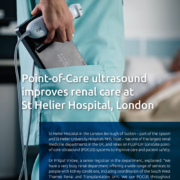
Point-of-Care ultrasound improves renal care at St Helier Hospital, London

, 26 August 2020/in Featured Articles /by 3wmedia

St Helier Hospital in the London Borough of Sutton – part of the Epsom and St Helier University Hospitals NHS Trust – has one of the largest renal medicine departments in the UK, and relies on FUJIFILM SonoSite pointof- care ultrasound (POCUS) systems to improve care and patient safety.
Dr Pritpal Virdee, a senior registrar in the department, explained: “We have a very busy renal department offering a wide range of services to people with kidney conditions, including coordination of the South West Thames Renal and Transplantation Unit. We use POCUS throughout the department for both patient assessment and ultrasound-guided interventions – such as line or drain insertions, aspirations, biopsies – performing over 1,000 procedures every year.”
Better for clinicians and patients
“Ultrasound allows you to visualise the target and surrounding structures during these procedures, making them faster and safer. We use POCUS extensively for patient management – for example, during cannulation or when assessing where to position an anaesthetic or pleural drain – or taking biopsies. The systems give you much more confidence when carrying out these procedures, as you can see exactly where you are placing the needle, and that you are not going to cause any damage from positioning them incorrectly. This helps you to reassure the patients as well, especially during kidney biopsies. Physician satisfaction has increased significantly in the department since adopting POCUS for these techniques, as we are much happier with the quality of work that we are now able to perform.”
Investigating the unknown
“POCUS is the ideal partner for quickly scanning a patient with unknown aetiology; I often use the systems for looking at renal patients that come in with acute kidney problems; if there is a delay to get a formal ultrasound examination, I can easily perform a scan myself. In this way, I can identify whether there is an obstruction or dilation of the kidney as soon as possible. It’s also useful for investigating the bladder, as sometimes the bladder scanners can be unreliable. I can simply select the appropriate probe and have a look; it’s really helpful in streamlining the process and providing a better clinical picture.”
“We also have a programme of medical insertion of peritoneal dialysis (PD) catheters under ultrasound guidance, which is quite unusual, and clinicians from other renal departments are now coming here to train in the procedure. This is another key area that POCUS is able to significantly improve as, without ultrasound guidance, there’s a high risk of perforating the bowel or causing injury to another structure. Having a good view of the peritonea shows you exactly what you are aiming for, and we now only perform this procedure with ultrasound.”
Ultrasound is a necessity
“We have always used SonoSite ultrasound systems in the department and have recently acquired two of the latest generation X-Porte systems. Buying the extra systems was a necessity, as we now use ultrasound for so many of our procedures. These instruments offer exceptional image quality and, crucially, they are very straightforward to operate; you can get a clearer view much quicker making each procedure even easier. When we first purchased the systems, we received a brief demo on how to operate them, and everyone started happily using them straightaway. This user-friendliness has been very popular with clinical staff, and the X-Portes have rapidly become our primary POCUS systems in our procedure rooms. This has allowed us to move our older systems onto the wards, which is very convenient as the department is spread over quite a large area. We are always open to learning about new ultrasound applications and, as the physicians here develop more specific interests, we will likely use the built-in tutorials that are supplied with the systems to further develop our skills.”
No turning back
“The X-Porte certainly gives us a level of detail that we’ve never seen before; you can easily visualise tiny blood vessels, or observe any bleeding that occurs during a biopsy procedure – it’s incredible. I have been working with ultrasound since 2010 and, when we first acquired the new instruments, it still took a little time to get used to the resolution available. Until you’ve used a system like the X-Porte, you don’t realise what can be achieved with ultrasound, but we wouldn’t go back now,” Pritpal said.

https://interhospi.com/wp-content/uploads/sites/3/2020/08/AD_SONOSITE_1.jpg 1415 1000 3wmedia https://interhospi.com/wp-content/uploads/sites/3/2020/06/Component-6-–-1.png 3wmedia2020-08-26 14:16:482021-01-08 12:29:42Point-of-Care ultrasound improves renal care at St Helier Hospital, London
Page 100 of 103«‹9899100101102›»

Latest issue of International Hospital

April 2024

2 June 2026

DeepHealth achieves multiple regulatory milestones for Neuro, Prostate and LumbarMR

7 January 2026

Gulf Aorta Summit 2026 Returns to Dubai with a Global Lineup of Aortic Experts

17 December 2025

GE HealthCare receives CE mark for 128cm total body PET/CT

Digital edition
All articles Archived issues

Free subscription

View more product news

Get our e-alert

The medical devices information portal connecting healthcare professionals to global vendors

Sign in for our newsletter
  • News
    • Featured Articles
    • Product News
    • E-News
  • Magazine
    • About us
    • Archived issues
    • Media kit
    • Submit Press Release

Prins Hendrikstraat 1
5611HH Eindhoven
The Netherlands
info@interhospi.com

PanGlobal Media IS not responsible for any error or omission that might occur in the electronic display of product or company data.

Scroll to top

This site uses cookies. By continuing to browse the site, you are agreeing to our use of cookies.

Accept settingsHide notification onlyCookie settings

Cookie and Privacy Settings


How we use cookies

We may ask you to place cookies on your device. We use cookies to let us know when you visit our websites, how you interact with us, to enrich your user experience and to customise your relationship with our website.

Click on the different sections for more information. You can also change some of your preferences. Please note that blocking some types of cookies may affect your experience on our websites and the services we can provide.

Essential Website Cookies

These cookies are strictly necessary to provide you with services available through our website and to use some of its features.

Because these cookies are strictly necessary to provide the website, refusing them will affect the functioning of our site. You can always block or delete cookies by changing your browser settings and block all cookies on this website forcibly. But this will always ask you to accept/refuse cookies when you visit our site again.

We fully respect if you want to refuse cookies, but to avoid asking you each time again to kindly allow us to store a cookie for that purpose. You are always free to unsubscribe or other cookies to get a better experience. If you refuse cookies, we will delete all cookies set in our domain.

We provide you with a list of cookies stored on your computer in our domain, so that you can check what we have stored. For security reasons, we cannot display or modify cookies from other domains. You can check these in your browser's security settings.

.

Google Analytics Cookies

These cookies collect information that is used in aggregate form to help us understand how our website is used or how effective our marketing campaigns are, or to help us customise our website and application for you to improve your experience.

If you do not want us to track your visit to our site, you can disable this in your browser here:

.

Other external services

We also use various external services such as Google Webfonts, Google Maps and external video providers. Since these providers may collect personal data such as your IP address, you can block them here. Please note that this may significantly reduce the functionality and appearance of our site. Changes will only be effective once you reload the page

Google Webfont Settings:

Google Maps Settings:

Google reCaptcha settings:

Vimeo and Youtube videos embedding:

.

Privacy Beleid

U kunt meer lezen over onze cookies en privacy-instellingen op onze Privacybeleid-pagina.

Privacy policy
Accept settingsHide notification only

Sign in for our newsletter

Free subscription