
Point-of-Care ultrasound improves renal care at St Helier Hospital, London
St Helier Hospital in the London Borough of Sutton – part of the Epsom and St Helier University Hospitals NHS Trust – has one of the largest renal medicine departments in the UK, and relies on FUJIFILM SonoSite pointof- care ultrasound (POCUS) systems to improve care and patient safety.
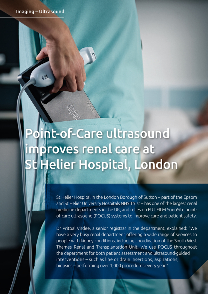
Dr Pritpal Virdee, a senior registrar in the department, explained: “We have a very busy renal department offering a wide range of services to people with kidney conditions, including coordination of the South West Thames Renal and Transplantation Unit. We use POCUS throughout the department for both patient assessment and ultrasound-guided interventions – such as line or drain insertions, aspirations, biopsies – performing over 1,000 procedures every year.”
Better for clinicians and patients
“Ultrasound allows you to visualise the target and surrounding structures during these procedures, making them faster and safer. We use POCUS extensively for patient management – for example, during cannulation or when assessing where to position an anaesthetic or pleural drain – or taking biopsies. The systems give you much more confidence when carrying out these procedures, as you can see exactly where you are placing the needle, and that you are not going to cause any damage from positioning them incorrectly. This helps you to reassure the patients as well, especially during kidney biopsies. Physician satisfaction has increased significantly in the department since adopting POCUS for these techniques, as we are much happier with the quality of work that we are now able to perform.”
Investigating the unknown
“POCUS is the ideal partner for quickly scanning a patient with unknown aetiology; I often use the systems for looking at renal patients that come in with acute kidney problems; if there is a delay to get a formal ultrasound examination, I can easily perform a scan myself. In this way, I can identify whether there is an obstruction or dilation of the kidney as soon as possible. It’s also useful for investigating the bladder, as sometimes the bladder scanners can be unreliable. I can simply select the appropriate probe and have a look; it’s really helpful in streamlining the process and providing a better clinical picture.”
“We also have a programme of medical insertion of peritoneal dialysis (PD) catheters under ultrasound guidance, which is quite unusual, and clinicians from other renal departments are now coming here to train in the procedure. This is another key area that POCUS is able to significantly improve as, without ultrasound guidance, there’s a high risk of perforating the bowel or causing injury to another structure. Having a good view of the peritonea shows you exactly what you are aiming for, and we now only perform this procedure with ultrasound.”
Ultrasound is a necessity
“We have always used SonoSite ultrasound systems in the department and have recently acquired two of the latest generation X-Porte systems. Buying the extra systems was a necessity, as we now use ultrasound for so many of our procedures. These instruments offer exceptional image quality and, crucially, they are very straightforward to operate; you can get a clearer view much quicker making each procedure even easier. When we first purchased the systems, we received a brief demo on how to operate them, and everyone started happily using them straightaway. This user-friendliness has been very popular with clinical staff, and the X-Portes have rapidly become our primary POCUS systems in our procedure rooms. This has allowed us to move our older systems onto the wards, which is very convenient as the department is spread over quite a large area. We are always open to learning about new ultrasound applications and, as the physicians here develop more specific interests, we will likely use the built-in tutorials that are supplied with the systems to further develop our skills.”
No turning back
“The X-Porte certainly gives us a level of detail that we’ve never seen before; you can easily visualise tiny blood vessels, or observe any bleeding that occurs during a biopsy procedure – it’s incredible. I have been working with ultrasound since 2010 and, when we first acquired the new instruments, it still took a little time to get used to the resolution available. Until you’ve used a system like the X-Porte, you don’t realise what can be achieved with ultrasound, but we wouldn’t go back now,” Pritpal said.
