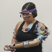
Researchers at Cleveland Clinic have engineered a first-of-its-kind bionic arm for patients with upper-limb amputations that allows wearers to think, behave and function like a person without an amputation, according to new findings published in Science Robotics.
https://interhospi.com/wp-content/uploads/sites/3/2021/11/CCF-Robotic-arm1.jpg23822100panglobalhttps://interhospi.com/wp-content/uploads/sites/3/2020/06/Component-6-–-1.pngpanglobal2021-11-04 12:30:012021-11-04 12:30:01Researchers develop bionic arm with intuitive control and touch sensation
The prevalence of obesity around the world has tripled over the past 40 years, and, along with that rise, dieting and attempts to lose weight also have soared. But according to a review article published September 20 in the journal iScience [1], when it comes to getting healthy and reducing mortality risk, increasing physical activity […]
https://interhospi.com/wp-content/uploads/sites/3/2021/11/walking-scaled.jpg20482560panglobalhttps://interhospi.com/wp-content/uploads/sites/3/2020/06/Component-6-–-1.pngpanglobal2021-11-04 12:26:102021-11-04 12:26:10Researchers call for a focus on fitness over weight loss for obesity-related behavioural change
According to psychologists, in addition to our physiological immune system we also have a behavioural one: an unconscious code of conduct that helps us stay disease-free, including a fear and avoidance of unfamiliar – and so possibly infected – people.
https://interhospi.com/wp-content/uploads/sites/3/2021/11/Donald_Trump.jpg17072560panglobalhttps://interhospi.com/wp-content/uploads/sites/3/2020/06/Component-6-–-1.pngpanglobal2021-11-04 12:21:242021-11-04 12:21:24Rates of infectious disease linked to authoritarian attitudes and governance
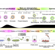
For species that rely on sexual reproduction, including mice and men, offspring can only happen if sperm from the male fertilize eggs from the female. Even artificial fertilization techniques depend on donors for both of these cells. However, a new study led by researchers from ASHBi (ASHBi Institute for the Advanced Study of Human Biology […]
https://interhospi.com/wp-content/uploads/sites/3/2021/11/sperm-cells.jpg9101624panglobalhttps://interhospi.com/wp-content/uploads/sites/3/2020/06/Component-6-–-1.pngpanglobal2021-11-04 12:13:022021-11-04 12:13:02Scientists make sperm from mouse pluripotent stem cells, produce healthy offspring
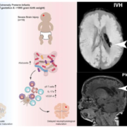
Extremely premature infants are at a high risk for brain damage. Researchers at the University of Vienna and the Medical University of Vienna have now found possible targets for the early treatment of such damage outside the brain: Bacteria in the gut of premature infants.
https://interhospi.com/wp-content/uploads/sites/3/2021/11/gut_bacteria_brain.jpg484848panglobalhttps://interhospi.com/wp-content/uploads/sites/3/2020/06/Component-6-–-1.pngpanglobal2021-11-04 12:08:062021-11-04 12:08:06Researchers find biomarkers in gut microbiome indicate early brain injury in extreme premature infants
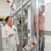
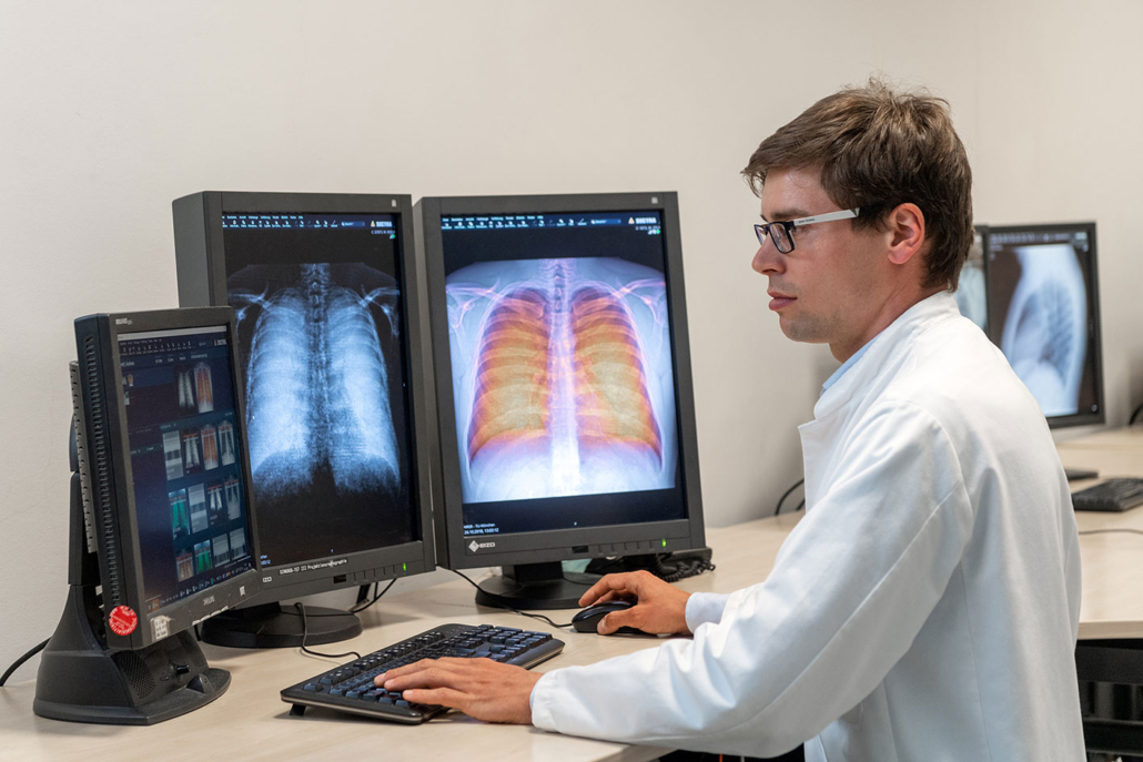
For the first time, researchers at the Technical University of Munich (TUM) have successfully used a new X-ray method for respiratory diagnostics with patients. Dark-field X-rays visualize early changes in the alveolar structure caused by the lung disease COPD and require only one fiftieth of the radiation dose typically applied in X-ray computed tomography. This permits broad medical application in early detection and treatment follow-up of respiratory ailments.
There are millions of cases in which serious respiratory system illnesses place limitations on quality of life. Every year more than four million people die of serious respiratory ailments worldwide. Partially destroyed alveoli and an over-inflation of the lungs (emphysema) are typical of the life-threatening ailment Chronic Obstructive Pulmonary Disease (COPD).
However, the fine distinctions between healthy and diseased tissue are barely visible on conventional chest X-rays. Detailed diagnostic information is only available using three-dimensional computed tomography approaches, in which the computer assembles many individual images. Until now there has been no fast and cost-effective option for early detection and follow-up examinations with a low radiation exposure as used in plain chest X-rays.
A procedure developed at the Technical University of Munich could now fill this gap: dark-field chest X-rays. In the November 1, 2021 issue of The Lancet Digital Health a research team led by Franz Pfeiffer, Professor for Biomedical Physics and Director of the Munich Institute of Biomedical Engineering at TUM, present the results of an initial clinical patient study, which used the new X-ray technology for the diagnosis of the lung disease COPD.
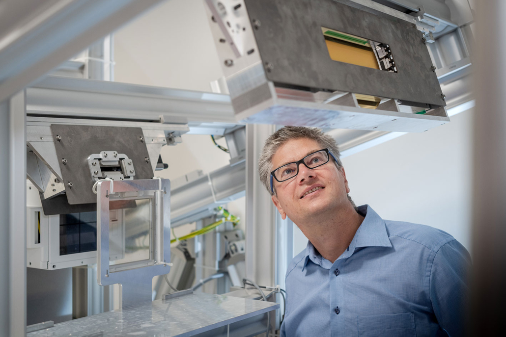
The wave character of X-rays is the key
Conventional X-ray imaging is based on the attenuation of X-rays on their way through the tissue. Dark-field technology on the other hand use the wave nature of X-ray light, which is discarded in conventional X-ray imaging.
The new method thus uses the physical phenomenon of scattering in a manner similar to the long-known principle of dark-field microscopy with visible light. This allows to visualize the structure of objects that are for the most part transparent. These structures appear in the microscope as bright images on a dark background, which has given the method its name.
“The X-ray dark-field signal is particularly strong for interfaces between air and tissue,” Prof. Pfeiffer points out. “This makes it possible for a dark-field X-ray image of the lung to clearly distinguish between intact alveoli, i.e. those filled with air, and regions in which less intact alveoli exist.”
The dark field X-ray method visualizes early changes in the alveolar structure as a result of the lung disease COPD. Franz Pfeiffer, Professor for Biomedical Physics, hopes that this will significantly improve the early detection of lung diseases.
Lower radiation dose
In addition, an examination using dark-field chest X-ray technology involves a significantly lower radiation dose than presently used computed tomography. This is because dark-field chest X-rays require only one exposure per patient, as compared to the large number of individual images taken from different directions which are necessary in computed tomography.
“We expect the radiation exposure to be reduced by a factor of fifty,” says Prof. Pfeiffer. Furthermore, the first clinical results have confirmed that the dark-field X-rays provide additional image information on the underlying microstructure of the lung.
“Given the close connection between the alveolar structure and the functional condition of the lung, this ability is of great significance for pulmonary medicine,” explains Dr. Alexander Fingerle, senior physician at TUM’s university hospital Klinikum rechts der Isar’s Department of Diagnostic and Interventional Radiology. “In the future dark-field X-rays could help improve early detection of COPD and other respiratory ailments.”
Better X-ray equipment for early detection
Prof. Pfeiffer hopes these initial clinical results with patients will accelerate the execution of further clinical studies and the development of marketable devices that use the dark-field method.
“Dark-field chest X-rays are currently giving us a chance to significantly improve the early detection of lung diseases and at the same time to implement it on a wider basis than before,” Prof Pfeiffer notes.
Since dark-field imaging is not limited to COPD, further translational studies with other pulmonary pathologies such as pulmonary fibrosis, pneumothorax, lung cancer and pneumonia, including COVID-19, are of great interest.
Reference:
K. Willer, et. al. X-ray dark-field chest imaging for detection and quantification of emphysema in patients with chronic obstructive pulmonary disease: a diagnostic accuracy study. The Lancet Digital Health. November 1, 2021. doi: https://doi.org/10.1016/S2589-7500(21)00146-1
The International Hospital Federation (IHF) and Africa Healthcare Federation (AHF) have entered into a partnership to increase the participation of healthcare leaders from African countries in the IHF and other global hospital and healthcare initiatives.
Owlstone Medical, a global leader in breath biopsy for applications in early disease detection and precision medicine, closed its Series D financing round after securing $58 million in funding, exceeding its $50 million target after it was over subscribed. This brings the total raised by the company to more than $150 million since founding in […]
https://interhospi.com/wp-content/uploads/sites/3/2021/11/owlstone-breat-biopsy.png570520panglobalhttps://interhospi.com/wp-content/uploads/sites/3/2020/06/Component-6-–-1.pngpanglobal2021-11-04 08:43:382021-11-04 08:43:38Owlstone Medical secures $58 million to advance breath biopsy
A ground-breaking regional anaesthesia device, invented by clinicians at The Queen Elizabeth Hospital King’s Lynn NHS Foundation Trust (QEH) and developed in conjunction with medical device company Medovate, has been awarded ‘Patient Safety Innovation of the Year’ at this year’s HSJ Patient Safety Awards.
https://interhospi.com/wp-content/uploads/sites/3/2021/11/Medovate-scaled.jpg17062560panglobalhttps://interhospi.com/wp-content/uploads/sites/3/2020/06/Component-6-–-1.pngpanglobal2021-11-04 08:41:302021-11-04 08:41:30SAFIRA regional anaesthesia device awarded HSJ Patient Safety Innovation of the Year
Brooks Automation and Cleveland Clinic have opened a new 22,000-square-foot biospecimen sample management and repository facility on Cleveland Clinic’s main campus in Cleveland, Ohio. The two-story biorepository is managed by Azenta Life Sciences, Brooks’ recently re-branded life sciences division, and includes ultra-cold and cryogenic storage. The new facility increases biobanking capacity at Cleveland Clinic and […]
We may ask you to place cookies on your device. We use cookies to let us know when you visit our websites, how you interact with us, to enrich your user experience and to customise your relationship with our website.
Click on the different sections for more information. You can also change some of your preferences. Please note that blocking some types of cookies may affect your experience on our websites and the services we can provide.
Essential Website Cookies
These cookies are strictly necessary to provide you with services available through our website and to use some of its features.
Because these cookies are strictly necessary to provide the website, refusing them will affect the functioning of our site. You can always block or delete cookies by changing your browser settings and block all cookies on this website forcibly. But this will always ask you to accept/refuse cookies when you visit our site again.
We fully respect if you want to refuse cookies, but to avoid asking you each time again to kindly allow us to store a cookie for that purpose. You are always free to unsubscribe or other cookies to get a better experience. If you refuse cookies, we will delete all cookies set in our domain.
We provide you with a list of cookies stored on your computer in our domain, so that you can check what we have stored. For security reasons, we cannot display or modify cookies from other domains. You can check these in your browser's security settings.
.
Google Analytics Cookies
These cookies collect information that is used in aggregate form to help us understand how our website is used or how effective our marketing campaigns are, or to help us customise our website and application for you to improve your experience.
If you do not want us to track your visit to our site, you can disable this in your browser here:
.
Other external services
We also use various external services such as Google Webfonts, Google Maps and external video providers. Since these providers may collect personal data such as your IP address, you can block them here. Please note that this may significantly reduce the functionality and appearance of our site. Changes will only be effective once you reload the page
Google Webfont Settings:
Google Maps Settings:
Google reCaptcha settings:
Vimeo and Youtube videos embedding:
.
Privacy Beleid
U kunt meer lezen over onze cookies en privacy-instellingen op onze Privacybeleid-pagina.