
Three-dimensional echocardiography in clinical practice
Echocardiography has become an integral part of modern cardiology. Being non-invasive, it is useful in assessing ventricular size and function, diagnosing and evaluating valvular disease, and investigating chest pain, possible cardiac emboli and congenital heart disease. Recent advances in non-invasive imaging of the heart with three-dimensional echocardiography (3DE) have simplified our understanding and management of heart diseases. Transthoracic 3DE provides an easier, accurate and reproducible interpretation of the complex cardiac anatomy. It provides unprecedented views of cardiac structures from any perspective in the beating heart helping in clinical assessment of cardiac pathology. One major advantage of the third dimension is the improvement in the accuracy and reproducibility of chamber volume measurement by eliminating geometric assumptions and errors caused by foreshortened views. Another benefit of 3DE is the realistic en face views of heart valves, enabling a better appreciation of the severity and mechanisms of valve diseases in a non-invasive manner.
by Dr Shantanu P. Sengupta
Real time three-dimensional echocardiography (RT3DE) has been a major advancement in the field of cardiac imaging. Advances in the acquisition, storage and analysis of RT3DE images have made its use increasingly common in echocardiography laboratories, not only for research purposes, but also in daily clinical practice. The technique provides a good spatial and temporal resolution of images of the heart. Also, adding the fourth (time) and the fifth dimension (functional assessment of the cardiac structures) is now possible due to recent advances in this field of cardiac ultrasound.
Usefulness of 3D echocardiography
The first 3DE images of the heart were obtained by Dekker et al in 1974 [1]. Since then, various 3D systems have been developed based on a reconstruction of acquired two-dimensional images (2DE) synchronised to the electrocardiogram and respiratory motion. This tool has contributed valuable information on cardiac anatomy and function. However, due to the lengthy image-processing time required, its earlier use was clinically limited to a few echocardiography laboratories and the research arena.
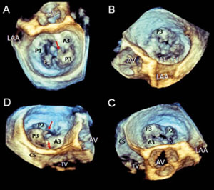
The recent development of matrix transducers with more than 3000 crystals along with new processors has led to the acquisition of real-time images without the need for off-line reconstruction. These new advances have allowed the application of 3DE to daily clinical practice [Figure 1, 2].
Three types of images can be acquired with 3DE: near real time, full volume images; real time (
