
The gamma knife – a new tool against epilepsy ?
The gamma knife is the best known system for radio surgery (RS). It allows non-invasive brain surgery to be performed in one session, with extreme precision. Based on preoperative radiological examinations, such as CT or MR scans and angiography, the gamma knife provides highly accurate irradiation of deep-seated targets in the brain, using a multitude of collimated beams of ionizing radiation with scalpel-like precision.
No surgical incision, no anesthesia
The uniqueness of the gamma knife (and RS surgery in general) is that no surgical incision is required. This serves to minimize risk to adjoining tissue, reduce the risk of surgical complications. It also eliminates the side effects and dangers of general anesthesia, which would be indispensable for the type of medical conditions it is used to target.
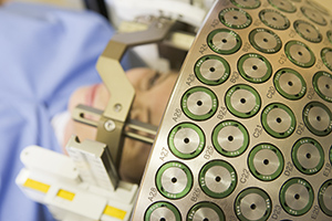
A gamma knife typically contains 201 cobalt-60 sources. Each is mounted in a circular array within a shielded system. The device aims gamma rays via a specialized helmet surgically fixed to the patient’s skull to a target point in the brain. The ‘blades’ of the gamma knife are the beams of gamma radiation programmed to target the lesion at the point where they intersect. In a single treatment session, beams of gamma radiation focus precisely on the lesion. Over time, most lesions slowly decrease in size and dissolve. The exposure is brief and only the tissue being treated receives a significant radiation dose, while the surrounding tissue remains unharmed.
Revolution for brain surgery
The gamma knife has revolutionized brain surgery. Over the last three decades, it has changed the landscape of neurosurgery – treating a range of conditions from brain tumours to vascular malformations with an unmatched level of accuracy. The gamma knife enables patients to undergo a non-invasive form of brain surgery without surgical risks, a long hospital stay or subsequent rehabilitation.
The gamma knife was officially named the Leksell gamma knife, after its lead inventor Lars Leksell, who developed the system in 1967 at the Karolinska Institute in Stockholm. Other key team members included Ladislau Steiner, a Romanian-born neurosurgeon and Borje Larsson, a radiobiologist from Sweden’s Uppsala University.
The CyberKnife
1990 saw the launch of another form of radio-surgical system based on linear accelerators. The best known of these is the CyberKnife, invented in the US by John R. Adler, a Stanford University Professor of Neurosurgery and Radiation Oncology. Unlike the gamma knife, the CyberKnife does not use radioisotopes. Instead, it uses a linear accelerator mounted on a moving arm to deliver X-rays, once again, to a very precise area. The CyberKnife does not use a frame to secure the patient. Instead, a computer monitors a patient’s position during treatment, using fluoroscopy. In other words, the CyberKnife allows for tracking a tumour, rather than fixing the patient. As it does away with a frame, its targets go beyond the brain.
Gamma knife and CyberKnife: Indications
Typically, a gamma knife is used to treat cancer that has metastasized to the brain from another part of the body, acoustic neuroma (a slow-growing tumour of the nerve connecting the ear and brain, pituitary tumours and non-cancerous brain tumours. Its application has also been extended to include certain blood vessel malformations, and fistulas, neuralgia and tremors due to Parkinson’s disease.
On its part, the different design of the CyberKnife allows it to also treat a host of other cancers (breast, kidney, liver, lung, pancreas, prostate and certain skin cancers. The CyberKnife is however, generally not used to treat non-cancerous brain tumours such as chordoma and meningioma.
Gamma knife and epilepsy: a European initiative
In recent years, the gamma knife has drawn attention due to its showing ‘some promise’ for treating certain types of epilepsy.
Attention to such possibilities however date back to 1993, when the first gamma knife treatment for temporal lobe epilepsy was performed at the Hopital Timone in Marseille, France. Just over 5 years later, Na Homolce Hospital in Prague followed with a four-year evaluation on the use of gamma knife in 14 mesial temporal lobe epilepsy (MTLE) patients.
Encouraging results from first study
A pioneering study on gamma knife and epilepsy at France’s Hopital Timone was published in 2000. It covered 25 patients with drug-resistant MTLE with 16 followed up for a period of over 24 months. Thirteen (81%) were seizure free, with two improved. The median latent interval from the gamma knife intervention to seizure cessation was 10.5 months (varying from 6 to 21 months), with two patients immediately becoming seizure free. No cases of permanent neurological deficit (except three cases of non-symptomatic visual field deficit), or morbidity, or mortality were observed.
Although the authors concluded that the ‘optimal parameters for treatment’ remain to be defined, as do studies on ‘dose-related efficacy, effectiveness over longer follow-up periods, and neuropsychological effects’, gamma knife interventions could be ‘a reasonable option,’ and its introduction into epilepsy treatment can reduce the invasiveness and morbidity.’
First and second follow ups to French study
The first five-year follow up to the above released its findings from France in 2004. It found a reduction in median seizure frequency, from 6.16 the month before treatment to 0.33 at 2 years after treatment. In two years, as many as 65% of patients (13 of 20) were seizure free. Five patients reported transient depression, headache, nausea, vomiting, and imbalance. There was ‘no permanent neurological deficit reported except nine visual field deficits.’ Finally, no neuropsychological deterioration was observed two years after treatment and the ‘quality of life was significantly better than that before surgery.’
A second follow-up, in 2008, noted that the gamma knife was ‘an effective and safe treatment for mesial temporal lobe epilepsy.’ Results, it found were ‘maintained over time with no additional side effects. Long-term results compare well with those of conventional surgery.’ The findings remained encouraging, with the mean delay for appearance of the first neuroradiological changes at 12 months. However, all patients who had been initially seizure free experienced a relapse of isolated aura or complex partial seizures during the crucial tapering of the antiepileptic drug. Restoration of medication resulted in good control of seizures.
Efforts in the US: focus on caution
In 2009, one of the first major multi-centric US studies on the gamma knife and epilepsy, led by a team from the University of California, San Francisco, reported three-year outcomes using radiosurgery (RS) for unilateral MTLE.
The authors found seizure remission rates comparable with those reported for open surgery. There were also ‘no major safety concerns with high-dose RS compared with low-dose RS.’ However, they called for additional research to determine whether RS ‘may be a treatment option for some patients with mesial temporal lobe epilepsy.’
Caution was again urged the next year when the US research group noted that RS was a promising treatment for intractable MTLE. However, they also observed ‘that the basis of its efficacy is not well understood…’ The researchers, however, minced no words in their observation that ‘Temporal lobe stereotactic radiosurgery resulted in significant seizure reduction in a delayed fashion which appeared to be well-correlated with structural and biochemical alterations observed on neuroimaging. Early detected changes may offer prognostic information for guiding management.’
Growing interest and availability in US
Nevertheless, there is growing interest across the US in using the gamma knife for epilepsy.
Its potential is highlighted (albeit, to varying degrees) by top facilities such as the Mayo Clinic and other leading hospitals like the University of California at San Francisco. On the other side, the University of Pittsburgh Medical Center explicitly specifies the gamma knife for treatment-resistant epilepsy. An active programme of use is also announced by St. Louis Children’s Hospital, for ‘certain epileptogenic lesions,’ corpus callosotomies as well as hypothalamic hamartomas – a benign plume-like malformation that causes a syndrome characterized by treatment-resistant epilepsy.
Some smaller centres in the US are also describing the Gamma Knife as ‘giving patients with epilepsy another option for treatment.’
Europe seemingly lags US
Although France pioneered studies into the use of the gamma knife in epilepsy, interest in Europe still lags that being shown in the US. One reason may also be that other efforts in Europe have been evidently unsuccessful. For example, a four-year study in the late 1990s in the Czech Republic on using the gamma knife in epileptic patients concluded: ‘Radiosurgery with 25, 20, or 18-Gy marginal dose levels did not lead to seizure control in our patient series, although subsequent epilepsy surgery could stop seizures.’ On the other hand, higher doses were associated with the risk of brain edema, intracranial hypertension, and a temporary increase in seizure frequency.
The ROSE study
Both in the US and Europe, the outlook on using Gamma Knife in MTLE is clearly one of cautious optimism.
Trials conducted to date seem to show mixed results, or do not provide researchers enough conviction, as yet.
For the moment, attention remains focused on an ongoing multi-centre trial called ROSE (Radiosurgery or Open Surgery for Epilepsy). The randomized, double blind trial is funded by the US National Institutes of Health, and is being conducted at 13 centres in the US and the prestigious All India Institute of Medical Sciences in New Delhi.
The trial takes up the hypothesis ‘that radiosurgery is as safe and effective as temporal lobectomy in treating patients with seizures arising from the medial temporal lobe.’ It randomizes patients to either technique and is due to compare seizure remission, cognitive outcomes, and cost. The trial will not only measure outcomes (determined during the course of the final year of a 3-year follow-up period). It will also pay attention to interim measures concerning patient safety, quality of life etc., and compare these between the two groups. The eventual aim is to guide physicians to direct patients between traditional and RS techniques matched to patient characteristics.
