
The coming of age of intravascular ultrasound
Intravascular Ultrasound (IVUS) provides images of the insides of blood vessels for the diagnosis and treatment of cardiovascular disease. Although the roots of IVUS date back a quarter century, it is only recently that physicians have begun to take serious cognizance of its potential.
Improving PCI outcomes
The growth in interest in IVUS is being driven by changes in the way for treating acute coronary syndrome (ACS). ACS is a common complication of coronary heart disease, which is the leading cause of death in industrialized countries.
The typical approach to treat ACS consists of percutaneous coronary intervention (PCI): catheter angiography, balloon angioplasty and the use of stents. However, the proponents of IVUS have been making a strong case about its utility to improve PCI outcomes, including those offered by the latest generation of drug-eluting stents (DES).
IVUS consists of a miniaturized ultrasound imaging device. This is mounted on a catheter which is threaded across an artery to provide both cross-sectional and longitudinal views. It thus reveals not only the shape and amount of plaque but the deployment and expansion of a stent, too. Such information is not accessible via standard angiography. The main area for application is coronary arteries. Growing (if still far smaller) areas consist of peripheral arteries as well as intra-cardiac imaging.
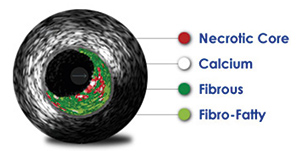
State-of-the-art IVUS systems use spectral backscatter and frequency analysis to classify plaque (distribution, burden and calcification) and are claimed to achieve up to 97% accuracy. They are also used to plan for treatment with stents
