
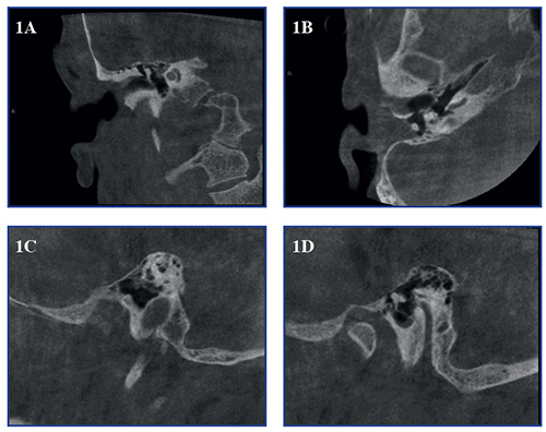
Temporal bone imaging with CBCT
The Cone Beam CT (CBCT) technique is normally used in head and neck area for dento-maxillofacial, sinonasal and cervical spine imaging. Until recently, temporal bone imaging has typically been done with multidetector CT (MDCT). An issue related to CBCT in this anatomical area is the thick bony mass that surrounds the small details of the middle and inner ear. When the narrow cone shaped beam is localized to the small field-of-view (FOV) , this bony mass, which is outside of the target area, significantly attenuates the X-rays before and after they pass the region of interest and therefore pose challenges to the image formation. The MDCT technique covers axially the whole head and hence does not have this problem, but at the expense of a considerably higher radiation dose. The usefulness and diagnostic capability of the CBCT technique in the temporal bone area is demonstrated in the following three case reports.
Case reports
The CBCT unit used in this study is SCANORA
