
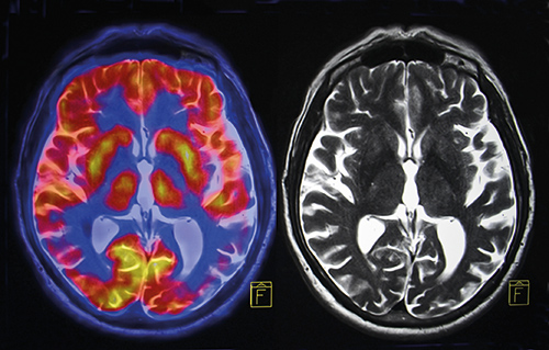
PET/MRI: enabling combined metabolic-anatomical imaging
PET/MRI is a hybrid combination of two imaging modalities. The first consists of positron emission tomography (PET) for ultra-sensitive imaging of metabolism and tracking uniquely labelled cell types or cell receptors. The second consists of the structural and functional characterization of tissue provided by magnetic resonance imaging (MRI).
The combination of metabolic and anatomical imaging provides superior diagnostic capabilities in certain clinical situations – above all, for cancer.
PET: 18F-FDG and tumour response
PET has a uniquely high sensitivity, in the picomolar range, and uses radio-labelled tracers which provide molecular information for characterizing tumours and metastases. The tracers are injected in minute, non-pharmacological doses, and 3-D images are subsequently reconstructed by computer to show the concentration and location of specific tracers.
PET rapidly began to gain traction in the early 2000s, after it was realized that the imaging of specific molecular targets associated with cancer would permit earlier diagnosis and better management of oncology patients. A review published in 2002 foresaw PET becoming increasingly important in cancer imaging over the decade. Since then, PET has offered a way out of some of the key limitations in anatomical approaches for imaging cancer biology.
More recently, PET has begun to gain widespread acceptance for assessments of tumour response to chemotherapy and radiotherapy, when combined with the 18F-fluorodeoxyglucose (18F-FDG), glucose analogue. The commercial availability of the latter catalysed widespread introduction of PET in smaller hospitals and medical centres. Although quantitative analysis of 18F-FDG uptake is required for predicting tumour response early in therapy, according to some a visual interpretation of scans is often sufficient to assess response after the completion of therapy.
PET/CT seeks to address anatomical information deficit
PET images, however, still lack detailed anatomical information. An effort to address such a lack was to fuse PET data with high-resolution, three-dimensional morphological images from computer tomography (CT), which have been achieving sub-millimetre spatial resolution for well over a decade. Since the mid-2000s, PET examinations have indeed been performed in combination with CT and hybrid PET/CT systems have shown far greater accuracy in data registration from the two modalities than achievable by software fusion of separate images. The hybrid data has of course also proven to be of higher diagnostic value than either PET or CT on their own – apart from also simplifying the logistics of patient management. New PET installations are now almost exclusively comprised of combined PET/CT scanners
Key advantages of the combination, according to a 2009 study by Vanderbilt University Medical Center in the US include superior lesion detection, improvement in the localization of foci of uptake resulting in
