
New dark-field X-ray technology improves diagnosis of pulmonary disease
For the first time, researchers at the Technical University of Munich (TUM) have successfully used a new X-ray method for respiratory diagnostics with patients. Dark-field X-rays visualize early changes in the alveolar structure caused by the lung disease COPD and require only one fiftieth of the radiation dose typically applied in X-ray computed tomography. This permits broad medical application in early detection and treatment follow-up of respiratory ailments.
There are millions of cases in which serious respiratory system illnesses place limitations on quality of life. Every year more than four million people die of serious respiratory ailments worldwide. Partially destroyed alveoli and an over-inflation of the lungs (emphysema) are typical of the life-threatening ailment Chronic Obstructive Pulmonary Disease (COPD).
However, the fine distinctions between healthy and diseased tissue are barely visible on conventional chest X-rays. Detailed diagnostic information is only available using three-dimensional computed tomography approaches, in which the computer assembles many individual images. Until now there has been no fast and cost-effective option for early detection and follow-up examinations with a low radiation exposure as used in plain chest X-rays.
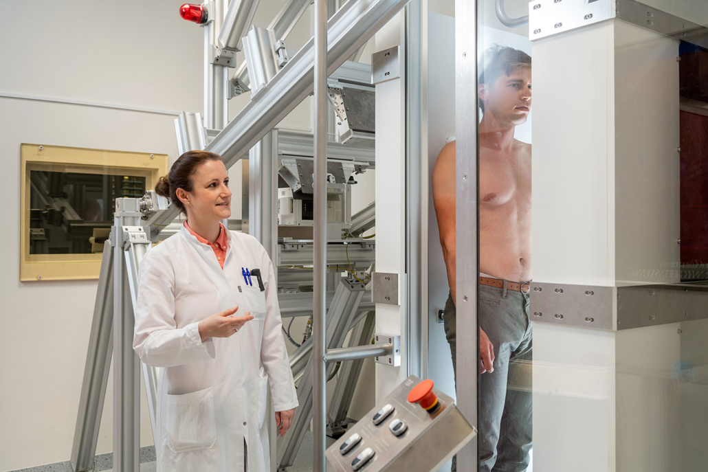
A procedure developed at the Technical University of Munich could now fill this gap: dark-field chest X-rays. In the November 1, 2021 issue of The Lancet Digital Health a research team led by Franz Pfeiffer, Professor for Biomedical Physics and Director of the Munich Institute of Biomedical Engineering at TUM, present the results of an initial clinical patient study, which used the new X-ray technology for the diagnosis of the lung disease COPD.
The wave character of X-rays is the key
Conventional X-ray imaging is based on the attenuation of X-rays on their way through the tissue. Dark-field technology on the other hand use the wave nature of X-ray light, which is discarded in conventional X-ray imaging.
The new method thus uses the physical phenomenon of scattering in a manner similar to the long-known principle of dark-field microscopy with visible light. This allows to visualize the structure of objects that are for the most part transparent. These structures appear in the microscope as bright images on a dark background, which has given the method its name.
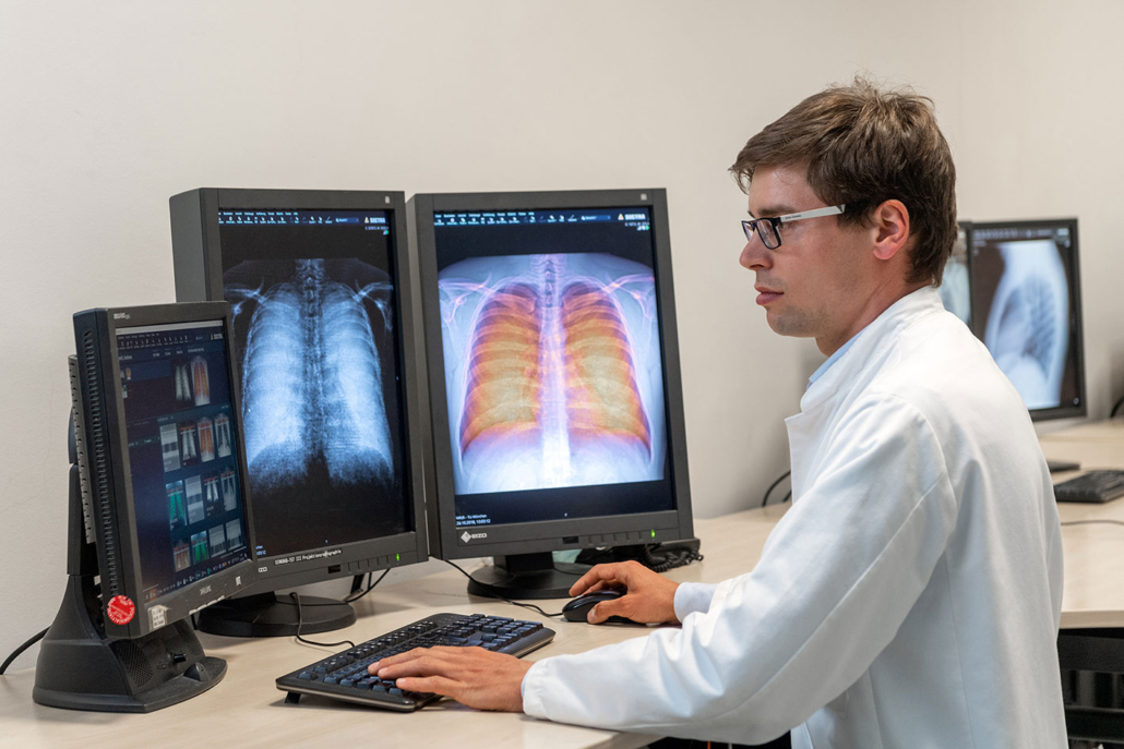
“The X-ray dark-field signal is particularly strong for interfaces between air and tissue,” Prof. Pfeiffer points out. “This makes it possible for a dark-field X-ray image of the lung to clearly distinguish between intact alveoli, i.e. those filled with air, and regions in which less intact alveoli exist.”

The dark field X-ray method visualizes early changes in the alveolar structure as a result of the lung disease COPD. Franz Pfeiffer, Professor for Biomedical Physics, hopes that this will significantly improve the early detection of lung diseases.
Lower radiation dose
In addition, an examination using dark-field chest X-ray technology involves a significantly lower radiation dose than presently used computed tomography. This is because dark-field chest X-rays require only one exposure per patient, as compared to the large number of individual images taken from different directions which are necessary in computed tomography.
“We expect the radiation exposure to be reduced by a factor of fifty,” says Prof. Pfeiffer. Furthermore, the first clinical results have confirmed that the dark-field X-rays provide additional image information on the underlying microstructure of the lung.
“Given the close connection between the alveolar structure and the functional condition of the lung, this ability is of great significance for pulmonary medicine,” explains Dr. Alexander Fingerle, senior physician at TUM’s university hospital Klinikum rechts der Isar’s Department of Diagnostic and Interventional Radiology. “In the future dark-field X-rays could help improve early detection of COPD and other respiratory ailments.”
Better X-ray equipment for early detection
Prof. Pfeiffer hopes these initial clinical results with patients will accelerate the execution of further clinical studies and the development of marketable devices that use the dark-field method.
“Dark-field chest X-rays are currently giving us a chance to significantly improve the early detection of lung diseases and at the same time to implement it on a wider basis than before,” Prof Pfeiffer notes.
Since dark-field imaging is not limited to COPD, further translational studies with other pulmonary pathologies such as pulmonary fibrosis, pneumothorax, lung cancer and pneumonia, including COVID-19, are of great interest.
Reference:
K. Willer, et. al. X-ray dark-field chest imaging for detection and quantification of emphysema in patients with chronic obstructive pulmonary disease: a diagnostic accuracy study. The Lancet Digital Health. November 1, 2021. doi: https://doi.org/10.1016/S2589-7500(21)00146-1
