
New approaches to dose reduction in interventional CT imaging
Imaging in the interventional suite has enabled the use of minimally invasive treatment in cases that were traditionally facing surgery. Nowadays stenoses in the coronary arteries or cerebral malformations can be treated without having to open the thorax or skull and microscopic devices can be implanted in an endovascular manner. The morbidity and mortality of this kind of therapy dropped steadily over the past decade because of advances in technology [1].
by Dr Andreas Maier
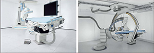
The state-of-the-art in imaging for most cardiac and neurological treatment options is X-ray fluoroscopy using angiographic C-arm systems. Scanners [Figure 1] are commonly used for such purposes in the interventional room. Using X-rays for imaging has many advantages, such as imaging in real-time which allows direct feedback in catheter guidance, high image quality and few additional constraints on the interventional workflow. However, the use of X-rays always implies a certain radiation dose for the patient.
In the case of two dimensional (2D) or fluoroscopy imaging, several measures to reduce the dose for the patient and the interventionalist are taken. Often patient and operator are shielded using lead. Another very common method is to focus the irradiation only on the region that is of interest at the current state of the intervention. This region of interest (ROI) imaging is achieved using a lead collimator that is inserted into the X-ray beam. As a result, only the ROI is visualised.
In 2006, computed tomography (CT) volumetric images that could show soft-tissue contrast became also available in the interventional suite with the introduction of flat-panel detectors [2] . This new kind of imaging enabled one to visualise complex anatomy in three dimensions (3D). In neurosurgery, 3D digital subtraction angiography (DSA) is able to show the complete arterial vessel tree. This kind of visualisation can be used to improve guidance in minimally invasive surgery. The dose of a full head scan is moderate with about 2.7 mSv. This is comparable to a diagnostic head CT scan.
The main advantage of the 3D technique is its ability to distinguish small differences in contrast that are usually not visible in the 2D projection. Figure 2 shows the 3D reconstruction of an implanted flow diverter stent. Note that the carrier mesh structure can be recognised in this high resolution image.
From the 3D scan, the spatial position of the implanted device is also known. Thus, this information can be used as an overlay for the 2D projection image to improve the visibility of the object of interest. Thus, objects that are usually not visible in the projection image can now be shown. Figure 3 shows a clinical example with an overlay for 2D DSA.
Dose reduction for 3D imaging
For the above-mentioned scenario, it is sufficient to scan only a limited volume that contains the implanted device. Thus, it would be sufficient to scan only a volume of interest (VOI). While this goal seems easy to achieve, the limited field of view poses a major challenge to the reconstruction algorithm. For common reconstruction methods, a full view of the object is required. Otherwise, severe artefacts appear that render the image clinically unusable.
An exact reconstruction is only possible using prior information [3] or a scanning procedure with a slight increase in dose [4], an approximate method is required to be able to reconstruct without artificial structures in the image. This can either be achieved by heuristic methods which model the outside of the field of view by extrapolation [5] or by a clever combination of different filtering techniques [6]. In either scenario, the reconstruction is visually correct, but the reconstructed gray values measured in Hounsfield-Units (HU) are only known up to a scaling factor. For the interventional scenario, this information is sufficient as the main purpose of the image is to display structural information during the surgery. The absolute HU value of the tissue is currently only used in diagnostic scenarios.
Initial results show that the effective dose can be reduced efficiently using such VOI scanning techniques [cf. Table 1]. The use of the full detector with 30 cm x 40 cm results in an effective dose of 2.7 mSV for the patient. If the area is reduced to 5 cm x 4 cm to image a small device, the effective dose is reduced by a factor of 27. Note that this dose is comparable to taking about two chest X-ray images and that the annual average dose by background radiation is about 4 mSv.
Reducing dose, reducing view
The use of VOI imaging allows one to reduce the dose in interventional applications dramatically. The reduction in dose, however, also comes at the cost of a reduced field of view. Thus, dose savings will only be applicable to certain applications. Especially the implantation of micro devices can be supported with this technique. In these cases, repeated scans of the same volume are possible. In particular, difficult cases will benefit from this imaging technique, as the interventionalist would be able to repeat 3D scans about 30 times at the dose of a normal head CT scan. Thus, it gets possible to observe the expansion of a stent in multiple stages during the implantation before the placement is final and can no longer be revised.
Unfortunately, the presented methods are mostly only applicable to interventional CT as angiography systems have highly flexible collimators. For diagnostic CT scanners which have gantries that do a full rotation three times a second, additional efforts have to be made in collimator design before these methods will be applicable.
Conclusion
The use of volume of interest scanning provides an opportunity to scan small volumes at very small doses. Using such scanning protocols will enable new workflows in the clinical routine and will increase the safety of minimally invasive procedures further. Fortunately, major vendors of angiographic scanners are already working towards establishing such protocols in clinical practise.
Acknowledgements
The author would like to thank Siemens AG, Healthcare Sector for the permission to use images of their scanners. Furthermore, the author also would like to thank Dr Mawad at St. Luke
