
Leading hospital in Cyprus offers Digital Breast Tomosynthesis
YGIA Polyclinic’s Radiology Department has been operating for 27 years and its staff is actively involved in ongoing clinical research and training to ensure the best possible services to all patients. A year ago, it embarked on setting up a breast imaging unit equipped with state-of-the-art technology, culminating in the installation of the Hologic Selenia Dimensions DBT system.
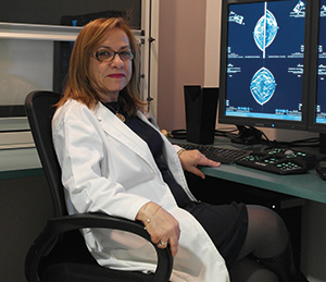
Dr. Annie Papoutsou, Head of the X-ray Department gives us the full picture.
How has your hospital expanded in recent years?
In 2007, after major renovations and an extension of its building facilities, the YGIA Polyclinic private hospital boosted its capacity to 152 beds, extended the number of operating theatres to 12, established and extended the capabilities of its Clinical Laboratory Department, Radiology Department (X-ray, Mammog, Fluoroscopy, MRI, CT, Ultrasound), produced a multi-dynamic Intensive Care Unit (ICU), Obstetrics & Gynecology, and Pediatrics Departments. Furthermore, from mid June 2012, a state-of-the-art Cardio-Vascular Catheterizations Centre was established at the hospital offering the only 24-hour acute percutaneous coronary intervention (PCI) service in Cyprus. Moreover, the hospital has a range of fully equipped ambulances working 24 hours in order to be able to best respond to emergencies.
What type of equipment is used by your department?
Most of our X-ray rooms use the latest DR digital detectors providing superior quality images almost instantly, and are linked to an enterprise-wide fully integrated RiS/PACS.
Last year we organized a breast imaging unit, equipped with the latest technology, FFDM Hologic Selenia Dimensions, a GE ultrasound with strain and shearwave elastography and a Hitachi ultrasound with high frequency linear probe.
In our department we performed more than 90,000 exams yearly.
What was the rationale for selecting the Hologic Selenia Dimensions system?
The decision was based on the special features offered by the Selenia Dimensions; these include:
- 2D imaging or combo mode (2D+3D) imaging in the same compression.
- Exceptionally sharp images with minimal dose.
- Streamlined workflow.
- Ergonomic design for comfort and ease of operation.
- We believe that it offers the best technology available.
What are the advantages of Digital Breast Tomosynthesis?
The use of Digital Breast Tomosynthesis in breast screening enables us to find more invasive cancers than conventional 2D mammography alone.
The masses, distortions and asymmetric densities are better visualized with the Selenia Dimension.
But also it can reduce the costs associated with unnecessary recalls and it can reduce the incidence of negative biopsies.
Has the number of exams been affected by the adoption of DBT?
Since the installation the number of mammography exams has increased up to 50%.
The recalls rate has decreased and also additional views have decreased.
When did you decide to acquire the C-View software and what are the benefits?
C-View software was installed from the first day of the equipment installation, giving us the possibility of eliminating the need for conventional 2D exams after 6 months. The combined tomosynthesis and C-View exam makes lower patient radiation dose possible. Tomosynthesis exams with C-View software offer a patient dose similar to a 2D only exam with superior clinical performance for all breast types.
You are also using the Affirm biopsy device –what is the typical biopsy procedure followed in your department?
Up until now we are using the Affirm stereotactic biopsy device and in the next few days we are going to install the Affirm stereotactic tomosynthesis biopsy device to target lesions seen only with tomosynthesis. The typical biopsy procedures followed in our department are the biopsy under the stereotactic guidance and hookwire localization for subtle masses clusters of microcalcifications and architecture distortion.
What do you see as the next step for improving the performance of your department?
Installing I-view with the contrast media.
I-View is a contrast-enhanced mammography technique that may be a viable alternative to breast MRI in performing contrast agent breast imaging. It offers certain advantages over MRI, including reduced cost and shorter procedure times. The imaging combination of contrast enhanced 2D imaging (CE2D) along with a 3D tomo scan, gives additional information beyond a CE2D examination alone, and may allow localization and morphologic evaluation of an enhancing lesion, further increasing the value of the CE2D procedure.
