Investment in training and integrated solutions improves detection and characterization of gastrointestinal lesions
In the fight against colorectal cancer, great strides have been taken to protect patients from undetected abnormalities. However, up to 26% of lesions are still missed in examinations. [1] The greatest diagnostic challenge in endoscopy is to improve dysplasia detection and increase detection rates of gastrointestinal lesions.
Incorporating artificial intelligence (AI) systems in endoscopy procedures is a step forward in overcoming this challenge. However, there is also a need to strengthen the characterization of these lesions. The solution relies on reliable detection skills, effective equipment and enhanced knowledge through investment in training. With these steps, it will be possible to characterize these lesions with the assistance of existing AI tools and reduce the number of lesions that go undetected.
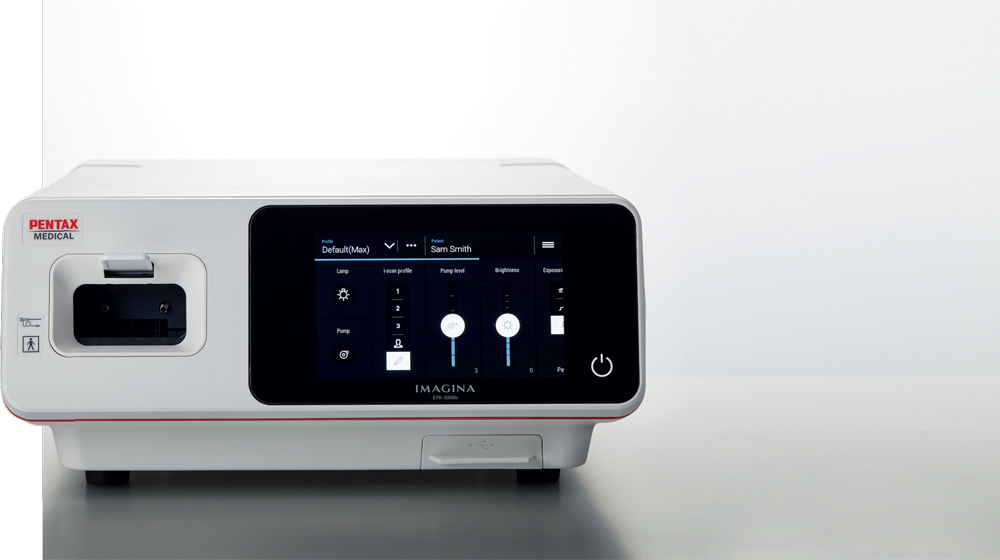
Greater training can empower endoscopists to improve their ability to detect gastrointestinal lesions. Supplementing this investment in human capital with integrated solutions that consist of complementary technologies, such as PENTAX Medical i-scan, can support endoscopists in overcoming the challenge of lesion detection and characterization.
Setting endoscopists up for success: investing in training
Investing in training of endoscopists is an important factor to improve detection of gastrointestinal lesions, and ultimately improving patient outcomes. Endoscopic detection and characterization of lesions are essential elements on the diagnostic pathway. Yet, characterization is only valuable when endoscopists can first detect the polyp. If a physician is not sufficiently trained, filters provided by characterization tools cannot help determine whether the polyp must be removed.
In an effort to overcome this challenge, manufacturers should support physicians in understanding and learning to use the latest innovative technologies and techniques.
Professor Ralf Kiesslich of HELIOS Dr Horst Schmidt Kliniken (HSK) Wiesbaden, Germany explained: “Detection and characterization are the most important steps during routine endoscopy. Firstly, all suspicious areas have to be identified. Subsequently, the endoscopist has to decide if an endoscopic intervention is necessary. The combination of virtual and optical chromoendoscopy offers a clear clinical pathway for endoscopic evaluation of GI tract pathologies.”
Enhanced detection and characterization with PENTAX Medical i-scan technology
Colorectal cancer prevention is the primary goal of screening and diagnostic colonoscopy. At least 50% of all interval carcinomas arise from undetected lesions during colonoscopy. [2] In combination with sufficient training, physicians should be equipped with integrated solutions that support each step of the clinical diagnostic pathway. PENTAX Medical i-scan, an existing tool in the field of endoscopy, can support physicians to overcome the challenge of lesion detection and characterization. Together with PENTAX Medical’s advanced HD+ endoscopes, PENTAX Medical i-scan has been proven to increase adenoma detection rates to help physicians meet quality measurement standards. [3]
PENTAX Medical i-scan optical enhancement
PENTAX Medical offers the unique combination of digital and optical image enhancement to further support a physician’s ability to detect and characterize lesions. PENTAX Medical developed the i-scan Optical Enhancement (OE), an optical filter that incorporates technology combining bandwidthlimiting light with digital image processing. [3]
PENTAX Medical i-scan image processing includes three AI algorithms: Surface Enhancement (SE), Contrast Enhancement (CE), and Tone Enhancement (TE).
Endoscopists must be equipped with the knowledge, practice and technology to accurately detect and characterize gastrointestinal lesions. To this end PENTAX Medical i-scan has supported endoscopists in detection and characterization for more than 13 years.
Professionals, such as Prof Kiesslich, confirm their confidence in using PENTAX Medical i-scan.
PENTAX Medical DISCOVERY
In addition, PENTAX has other tools to support clinicians. Endoscopists may be tired after a long day of procedures or may be distracted by external factors which can play a role in complicating lesion detection. In such instances, PENTAX Medical DISCOVERY™ can come into play. This innovative AI device provides highly focused clinical diagnostic support in the detection of lesions.
“The PENTAX Medical DISCOVERY is trained by advanced artificial intelligence to highlight the presence of pre-cancerous lesions with a visual marker in real-time – serving as an ever vigilant second observer,” explained Dr Sergey Kashin, Head of Endoscopy Department, Yaroslavl Regional Cancer Hospital, Russia.
Technology serves an important role in supporting endoscopists to improve their ability to detect and characterize gastrointestinal lesions. Yet despite technological advances, there is no substitute for thorough investment in training of endoscopists. PENTAX Medical provides outstanding medical courses and master classes of the highest quality tailored to meet individual needs. Through sharing knowledge, improving expertise and skills, physicians can be better equipped to protect patient safety and improve outcomes.
PENTAX Medical i-scan and PENTAX Medical DISCOVERY are valuable tools to complement and support endoscopists in overcoming the challenge of lesion detection and characterization and should be used by endoscopists in this field.
Reference
- van Keulen, K., Soons, E. & Siersema, P. The Role of Behind Folds Visualizing Techniques and Technologies in Improving Adenoma Detection Rate. Curr Treat Options Gastro 17, 394–407 (2019). doi.org/10.1007/s11938-019-00242-5
- Pohl H, Robertson DJ. Colorectal cancers detected after colonoscopy frequently result from missed lesions. Clin Gastroenterol Hepatol 2010;8:858-64.
- Bowman, E. A., & E. (2015). High Definition Colonoscopy Combined with i-SCAN Imaging Technology Is Superior in the Detection of Adenomas and Advanced Lesions Compared to High Definition Colonoscopy Alone. P. J. O’Dwyer, 2015. doi:10.1155/2015/167406


