
Fluence field modulated CT: A novel approach for noise and dose management in CT
CT scans produce high resolution, three-dimensional (3D) images routinely used in medical diagnostic and image guidance procedures. The rapid advances in CT, including faster acquisition times and enhanced tissue discrimination capabilities, have also led to a broader range of applications for CT. Recently, however, there has been increased concern regarding the radiation dose received by patients during CT scans. Risks due to radiation are present because CT generates 3D images from a set of X-ray radiographs (or projections), which are recorded at different angles as the X-ray source rotates about the patient.
by Steven Bartolac and Dr David Jaffray
The concerns regarding radiation have been largely stimulated by a number of publications produced over the last five years that have indicated that the number of CT scanning procedures is on the rise (on the order of about 10% per year [1]), and that the increased risk of cancer due to radiation doses received from CT scans may be non-negligible [2], especially in patients receiving multiple CT scans [3]. Motivated by these concerns, fluence field modulated computed tomography (FFMCT) has been proposed as a new approach for CT imaging that promises better management of dose to the patient [4-6] without sacrificing image quality [See Fig. 1].
The tradeoff between image quality and radiation exposure
Though image quality is dependent on many factors, including image blur, and distortions that can arise from poor modelling of the X-ray physics, image noise is often a key determinant of image quality and the utility of CT scans, and is most directly related to the imaging dose. Image noise refers to random fluctuations in the image, which, when large, can obscure the details of interest. In CT images, the largest source of noise is due to inherent statistical fluctuations associated with photon counting (i.e. Poisson noise). The magnitude of this noise is inversely proportional to the average number of photons that reach the detector. Increasing the number of incident photons, or the X-ray fluence (i.e. photons/unit area) can help limit noise in the image but also results in increases in dose. Conversely, attempting to reduce the dose is associated with increased noise. Managing this tradeoff requires choosing the most appropriate scan settings considering the imaging task, and individual factors including patient age and size.
Conventional strategies for dose management
Non-uniformity of noise in CT images (i.e. image noise which changes in magnitude at different positions within the image) occur because different regions of the patient attenuate the X-ray fluence to varying degrees. Generally, a longer path length through the patient suggests greater attenuation of the beam, and greater noise associated along that X-ray path. If the patient is modelled as an elliptic cylinder, the path length is longer near the centre of the patient than near the periphery. To compensate for these changes, bowtie filters are typically included in conventional scanners to limit the incident fluence at the peripheries of the patient and allow higher fluence near the centre, creating a more uniform exposure at the detector. In this way, overexposure to the peripheries is prevented when attempting to limit noise near the centre of the patient. In the elliptical model, some views of the patient are also more greatly attenuating than others. Tube current modulation (TCM) is therefore used in conjunction with bowtie filters, which allows the overall fluence to be increased or decreased depending on the view. [See Fig. 2 for schematic illustrations of bowtie filtration and TCM.]
FFMCT: a new paradigm for CT imaging
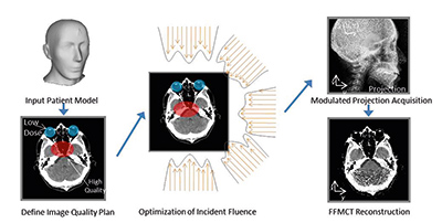
Though bowtie filters and TCM greatly aid in decreasing dose to patients, patient anatomy is inherently more complex than the representation by a simple elliptical shape. The presence of bony and lung tissue, for example, introduces large variations in beam attenuation across the field of view. The pattern of beam attenuation also depends greatly on the incident angle of the X-rays. Contrary to conventional approaches that use a fixed filter in place throughout the scan, FFMCT proposes to use a dynamic modulator allowing the fluence to change freely across the field of view and for different projection angles, such that each projection may have a distinct incident fluence pattern [See Fig. 1].
Increased flexibility in the delivery of X-ray fluence suggests better management of dose since higher exposures can be reduced where not required for maintaining the desired image quality. In the proposed methodology for FFMCT, an input model of the patient could be used to define an image quality plan defining the desired image quality for the given task [See Fig. 1]. The plan could further define specific regions where low dose is a priority. An optimization algorithm can then be used to search for a modulation pattern that comes as close to the planned objectives as possible. As many patients undergo multiple CT scans, a previous CT scan could potentially be used as the model; alternatively, a population based model could be used.
Achieving task-based, user-defined image quality
In many cases, the desired image quality may vary within the image. For example, one might desire higher image quality in a small region of interest (ROI) near a suspicious lesion in a repeat CT scan; in another case, one might only be interested in the region of the heart in a cardiac CT scan; in an image-guided surgical procedure, the ROI may be restricted to a localized region surrounding a surgical instrument. In these cases, allowing the image quality to be reduced outside the ROI may be advantageous, since it suggests a reduction in total dose to the patient. Initial research [5, 6] has suggested that fluence modulation patterns can be found that allow for better uniformity of image quality in target ROIs than afforded by conventional means, while allowing for image quality elsewhere to be reduced. [See Figs. 3 and 4.]
Dose reduction
The amount of dose reduction possible compared to conventional approaches using fluence field modulation depends highly on the patient and the task. Preliminary research using a simulated thorax phantom [5, 6] suggests that integral dose reduction across a single image slice (in Joules) could range up to 50% or higher for applications where the region of interest is well localized [5]. Local dose reductions (in cGy) outside the ROIs can approach 60-80%. Research is ongoing for evaluation of dose benefits to other sites.
Technology advances towards FFMCT
Currently, no device has been introduced in modern scanners that can offer the unconstrained, flexible modulation patterns proposed for fluence field modulation. Design challenges include speed demands on dynamic modulators given the very rapid gantry speeds of conventional scanners, and changes to the energy spectrum of the incident beam that might occur as a by-product of modulation using a dynamic filter. However, several simplified approaches for modulator designs have been proposed that make significant steps towards achieving fluence field modulation in clinical scanners, including a series of sliding wedges [7], dynamically moving discrete apertures [8, 9], and multiple sources in inverse geometry CT [10] (e.g. the
