
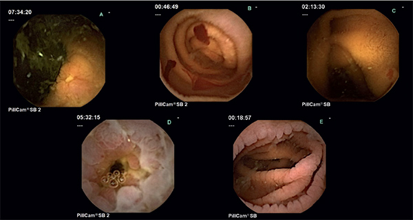
Endoscopic imaging of the small intestine
Historically, endoscopic investigation of the small intestine was limited due to inferior or invasive techniques. However, the emergence of video capsule endoscopy and device-assisted enteroscopy has revolutionised the management of small bowel diseases over the last ten years. These new endoscopic techniques are complemented by advances in radiology, particularly small bowel enterography. Future goals for development include steerable capsules and devices that improve ease and depth of insertion of the enteroscope to improve diagnosis and therapy of small bowel disease.
by Dr Christina A. Tennyson and Dr Carol E. Semrad
The small intestine constitutes the largest surface area of the gastrointestinal tract. Until recently, endoscopic exploration has been limited to its most proximal and distal ends because of its length (3-6 meters in adults) and coiled anatomy in the abdominal cavity. This has resulted in delayed diagnosis of small bowel tumours and limited options when having to treat small bowel bleeding, typically invasive intra-operative enteroscopy. Over the last ten years, however, the emergence of new endoscopic technologies, such as video capsule endoscopy and device-assisted enteroscopy, has revolutionised diagnosing and managing small bowel diseases, e.g. obscure bleeding, Crohn
