
Carotid artery stenting – the challenge of plaque protrusion
Although not commonplace, carotid artery stenting (CAS) is occasionally accompanied by the protrusion of plaque into the stent lumen, and a variety of ischemic complications during intra- and post-operative periods. Among these complications, plaque protrusion (PP) into the stent and thrombus on the stent after CAS are some of the most worrying for clinicians.
The correction of PP is achieved by additional post-dilations or stent-in-stent implantation.
First noticed in mid-1990s
The first observations of PP date over two decades and provided the impetus for embolic protection devices. In a paper published in the mid-1990s, a team led by Frank Veith, M.D (from the Mayo Clinic) suggested that restoration of flow and removal of protection devices might lead to the continuing break off of protrusions and provoke some delayed strokes.
Today, apart from PP, key causes of late in-stent stenosis seen in CAS include neo-intimal proliferation due to self-expanding stents as well as restricted post-procedural stent dimensions from inefficient balloon dilation.
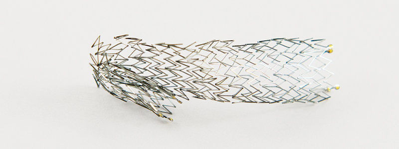
The covered stent
One way to prevent PP is by covering the plaque with a stent graft. Covered stents also offer advantage in usability, without the need for distal protection devices in difficult internal carotid arteries, where a protection filter may be near-impossible to place. However, covered stents are accompanied by a high rate of restenosis. In one randomized trial in the mid-2000s, researchers at Vienna Medical University reported a 38% restenosis rate in patients treated with covered stents for carotid artery stenosis, while restenosis was absent in the bare stent group. The precise causes of high restenosis rates with stent grafts require further research. One suspected factor is the buckling of a covered stent’s proximal and distal ends and the prevention of endothelization.
Both symptomatic and asymptomatic events
During stent implantation, plaque disruption and distal migration of plaque particles may cause symptomatic or asymptomatic ischemic events, in spite of protection devices. These can be viewed on diffusion-weighted (DW) magnetic resonance imaging (MRI).
In some CAS cases, physicians encounter plaque particles filling filters leading to symptomatic cerebral embolism. This is particularly true with ulcerated plaque and severe stenosis. What is now of growing concern is that, after stent implantation, plaque protrusion into the lumen can lead to peri-procedural stent thrombosis, 30-day stroke, and late in-stent stenosis.
Stroke biggest complication
The biggest complication with carotid artery stenting is stroke. This can occur during CAS and for up to 30 days after the procedure. Although the cause of late stroke after CAS remains unknown, PP is generally suspected to be a key cause.
PP incidence is evaluated by IVUS (intra-venous ultrasound) and angiography, although as we shall see, there are variations in results based on methodology. The prognosis of PP (over a 30-day period) and the incidence of ischemic lesions (48 hours after CAS) are usually assessed by diffusion-weighted images.
The Tokai study
In recent years, one oft-cited report concerns a study by the Department of Cardiology at Tokai University School of Medicine (Isehara). The findings were published in October 2014 by the ‘Journal of Stroke and Cerebrovascular Disorders’. During their study, the Tokai researchers evaluated 77 CAS procedures, which were performed consecutively with IVUS between May 2008 and December 2012. All cases were distally protected with filter devices. The rate of PP was assessed at the end of each procedure using IVUS and angiography.
Six plaque protrusions (7.8%) through the stent struts were detected by IVUS but only two (2.6%) by angiography. One of the major predictors of PP was pre-procedural severe stenosis with flow delay. Overall stroke rate was 2.6% (major 0%, minor 2.6%), and these occurred in the catheterization laboratory. However, no late stroke was observed at 30 days after procedure.
One of the key outcomes of the Tokai study was that IVUS seems to detect plaque protrusion better than angiography. Since the adequate management of plaque protrusion is considered as a means to reduce stroke complications, IVUS usage is worth considering.
The Yao-Nara study
In 2017, results of another, broader Japanese effort by researchers at Ishinkai Yao General Hospital (Yao) and Nara Medical University (Nara) suggested different conclusions. The study, which was published in the April 17 issue of ‘JACC: Cardiovascular Interventions’, sought to clarify the frequency and prognosis of plaque protrusions in CAS by analysing data on 328 patients treated under IVUS guidance in the period 2007-2016, using different types of stents and embolic protection devices.
At 30 days, the rate of ipsilateral ischemic stroke was 2.8% and the rate of transient ischemic attack was 2.6%. There were no patient deaths. Moreover, in most stroke cases, symptoms were observed immediately after dilatation. New ischemic lesions were found in 35.7% of patients within 48 hours of the procedure, based on diffusion-weighted imaging (DWI).
Lack in lesion variation, but stent type matters, as does evaluation method
One of the most intriguing conclusions was the lack of difference in the incidence of new ischemic lesions, in terms of stable versus unstable plaques. Analysis by stent type, however, did indicate difference. There were more ipsilateral ischemic lesions with open-cell stents as compared to closed-cell stents.
The authors suggest the findings indicate a necessity to minimize PP “to prevent periprocedural ischemic stroke” and that the placement of open-cell stents with high radial force may disintegrate unstable plaque, causing protrusions. One strategy mentioned by the authors to manage PP is to perform IVUS to check for large-volume protrusions. The latter are then sought to be differentiated as being either ‘convex’ or ‘non-convex’. For the former, stent-in-stent placement is performed using closed-cell stents until the disappearance of the protrusion. In the case of ‘nonconvex’ protrusions, the authors recommend 5-10 minutes of observation, followed again by stent-in-stent placement should the protrusion enlarge, or clinical follow-up within 30 days after CAS in case of no enlargement.
As we observed previously, there are differences in PP incidence based on whether it is evaluated by angiography or IVUS. One of the most significant limitations of the Japanese study above was the occurrence of 27 cases of plaque protrusion on IVUS, but just nine cases on angiography. The study protocol required confirmation by both modalities.
Limitations to Yao-Nara study
In an editorial accompanying the study, William A. Gray, MD (Lankenau Heart Institute, Wynnewood, PA), cautioned that this two thirds difference “will clearly affect many of the subsequent associations and conclusions.” Gray also underlined that by treating plaque protrusion with stent-in-stent placement in approximately half of the cases, the researchers might have potentially changed the clinical and imaging outcomes. Furthermore, he cautioned, the study was not core-lab controlled, with no routine use of MRI before and after procedures, and that the assessors were not blinded. Finally, they did not mandate use of specific stents or perform independent neurological assessment of clinical outcomes.
As a result, the association between stent type and plaque protrusion is ‘likely’. However, it may not be as strong as the authors contend.
Such shortcomings are likely to be addressed when the Japanese effort is paired with emerging data showing reductions in both plaque protrusion and ischemic lesions via the use of mesh-covered stents. Gray agrees that this is strengthening the case for “improvements in stent design.” Indeed, emerging micromesh stent designs are expected to contribute greatly to prevention of plaque protrusion and may become a new standard for CAS.
SCAFFOLD trial
In the United States, the SCAFFOLD trial, led by Peter A. Schneider, MD, of Kaiser Foundation Hospital at Honolulu (Hawaii) is completing evaluation of a mesh-covered, open-cell heparin coated stent in patients at high surgical risk. The objectives are to make the first 30 days safer, with the understanding that reduced cell size equates to less plaque prolapse and fewer delayed events.
Other similar trials using different mesh technologies are also under way, and more are imminent. In Italy, for instance, University of Roma La Sapienza has begun a positive-control study to analyze and compare the rate of off-table subclinical neurological events in two groups of patients submitted to CAS with a close-cell stent, and a new mesh-covered carotid stent called C-Guard.
Overall, new parameters are coming into place, via stents with differences in pore size, flexibility etc. The drivers for such efforts range from new materials to a broad range of cardiovascular conditions. In France, for example, University Hospital Grenoble is conducting trials with Mguard, a stainless-steel closed cell stent covered with an ultra-thin polymer mesh sleeve, to prevent distal embolization during percutaneous coronary intervention in ST-segment-elevation myocardial infarction.
Case selection and stenting success
One of the observations of SCAFFOLD was that case selection for stenting was a key “to good clinical results.”
So far, patient selection criteria for CAS is largely based on surgical risk related to other co-morbidities. The morphology of the atherosclerotic plaque is given little attention, although studies have demonstrated the existence of extensive variability, which in turn confers specific risks for plaque vulnerability. Overall, the detection of unstable plaque on MR plaque imaging and the use of open cell stent are considered to be significant predictive factors of PP.
In recent years, there have been growing calls for devising best practices in peri-procedural management and follow-up, and for continuous feedback from clinicians to industry to improve stent design.
In general, achieving better outcomes of CAS is seen as the best method to solidify its place as a frontline treatment of carotid vascular disease.
One promising approach for patient selection and identification of plaque, has been the use of virtual histology intravascular ultrasound imaging (VH IVUS). Researchers have suggested a strong correlation between VH IVUS plaque characterization and the true histological examination of plaque following endarterectomy, especially in ‘vulnerable’ plaque types.
The results of one of the earliest efforts in this area were published in October 2007 in ‘The Journal of Endovascular Therapy’. This followed a prospective, two-arm study by the Arizona Heart Hospital & Translational Research Center. The researchers enrolled 30 patients.
In the first arm of the study, 15 patients underwent VH IVUS examination of carotid plaque with a cerebral protection device. This was immediately followed by carotid endarterectomy (CEA), and the comparison of ‘virtual’ with true histology (classifying plaque type by VH IVUS and histopathology in a blinded study).
In the second arm, 15 patients undergoing CAS had a preliminary VH IVUS scan performed with cerebral protection. Debris collected from the filter following stenting was examined histologically and compared with the VH IVUS data.
The diagnostic accuracy of VH IVUS to agree with true histology in different carotid plaque types was 99.4% in thin-cap fibroatheroma, 96.1% for calcified thin-cap fibroatheroma, 85.9% in fibroatheroma, 85.5% for fibrocalcific, 83.4% in pathological intimal thickening, and 72.4% for calcified fibroatheroma.
