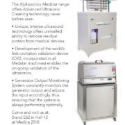


In the modern healthcare environment the demand towards CT goes beyond simple high throughput and accurate diagnosis. Efficient operator workflow, improved patient experience and easy installation into existing facilities are also main considerations. Speedia HD enables high-speed whole-body scanning with sub-millimetre slices, which is difficult to achieve on 16 slice CT systems. A single breath hold (approx. 14sec.), can produce high-resolution images in the range of 1100mm or more. Thereby allowing wide range, high resolution MPR images to be acquired as routine.
Speedia HD with its 40mm width detector and unique 3D reconstruction algorithm-CORE method, achieves the high-speed scan even when using a pitch of 1.58. Therefore, it enables a chest area of 320mm to be scanned in only 4.5sec and a thoraco-abdominal area of 570mm in just 7.5sec. This reduces the burden on the patients who have difficulty maintaining a still position or holding a breath for a long-time.
For more information please click here

This year at the European Congress of Radiology (ECR), Fujifilm displays its evolving portfolio of medical informatics and Enterprise Imaging innovations, presenting REiLI, its Artificial Intelligence (AI) technology initiative, and enhanced Synapse functions with SYNAPSE 3D CONSOLE MODE.
Fujifilm Medical Systems Europe will celebrate SYNAPSE’S 20-year anniversary and will present, REiLI the company’s global Medical Imaging and Informatics Artificial Intelligence (AI) technology initiative at the European Congress of Radiology (ECR) annual meeting to be held from February 27th to March 3rd, 2019 at the Austria Congress Center in Vienna, Austria.
Under the REiLI brand, Fujifilm is developing AI technologies that strongly support diagnostic imaging workflow, leveraging the combination of deep learning in its AI technology with the Company’s image processing heritage. Fujifilm’s artificial intelligence software is a work in progress and is not commercially available in Europe.
Applications currently in development include, but are not limited to: Region Recognition, an AI technology to accurately recognize and consistently extract organ regions, regardless of deviations in shape, presence or absence of disease, and imaging conditions; Computer Aided Detection, an AI technology to reduce the time of image interpretation and support radiologists’ clinical decision making; Workflow Support, using AI technology to realize optimal study prioritization, alert communications of AI findings, and report population automation. At Fujifilm’s in-booth AI Center, it will be possible to see live demonstrations of AI delivering enhanced workflows.
SYNAPSE 3D CONSOLE MODE is the powerful native Advanced Visualization workflow in Synapse PACS. Synapse 3D is designed to enhance visualization features in Synapse 5. It offers advanced 3D rendering in the Synapse PACS Viewer to perform fast and accurate extractions, stenosis measurements, brain perfusion CT, MRI, and more.
The Fujifilm Healthcare IT platform showcased at ECR includes also the comprehensive medical informatics and enterprise-imaging portfolio:
SYNAPSE 5 is our next generation PACS, Synapse is one of the fastest medical imaging solutions in the industry, offering sub second delivery of extremely large datasets. Its underlying architecture promotes significantly less bandwidth consumption and tighter security.
SYNAPSE VNA is the most secure, comprehensive application for ingesting, storing and providing access to the complete imaging record. It securely integrates more specialties, more devices, and more data than any other VNA.
SYNAPSE MOBILITY Enterprise Viewer uses the latest server-side rendering technology to stream imaging securely and quickly to any authorized user. It can be used within applications, directly from the EHR, or on our mobile device apps. Both within and outside of the Enterprise, giving access to imaging immediately and helping clinicians making the most informed and accurate decisions.
SYNAPSE 3D is an enterprise-wide solution for quickly accessing multiple Advanced Visualization processing tools (in excess of 50 modules). Designed for use across multiple specialties including radiology, cardiology, surgery and more. Full integration with Synapse PACS means one-click extremely fast image processing from any Synapse client.
SYNAPSE CWM, Clinical Workflow Manager, is the most advanced Radiology Information System on the market today. It continues to evolve to support the unique imaging and information needs in today’s radiology department. One platform can support acute care facilities, imaging centres, and radiology practices providing distributed diagnosis.
SYNCRO-DOSE is the Radiation Dose Index Monitoring system, compliant with the Directive 2013/59 / EURATOM of the European Union. Syncro-Dose is a comprehensive system for monitoring and managing patient radiation exposure at enterprise level across different imaging modalities and hospital facilities.
THE 20-YEAR ANNIVERSARY OF SYNAPSE: THE WORLD’S FIRST WEB- BASED PACS
In 1983, Fujifilm launched Fuji Computed Radiography (FCR), becoming the first company in the world to offer a digital X-ray diagnostic imaging system. Medical professionals quickly learned the merits of digital diagnostic images, including ease of storage and processing. They found that images from a variety of tests and procedures could be shared within and among facilities, and the images could even be used for remote diagnosis and consultation. Recognizing this trend, Fujifilm saw the opportunity to leverage the technologies it had developed for FCR and contribute to the evolution of connectivity within and among medical facilities. What made Fujifilm’s SYNAPSE concept different was that it used the emerging Internet and web technologies instead of private networks. It was, in essence, a Web-based PACS: the first in the world.
Offering outstanding medical connectivity based on the convenient and efficient sharing of information, SYNAPSE made possible initial diagnosis at a local clinic, followed by more complete testing and treatment at a larger medical facility, in turn followed by periodic monitoring at the original local clinic. SYNAPSE’s rapid rate of adoption was due in large part to its capability, to contribute significantly to the quality of medical care, including support for the important objective of informed consent. Nowadays 5000 Synapse PACS systems are installed in healthcare facilities around the world, earning the largest market share worldwide (estimation based on a set of data from multiple market research studies), and last September “SYNAPSE 3D” (also known as Synapse Vincent in some global markets) a 3D image analysis system, won the Red Dot Award: Communication Design 2018 – the prestigious international design award in recognition of superior design, outstanding performance, and excellent operability.

Over recent decades, the field of telemedicine has been witness to periods of great promise, relative stasis as well as overstretch and false starts.
However, there is one telemedicine application which has seen steady, consistent growth. This is emergency telemedicine, where application development, especially teleconsultation and teleradiology, has synchronized with increasing demand for A&E as well as the forward march of telecoms technology.
From the outset, the benefits of telemedicine were self-evident in emergency care in settings such as ski resorts, highway or rail accidents, and after natural disasters. Telemedicine enabled trauma specialists to interact with overtaxed field personnel on site, to gauge the severity of injuries and provide clinical assessments on treatment or evacuation. This aspect of telemedicine was spun out off one of its first movers, the military.
Telemedicine and travel
The roots of ‘serious’ telemedicine practice can be considered to date to the 1990s. Nevertheless, medical practitioners were aware of the enormous possibilities it afforded well before this.
In the 1930s, luxury liners used marine radio-telephones to communicate with physicians about urgent cases on board. Travellers were again the core target market for the first teleradiology consultations, conducted in the 1960s by Dr. Kenneth Bird, who used a two-way/interactive television system that connected Massachusetts General Hospital to Boston’s Logan Airport to provide emergency medical care.
1994: A&E referrals fall at Belfast hospital
Among the earliest case studies on modern emergency telemedicine is a 12-month review of a 1994 link between the Royal Victoria Hospital in Belfast and a minor treatment centre (MTC) in South Westminster, London. Over the study period, the telemedicine link was actively used in only about 0.5% of cases. However, the number of patients referred to a GP fell dramatically as did those referred to A&E. Another interesting observation was an increase in confidence of the nursing staff at the Westminster MTC.
Wembley study compares outcomes, radiologists vs. teleradiologists
At the beginning of 1996, Wembley Community Hospital in London established a minor accident treatment service (MATS), supported by an advanced telemedical link to Central Middlesex Hospital. The system was run by emergency nurse practitioners based on a set of clinical protocols which consisted of prompts advising the use of telemedicine.
Two years later, a paper evaluated six months activity at the MATS, covering all patients seen – a total of 2,843, with 150 teleconsultations. After an interval of three months, 99 per cent of telemedical and 95 percent of non-telemedical cases were followed up. Interestingly, while no further problems had arisen with the telemedical group, 26 of the non-telemedicine group had consulted their GP for the same problem. Another interesting finding was that A&E teleconsultants interpreting radiographs performed better than the consultant radiologist who subsequently interpreted the original films.
Head CT scans
Similar efforts were also made in the US and continental Europe. Other than minor injuries support, another application area with near-universal acceptance in A&E practice consisted of the transmission of head CT scans to a tertiary neurosurgical centre, in order to obtain an immediate expert opinion.
Economic impact of emergency telemedicine
By the late 1990s, rather than convenience alone, the first arguments about the economic impact of emergency telemedicine had begun to appear. A 1997 paper from Hong Kong found a significant reduction in unnecessary transfers, alongside a decrease in adverse events occurring during transfer. Another study from Austria during the same year concluded that though teleradiology for CT scans was more expensive than transferring the physical scans by taxi, it was considerably quicker, and much less expensive than transferring the patient.
Growing ER costs drive US interest in telemedicine
The acceleration of growth in mobile telecoms quality onwards from the late 2000s, along with sharp falls in cost, has intensified the case for emergency telemedicine. Alongside, increased demographic pressure on emergency rooms due to an ageing population and ER staff shortfalls have strengthened this further.
ER figures have been used to make the case for emergency telemedicine in the US. 130 million people visit ERs each year, up 36 percent from 97 million in 1995. In spite of this, the number of ERs in the US dropped by 11 percent over the period.
One leading healthcare provider, Cardinal Health, estimates that the average costs of a telehealth visit at USD 40-50, compared to USD 922 for an emergency room visit and that telemedicine could eliminate nearly 1 in 5 ER visits, which corresponds in numbers to almost two-thirds of those discovered to be non-urgent. Cardinal Health also states that 20% of ER visits require follow-up care for similar conditions, while only 6% of telehealth visits do. This echoes the spirit of the findings of the Wembley Community Hospital MATS study in 1996, mentioned previously.
Waiting times and demographic pressures
The problems with emergency medical care are similar in Britain. A&E waiting times have increased substantially over recent years, with many National Health Service (NHS) units failing to meet a four-hour standard for admission and discharge at national level. The number of people going to A&E has also risen substantially. In 2016/17 there were 23.4 million attendances at A&E departments – the equivalent of 63,000 attendances each day on average, and since 2011/12, this has been growing by 1.7 per cent each year – or the equivalent of an extra 5,100 each day.
These pressures have been exacerbated by closures. One in six A&E departments are being closed or downgraded, which corresponds to 33 casualty departments in hospitals in 23 areas of the UK.
The scourge of unnecessary visits
Unnecessary visits to A&E account for 16% of the total in England, but go over 50% in areas such as Durham and Darlington. From time to time, the media has a field day, citing lists from health officials about people going to A&E with broken false nails, splinters in their fingers, emergency contraception, as well as shaving and paper cuts.
The situation is similar in the US, where over 30% of visitors discover their case is not urgent – after being attended to. Some studies have estimated that 14 to 27 percent of ER visits could be treated at facilities like retail clinics or urgent care centres, with potential savings of USD 4.4 billion.
Telehealth to ‘redesign’ emergency medicine ?
A 2017 study from the University of Warwick calls for using telehealth to “redesign” emergency medical services. It chooses best of breed cases from different continents to make three cases:
• Specialists in underserved communities
• Pre-ambulance triage
• Ambulance-based triage
Providing patient access to remote specialists in underserved communities
In its early stages, emergency telemedicine applications were motivated by the need to provide more timely diagnosis and care to patients in underserved communities, in other words those lacking hospitals with full-time emergency medicine teams.
The Warwick study cites the Western Australia Emergency Telehealth Service (ETS, which comprises over 70 regional and remote hospital EDs as a “prominent example of this type of telehealth initiative.” The WA ETS makes specialist emergency medicine physicians available via videoconferencing to support regional hospital-based clinicians with the diagnosis and treatment of acute emergency patients. Another example in the Warwick study is the Cumbria and Lancashire Telestroke Network in Britain. This remote teleconsultation service connects 15 stroke consultants to provide ‘out-of-hours’ advice from their homes to hospital sites.
More recently, conclusive evidence about some of the above advantages has been obtained from another study at the University of Iowa’s Carver School of Medicine. The study found that telemedicine-equipped rural emergency departments provided patients with access to a clinician six minutes sooner than those in hospitals without the technology, regardless of whether or not telemedicine was used to intermediate the interactions. However, when telemedicine was used, as happened in 42% of the interactions, the door-to-provider time was shortened by nearly 15 minutes. This, according to lead author Nicholas Mohr, MD, an emergency physician and associate professor at the University, could change outcomes for patients with conditions like “severe trauma, stroke, myocardial infarction.”
Pre-ambulance triage, via teleconsultation with probable primary care patients
The second application highlighted by the Warwick researchers consists of pre-ambulance triage, via a system called ETHAN (Emergency Telehealth and Navigation). This was developed by the Houston (Texas) Fire Department in 2014, and combines teleconsultation, social services and alternative transportation. Its aim is to reduce the numbers of primary-care related patients being transported directly to the ED via fire-engine (although it could apply equally to ambulance). Apart from reducing ED patient loading, ETHAN makes substantial cost savings by eliminating unnecessary fire engine/ambulance journeys – estimated at USD 2500 per trip.
ETHAN equips EMS units with a Tablet to respond to patient initiated calls. Patients are connected via secure videoconferencing software to a hospital-based emergency physician who makes a diagnosis based on vital signs measured on scene by the field crew. After outlining treatment options, the physician then makes a final decision on whether the patient should be brought to the ED by fire engine/ambulance or via taxi, or taken by the latter to a primary care facility, or instructed on home care.
There is, however, little homogeneity in pre-ambulance triage, either in the US or elsewhere. In 2013, a systematic review of 120 publications by The Norwegian Knowledge Centre for the Health Services found that there was “a lack of scientific evidence about the effects of validated pre-hospital triage systems,” and called for further research.
Ambulance-based Triage
It has long been recognized that in-ambulance triage and care for an acute emergency patient during transportation to the ED, impact positively on patient outcomes, especially with time-critical conditions such as myocardial infarction and stroke. In several respects, Europe can be considered to be ahead of the US in this application. In Tucson (Arizona), a citywide ambulance telemedicine network, was shut down in 2011 due to budgetary problems and problems of reliability with the WiFi network.
On its part, the Warwick study reports on an ambulance-based telemedicine triage system with real-time bidirectional audio-video communication, carried out in Brussels. In 90 per cent of cases, preliminary pre-hospital diagnosis was formulated and was in agreement with in-hospital diagnoses. Failures, as had been the case in Arizona, resulted mainly from limited mobile connectivity.

In February last year, the US Food and Drug Administration (FDA) cleared the first medical device which uses artificial intelligence (AI) to provide clinical decision support for stroke. The Viz.AI Contact application uses an AI algorithm to identify a suspected stroke and notifies a specialist more quickly than was previously possible. Faster treatment, in turn, lessens the extent of a stroke or its progression. Subsequent FDA clearances and a recent decision to formalize regulations for such evaluations are likely to stimulate further innovation and acceptance of AI devices.
Saving time
Viz.AI Contact analyses CT images of the brain and sends a text notification by smartphone or tablet to a vascular neurologist or a neuro-interventional specialist, should a large vessel occlusion (LVO) be suspected. The algorithm automatically notifies the specialist at the same time that a review of the images is being conducted by a first-line provider. This is faster than the usual standard of care where patients wait for a radiologist to firstly review CT images and then notify a neurovascular specialist.
Retrospective study and real world data
Viz.AI, Inc., which developed the Contact application, submitted a retrospective study of 300 CT scans. This compared the performance of the image analysis algorithm and notification functionality against two trained neuro-radiologists.
Real-world evidence from a clinical study demonstrated quicker notification of a neurovascular specialist, in cases where blockage of a large vessel in the brain was suspected. In more than 95 percent of cases, the automatic notification was faster, saving an average of 52 minutes (with a range of between 6 and 206 minutes).
De Novo premarket review
The Viz.AI application was reviewed by the FDA through its De Novo premarket review process, a regulatory pathway for new types of medical devices that carry low to moderate risk, but lack a legally marketed predicate device to base a determination of equivalence. The FDA action creates a new regulatory classification, allowing other devices with the same medical imaging intended to obtain marketing authorization by 510(k) notification. One of the first areas to benefit from Viz.Ai will be AI or computer-aided triage devices, whose potential in fields such as emergency medicine is likely to be vast. Viz.AI, Inc., itself is developing Viz ICH, which uses AI to automatically detect intra-cerebral hemorrhages and triage the patient directly to the neurosurgeon on call.
Decision support for breast cancer screening
Nine months after FDA approval of Viz.AI, at the 2018 Radiological Society of North America (RSNA) annual meeting in November, Siemens Healthineers showcased the AI-based features of syngo.Breast Care, a mammography solution. syngo.Breast Care aims to provide interactive decision support for breast cancer screening.
Transpira, Siemens’ mammography reading software, is based on deep learning techniques, with training provided via over 1 million images. As a result, syngo.Breast Care’s AI-based algorithms evaluate and interpret individual lesions as well as 2-D mammograms and 3-D tomosynthesis. The system also sorts and scores cases on a 10-point scale, based on radiologist preferences of risk factors such as lesions, micro-calcifications and other abnormalities.
Siemens Healthineers aims to integrate interactive decision support into syngo.Breast Care, and reduce radiologists’ workload for the interpretation of mammograms. This has become especially challenging, given rapid growth in the use of techniques such as 3-D breast tomosynthesis.
Small firms also in play
Smaller firms have also targeted this area. ICAD’s ProFound AI, for example, also leverages AI to detect cancer in breast tomosynthesis. The software, which was FDA cleared less than a month after syngo.Breast Care was unveiled, examines every image in a tomosynthesis scan, detects malignant soft tissue densities and calcifications.
Profound AI estimates a ‘Certainty of Finding’ for each detection and, like the classification system in syngo.Breast Care, assigns Case Scores to each case to represent confidence that a detection or case is malignant. The scores are represented on a scale from 0 to 100 percent, with higher scores indicate high confidence levels in malignancy. This, in turn, is expected to improve detection, lead to fewer patient recalls and save mammographers time in reading images. This makes it geared toward screening, although it can evidently be used for diagnostic studies.
AI at inflection point
The above examples demonstrate that the use of AI is now close to an inflection point in terms of clinical decision support tools. These will provide physicians usable interactive and dynamic pathways which move beyond decision support to true evidence-based decision making, along with personalized care recommendations.
To many experts, AI seems to have been the missing link for tools that assist radiologists in improving appropriateness of follow-up recommendations for incidental findings, and thereby to enhance adherence to guidelines available at point of care. One of the consequences of such AI-assisted tools will be to reduce the variability in follow-up recommendations, as well as unnecessary imaging studies.
Diagnosis and decision support versus analysis and detection
Maximum attention to AI in imaging is currently on diagnosis and decision support. AI in areas such as quantitative analysis and assisted detection can be considered a spin-off from automation, which has been around for a longer period of time, but reinforced more recently by machine learning.
Automated quantification tools are now sufficiently mature and routinely accepted in the market. AI algorithms are used to make measurements from imaging exams and perform calculations which were previously manual and time-consuming. AI-driven quantitative analysis tools also are being used in data analytics for data mining electronic medical records, billing systems, patient scheduling and even in stand-alone scanners. Mined data range from radiation dose used by particular technologists for specific protocols to predictive analytics that pinpoint spikes in demand by day and time, and schedule back-up staff in the radiology department.
By contrast, the application of AI (and even automation) in medical fields such as computer-aided diagnosis and clinical decision support is very recent, and is likely to be some time before they become commonplace. The principal focus on AI use for image diagnosis is where timing is crucial – such as a heart attack or stroke (e.g. Viz.AI Contact). Closely related areas include tools to reduce review time for complex exams, and help triage patients needing more immediate care or other kinds of back-up.
Other new AI imaging applications
One exciting new entrant into AI in imaging is IcoMetrix, from Belgium’s IcoBrain. This FDA-cleared algorithm analyses CT scans to characterize traumatic brain injury, using deep learning to quantify the severity of such typically qualitative indicators of brain injury as hyperdense volumes, compression of the basal cisterns and midline brain shift.
Another FDA-cleared device is Cardio AIMR, which analyses MR images for cardiovascular blood flow. Its developer, Arterys, also has other AI tools to measure and track liver lesions and lung nodules, accelerate display of medical images, and interface with the common desktop Google Chrome browser to display mammograms.
The challenge of integration
Although the FDA is clearing the way for follow-on AI products, there are concerns that the process is constrained to highly specific medical imaging diagnostic reviews. Some radiologists are questioning the viability of new AI software systems, if they require scores of different contracts and integration into a hospital or enterprise imaging system – which would be a problem not only for hospital IT departments but also for legal review.
One of the ways forward is by reconfiguring approaches to enterprise imaging by streamlining workflow. Some vendors are developing bridges between different AI applications. One of the immediate goals is to have AI imaging dovetail into picture archive and communication systems (PACS) as well as vendor neutral archives. For example, Viz.ai software is designed to receive DICOM images directly from any CT scanner to a local virtual machine (VM) behind a network’s firewall.
Major firms nurture start-ups
Leading healthcare technology vendors are also starting to actively partner with smaller companies to provide a combination of in-house and third-party apps via a web-based AI app store platform. One good example of this is Siemens’ Digital Ecosystem, which offers an online menu of apps from Siemens and its partner, including some offering AI-enabled technology. Similar AI app store initiatives are also being taken by other vendors.
At RSNA 2018, where Siemens showcased syngo.Breast Care, IBM Watson said it would begin to partner with AI vendors to offer products on its new AI Marketplace, by offering standardized application programming interfaces (API) for building or integrating third party software and making it available through the IBM Cloud. Smaller vendors have seized such opportunities. French imaging agent vendor Guerbet, for instance, is working with IBM Watson Health to develop AI software to support liver cancer diagnosis and care.
IBM had initially planned to develop and launch its own AI solutions across the healthcare spectrum. However, it had to cope not only with delays in commercializing its own AI products, but small and nimbler start-ups, such as viz.AI getting ahead in obtaining FDA clearance. The biggest setback was MD Anderson ending its partnership on cancer imaging with IBM.
Other major players are also treading similar paths. GE Healthcare’s Edison platform is designed to help accelerate the development and adoption of AI and other new technologies, with clinical partners using Edison to develop and test algorithms and mate them to Edison applications and smart devices. On its part, at RSNA 2018, Philips Healthcare also launched its IntelliSpace Discovery 3.0 visualization and analysis platform to prepare patient data to train and validate deep learning algorithms. The platform is designed specifically to support imaging research.
FDA to formalize De Novo rules
Developments in AI-enabled clinical decision support, like broader AI healthcare applications, are likely to pick up after the FDA decided to formally establish regulations for the De Novo classification process in December 2018. Although the De Novo process is part of the Food and Drug Administration Modernization Act, the FDA Safety Innovation Act and the 21st Century Cures Act, it is currently not covered by any specific regulations. If finalized, the proposed rules are intended to provide clarity and transparency on the De Novo classification process.
Since its introduction in 1973, X-ray computed tomography (CT) has become a leading modality for diagnostic imaging. The advantages of CT are manifold. Above all, they include rapid scanning and small spatial resolution, which allows for relatively quick and accurate diagnosis of injuries and disease. CT has also been an imaging tool of choice for the staging and treatment follow-up of cancer.
Growth in use, but variations between countries
Overall, CT use has grown rapidly. The total number of scans in the US is estimated to be in the region of 80 million a year. In England, the National Health Service (NHS) reported 4.8 million CT scans in 2016/17, which is 40 percent more than the 3.4 million MRI scans done during that year. CT usage has also been growing rapidly – in England at about 8% annually, compared to just 1.5% for X-rays and 5% for ultrasound.
Nevertheless, there are significant variations between countries in the intensity of CT use. According to data from the Paris-based Organization for Economic Cooperation and Development (OECD), the annual rate of CT scans per 1,000 inhabitants ranges from a high of 225-230 in the US and Japan, to a low of 37 in Finland. The rate is about 80 in Italy, 90 in the Netherlands, 110 in Spain, 140 in Germany and 200 in Belgium and France.
Differences in radiation dosing practice
Though large, such divergences are considered to be less significant than differences in radiation dose to which patients are exposed, for the same condition. In December 2007, a study published in ‘European Radiology Supplement’ had found dosage could have been halved in many cases without impacting on image quality. Another study two years later revealed a 13-fold difference between the lowest and highest radiation doses used for identical CT procedures by four clinical sites in the neighbourhood of San Francisco.
Concerns about such issues have been dramatically highlighted after a major new international study, which attributes differences in dosage to the person doing the scanning rather than to patients or equipment. The study, published in ‘The British Medical Journal’ (BMJ) in January 2019, found that patient characteristics, make and model of scanner, and type of hospital where the CT scan was done had little effect on the amount of radiation used.
Analysis of 2 million CT scans in 151 institutions
The BMJ study was based on a massive effort by a research team led by Dr. Rebecca Smith-Bindman, a professor in the Department of Radiology and Biomedical Imaging at the University of California San Francisco (UCSF). The researchers analysed dose data for over 2 million CT scans of the abdomen, chest and head, at 151 institutions in seven countries.
Their findings are likely to resonate strongly, given the association of radiation with cancer. Although CT scans account for a minority of diagnostic radiologic procedures, they use large amounts of radiation per image. Some estimates suggest that CT contributes nearly half the US population’s radiation dose from all medical examinations. The figure in England is higher, at 68 percent, although plain radiography is used five times more often than CT in the country (22.9 million procedures in 2016/17 versus 4.8 million).
Cancer risks of CT
The association with cancer has been controversial, especially when predictions of the impact of CT scanning have been based on a linear-no-threshold dose-response model. Some have argued that CT radiation doses are too low to produce any health effect.
There is also uncertainty about how to calculate risk accurately. This is because of a host of factors. Firstly, radiologists are not necessarily familiar with CT radiation exposure descriptors (volume CT dose index and dose length product). Secondly, there have been a series of revisions about the relative sensitivity of organs to radiation. Finally, radiation dose in units such as millisieverts (mSv) are used to estimate population risks based on generic models, not individual patient calculated dose. Indeed, the radiation dose in a typical CT scan (1–14 mSv depending on the exam) is similar to the annual dose received from natural sources, such as radon and cosmic radiation – which typically varies from 1 to 10 mSv, depending on where a person lives.
Even small risks justify search for solutions
Nevertheless, the current consensus is that, even if the risk of cancer from CT imaging is small, the economic burden of treatment of the proportionately reduced number may well be significant, given the high prices of cancer treatment.
Neither does anyone question the logic of attacking even a small cancer risk. In December 2009, a report in ‘The Archives of Internal Medicine’ made a detailed assessment of projected cancer risks due to CT scans in the US. The study was conducted by a team from the Radiation Epidemiology Branch of the National Cancer Institute (NCI), and argued that changes in practice might help to avoid the possibility of reaching an attributable risk of 29,000 cancer cases based on CT scans in the year 2007. The authors also observed that the impact would be largest in abdomen, pelvis and chest CT scans in adults aged 35 to 54 years.
Unnecessary scans
One of the most vexatious issues concerns CT scans which are not medically necessary, especially when it concerns repeat imaging of a particular patient – and the ensuing enhancement of cancer risk. According to one estimate, unnecessary scans could account for as much as 30 percent of CTs in the US. In Europe, such a figure is also likely to be high in countries such as Belgium and France where per capita CT scan levels are close to those of the US.
Though the US state of California has passed a law requiring documentation in a patient’s medical record of radiation dose used for every CT scan, compliance has been inconsistent. Perspectives in Europe are problematic too. For example, the European Union collects dose levels in Europe, but there are major differences in definitions and data collection techniques.
Progress in pediatric dosing
Until the NCI study at the end of 2009, the emphasis on reducing CT cancer risks had largely been on pediatric scans. The authors of that paper noted there was evidence of pediatric doses being reduced as a result of social marketing campaigns such as Image Gently. The latter was launched in 2008 by the Alliance for Radiation Safety in Pediatric Imaging.
Lessons from the pediatric dose control campaign
One of the key recommendations of Image Gently was to promote standardization of pediatric dose measurements and display across vendor equipment.
This is precisely what the recent BMJ study proposes to do for all patients. The authors of the study assessed mean effective doses and proportions of high dose examinations (defined as CT scans with doses above the 75th percentile defined during a baseline period) for abdomen, chest, combined chest and abdomen, and head CT. These were classified by patient characteristics (sex, age, and size), type of institution (trauma centre, 24×7 care provision, academic and private hospital), practice volumes, machine manufacturer and model, country etc. The figures were adjusted for patient characteristics, using hierarchical linear and logistic regression.
For example, after taking into account patient factors, a fourfold range in radiation doses still existed in abdominal scans. Similar variations were found for chest and combined chest-and-abdomen scans.
Huge variations in dose
The BMJ study found that variations in radiation dose across institutions and countries were huge. For abdomen CT examinations, the mean effective radiation dose differed by a factor of four, with a 17-fold range in the share of high dose examinations (4 to 69%). Variations in mean effective dose for chest scans and combined chest plus abdomen scans were also close to four times, while the share of high dose exams varied from 1 to 26%, and 2 to 78%, respectively. For head CT, the differences were less spectacular (with the range of mean effective doses less than 1.5 times and the share of high dose exams ranging from 8 to 27%.
Achievable and universal standards
However, when the UCSF group adjusted for technical parameters, that is, in terms of the way CT scanners were used by medical staff, the variations in doses nearly disappeared.
The researchers conclude that it is possible to optimize doses to a “single set of achievable quality standards” and apply this “to all hospitals and imaging facilities.” They also noted that the choice of “appropriate CT protocol parameters might be less complex than widely believed.” The key to protocol optimization lies in updating physician awareness and recalibrating expectations about what constitutes a diagnostic CT scan. The latter will be based on a better alignment of CT protocol parameter choices with diagnostic image quality requirements.
One interesting finding was that institutions with lower average doses shared scanning approaches. These institutions tended to limit the number of protocols, with each relying on the minimum dose required to answer the clinical question. They used multiple CT scanning infrequently, had lower settings for tube current and tube potential, and used higher pitch for most, if not all, imaging indications.
The way ahead
The road to CT dose reduction and standardization will vary by type of institution and country. This is due to differences in the make and model of CT scanners as well as medical cultures, in terms of radiologist preferences and personnel support. There are case studies of protocol overhauls taking a year or more, and needing to be kept up-to-date with new CT software and scanner upgrades. Examinations with higher radiation exposure generally give more acceptable images than those where exposure is lower. The challenge is to optimize a ‘correct’ minimum dose for different patient sizes, ages and conditions. Continuing improvements in scanning technology will undoubtedly also be part of the process of optimizing protocols. On their side, some companies have been experimenting with artificial intelligence algorithms to position patients correctly in a CT scanner. Off-centre CT scans can expose patients to much higher levels of radiation
than necessary.
Artificial intelligence (AI) is the hot topic these days in a host of industries as well as in medicine and especially radiology. It certainly was the major theme at RSNA last November which opened with an insightful President address focusing on the rise of technologies such as AI and machine learning and their impact on the future of radiology practice. Naturally, ECR couldn’t be outdone on the subject, so AI will be at least as prominent in Vienna this year as it was in Chicago a few months ago. Indeed, interactive exhibits, trainings and a plethora of scientific sessions (a record number of 44 covering 317 presentations) will be devoted to AI at ECR 2019. In addition, the AIX Theatre – located in the heart of the AI Exhibition (AIX) inside the X1 hall – will feature interactive panel discussions and 8-minute pitches from over 20 innovative companies in the field. Thanks to the explosion in the stock of medical images in recent years, combined with the exponential growth in computing power as well as the availability of enormous storage capacities, AI is on the verge of gradually transforming, not just radiology but the entire practice of medicine in the coming decade. This is creating tremendous opportunities for the big vendors (the likes of Siemens, GE and Fujifilm to name but a few) but also for a larger number of smaller technology companies, some of which will enter into partnerships with the big players.
There are two opposing views about how the deployment of AI will affect the radiologist’s job. The conventional opinion is that AI will relieve radiologists from part of the burden – both physical and visual – of reading images of as many as 100 examinations per day (this translates into interpreting a new clinical image every 3 to 4 seconds), a busy radiologist being able to read about 20,000 studies a year. With the number of examinations continuing to grow far more rapidly than the number of radiologists in most countries, radiologists, thanks to AI, will become even more productive as they will no longer need to look at all the images but in many cases (especially for the most routine exams) will simply validate the outcomes provided by the algorithms. The other, more ambitious but potentially controversial view is that AI will radically transform the function of radiologists so that they will in future act as clinicians rather than pure image readers and spend more time in noninterpretive and more patient-centric activities such as interacting with colleagues in other specialties. The question is, are radiologists prepared for this new role?
Prins Hendrikstraat 1
5611HH Eindhoven
The Netherlands
info@interhospi.com
PanGlobal Media IS not responsible for any error or omission that might occur in the electronic display of product or company data.
