


Improvements in cell block processing: The Cell-Gel method
BACKGROUND: The ability to produce adequate cell blocks profoundly impacts the diagnostic usefulness of cytology specimens. Cell blocks are routinely processed from fine-needle aspiration specimens or concentrated fluid samples. Obtaining directed passes for the sole purpose of producing a cell block is common practice, particularly when the cytopathologist anticipates the need for ancillary immunocytochemical stains and/or molecular studies.
METHODS: The authors developed an effective and inexpensive process for producing cell blocks that consistently yields abundant cellular material, which they have termed the Cell-Gel method. This method can be simplified into 3 main steps: (1) preparing the sample; (2) constructing the cell block; and (3) processing the cell block. Highlights of the protocol include using a hemolytic fixative for sample preparation and disposable base moulds for cell block construction.
RESULTS: The cell block failure rate in the current study decreased from 18% with the HistoGel Tube method (January 2014 – December 2014) to 6% with the Cell-Gel method (January 2015 – December 2016). The authors evaluated 110 cell blocks processed with the HistoGel Tube method and 110 cell blocks processed with the Cell-Gel method, for a total evaluation of 220 cell blocks.
CONCLUSIONS: The authors have developed an effective and inexpensive protocol for producing cell blocks that consistently yields abundant cellular material. The Cell-Gel method uses a hemolytic fixative and disposable base moulds to produce adequate cell blocks. When the method was implemented, the cell block failure rate of the study laboratory decreased by approximately 67%.
Lung carcinoma predictive biomarker testing by immunoperoxidase stains in cytology and small biopsy specimens: advantages and limitations
CONTEXT: In the burgeoning era of molecular genomics, immunoperoxidase (IPOX) testing grows increasingly relevant as an efficient and effective molecular screening tool. Patients with lung carcinoma may especially benefit from the use of IPOX because most lung carcinomas are inoperable at diagnosis and only diagnosed by small tissue biopsy or fine-needle sampling. When such small specimens are at times inadequate for molecular testing, positive IPOX results still provide actionable information.
OBJECTIVE: To describe the benefits and pitfalls of IPOX in the detection of biomarkers in lung carcinoma cytology specimens and small biopsies by summarizing the currently available commercial antibodies, pre-analytic variables, and analytic considerations.
DATA SOURCES: PubMed.
CONCLUSIONS: Commercial antibodies exist for IPOX detection of aberrant protein expression due to EGFR L858R mutation, EGFR E746_A750 deletion, ALK rearrangement, ROS1 rearrangement, and BRAF V600E mutation, as well as PD-L1 expression in tumour cells. Automated IPOX protocols for ALK and PD-L1 detection were recently approved by the Food and Drug Administration as companion diagnostics for targeted therapies, but consistent interpretive criteria remain to be elucidated, and such protocols do not yet exist for other biomarkers. The inclusion of cytology specimens in clinical trials would expand patients’ access to testing and treatment, yet there is a scarcity of clinical trial data regarding the application of IPOX to cytology, which can be attributed to trial designers’ lack of familiarity with the advantages and limitations of cytology. The content of this review may be used to inform clinical trial design and advance IPOX validation studies.
DUX4 immunohistochemistry is a highly sensitive and specific marker for CIC-DUX4 fusion-positive round cell tumor
The histologic differential diagnosis of pediatric and adult round cell tumours is vast and includes the recently recognized entity CIC-DUX4 fusion-positive round cell tumour. The diagnosis of CIC-DUX4 tumour can be suggested by light microscopic and immunohistochemical features, but currently, definitive diagnosis requires ancillary genetic testing such as conventional karyotyping, fluorescence in situ hybridization, or molecular methods. We sought to determine whether DUX4 expression would serve as a fusion-specific immunohistochemical marker distinguishing CIC-DUX4 tumour from potential histologic mimics. A cohort of CIC-DUX4 fusion-positive round cell tumours harbouring t(4;19)(q35;q13) and t(10;19)(q26;q13) translocations was designed, with additional inclusion of a case with a translocation confirmed to involve the CIC gene without delineation of the partner. Round cell tumours with potentially overlapping histologic features were also collected. Staining with a monoclonal antibody raised against the C-terminus of the DUX4 protein was applied to all cases. DUX4 immunohistochemistry exhibited diffuse, crisp, strong nuclear staining in all CIC-DUX4 fusion-positive round cell tumours (5/5, 100% sensitivity), and exhibited negative staining in nuclei of all of the other tested round cell tumours, including 20 Ewing sarcomas, 1 Ewing-like sarcoma, 11 alveolar rhabdomyosarcomas, 9 embryonal rhabdomyosarcomas, 12 synovial sarcomas, 7 desmoplastic small round cell tumours, 3 malignant rhabdoid tumours, 9 neuroblastomas, and 4 clear cell sarcomas (0/76, 100% specificity). Thus, in our experience, DUX4 immunostaining distinguishes CIC-DUX4 tumours from other round cell mimics. We recommend its use when CIC-DUX4 fusion-positive round cell tumour enters the histologic differential diagnosis.
Role of quantitative p16INK4A mRNA assay and digital reading of p16INK4A immunostained sections in diagnosis of cervical intraepithelial neoplasia
Visual interpretation of cervical biopsies is subjective and variable, generally showing fair to moderate inter-reader agreement in distinguishing high from low grade cervical intraepithelial neoplasia (CIN). We investigated the performance of two objective p16 quantitative tests in comparison with visual assessment: (i) p16-mRNA assay and (ii) digital analysis of sections stained for p16 protein. The primary analysis considered 232 high-risk human papilloma virus positive (HPV+) samples from diagnostic cervical specimens. A p16 RT-qPCR (p16-mRNA assay) was run on mRNA extracted from formalin-fixed paraffin-embedded sections. Two p16 immunohistochemistry (IHC) readings, a visual read by a histopathologist (Visual IHC) and a digital read of a high-resolution scan (Digital IHC), were done on adjacent sections. The worst reviewed CIN grade (agreed by at least two histopathologists) from up to two biopsies and a loop excision was taken, with CIN2/3 as the primary endpoint. Visual IHC attained a specificity of 70% (95%CI 61–77) for 85% (95%CI 77–91%) sensitivity. The four-point Visual IHC staining area under the curve (AUC) was 0.77 (95%CI 0.71–0.82), compared with 0.71 (95%CI 0.64–0.77) for p16-mRNA and 0.67 (95%CI 0.60–0.74) for Digital IHC. Spearman rank-order correlations were: visual to p16-mRNA 0.41, visual to digital 0.49 and p16-mRNA to digital: 0.22. The addition of p16-mRNA assay to visual reading of p16 IHC improved the AUC from 0.77 to 0.84 (P=0.0049). p16-mRNA testing may be complementary to visual IHC p16 staining for a more accurate diagnosis of CIN, or perhaps a substitute in locations with a lack of skilled pathologists.
Biomarkers for pathology diagnosis of uterine cervix malignant glandular lesions
Immunohistochemistry is widely used to support a pathology diagnosis of cervical adenocarcinoma despite the absence of a systematic review and meta-analysis of the published data. This systematic review and meta-analysis was performed to investigate the sensitivity and specificity of immunohistochemistry biomarkers in the tissue-based diagnosis of cervical adenocarcinoma histotypes compared with normal endocervix and benign glandular lesions. The systematic review and meta-analysis used a PICOT framework and QUADAS-2 to evaluate the quality of included studies. The literature search spanned 40 years and ended June 30, 2015. Abstracts of identified records were independently screened by two of the authors who then conducted a full-text review of selected articles. Sensitivity and specificity of immunohistochemistry expression in malignant glandular lesions of the cervix classified per WHO 2003 compared with 5 benign comparators (normal/benign endocervix, and benign endocervical, endometrioid, gastric, and mesonephric lesions) were calculated. Of 902 abstracts screened, 154 articles were selected for full review. Twenty-five articles with results for 36 biomarkers were included. The only biomarker with enough studies for a meta-analysis was p16 and the definition of positive p16 staining among them was variable. Nevertheless, any positive p16 expression was sensitive, ranging from 0.94 to 0.98 with narrow confidence intervals (CIs), for adenocarcinoma in situ (AIS) and mucinous adenocarcinomas in comparison with normal/benign endocervix and benign endocervical and endometrioid lesions. Specificity for AIS and mucinous adenocarcinomas was also high with narrow CIs compared with benign endocervical lesions. The specificity was high for AIS, 0.99 (0.24, 1.0), and mucinous adenocarcinoma, 0.95 (0.52, 1.0), compared with normal/benign endocervix but with wider CIs, and low with very wide CIs compared with benign endometrioid lesions: 0.31 (0.00, 0.99) and 0.34 (0.00, 0.99), respectively. Results from single studies showed that p16, p16/Ki67 dual stain, ProExC, CEA, ESA, HIK1083, Claudin 18, and ER loss in perilesional stromal cells were useful with high (≥0.75) sensitivity and specificity estimates in ≥1 malignant versus benign comparisons. None of the biomarkers had highly useful sensitivity and specificity estimates for AIS, mucinous adenocarcinomas, or minimal deviation adenocarcinoma/gastric adenocarcinoma compared with benign gastric or mesonephric lesions or for mesonephric carcinoma compared with normal/benign endocervix, benign endocervical, endometrial, or mesonephric lesions. Any expression of p16 supports a diagnosis of AIS and mucinous adenocarcinomas in comparison with normal/benign endocervix and benign endocervical lesions. The majority of studies did not separate mosaic/focal p16 staining from diffuse staining as a distinct pattern of p16 overexpression and this may have contributed to the poor performance of p16 in distinguishing AIS and mucinous adenocarcinomas from benign endometrioid lesions. Single studies support further investigation of 8 additional biomarkers that have highly useful sensitivity and specificity estimates for ≥1 malignant glandular lesions compared with ≥1 of the 5 benign comparators.
GATA3 expression in triple-negative breast cancers
AIMS: GATA-binding protein 3 (GATA3) is a well-studied transcription factor found to be essential in the development of luminal breast epithelium and has been identified in a variety of tumour types, including breast and urothelial carcinomas, making it a useful immunohistochemistry marker in the diagnosis of both primary and metastatic disease.
METHODS AND RESULTS: We investigated GATA3 protein expression in a 106 primary triple-negative breast carcinomas (100 basal-like, six non-basal-like) using Cell Marque mouse monoclonal anti-GATA3 (L50-823). Reverse transcription-quantitative polymerase chain reaction (RT-qPCR) was used to quantify mRNA expression in 22 triple-negative breast cancers (TNBCs) (20 primary and two cell lines), four luminal (three primary and one cell line) and five human epidermal growth factor receptor 2 (HER2) (four primary and one cell line) amplified tumours. In 98 TNBCs where IHC was assessable, 47 (48%) had a 1+ or greater staining with 20 (21%) having high GATA3 expression when using a weighted scoring.
CONCLUSION: Our study has demonstrated that GATA3 expression is common in primary triple-negative breast carcinomas. It also suggests that although GATA3 is an estrogen receptor (ER) regulated gene, it still proves useful in differentiating between primary and metastatic tumours in patients with a history of breast cancer regardless of its molecular subtype.
Accuracy of fine needle cytology in histological prediction of papillary thyroid carcinoma variants: a prospective study
Fine needle cytology (FNC) is a crucial procedure in the preoperative diagnosis of thyroid tumours. Papillary thyroid carcinoma (PTC), in its classic variant (cPTC), is the most common malignant neoplasm of the thyroid. Several histological variants of PTC have been described, each one with its own characteristics and prognosis. The ability of FNC to identify the variants represents a challenge even for a skilled pathologist. The aim of this study was to evaluate the diagnostic cytological accuracy of FNC in PTC and to look for specific features that could predict the different variants. This was a single centre prospective study on 128 patients who received a diagnosis of PTC on FNC. The smears were blindly reviewed by two cytopathologists to create a frequency score (0, 1, 2, 3) of the features for each variant. The cytological parameters were divided into three groups: architectural, nucleo-cytoplasmic, and background features. Univariate analysis was performed by chi-square test with Yates correction and Fisher exact test as appropriate. Multiple regression analysis was performed among the variables correlated at the linear correlation. The correlation study between cytology and histology showed an accuracy of FNC in classic, follicular, and oncocytic PTC variants of 63.5, 87.5, and 87%, respectively. Familiarity with cytological features may allow an early diagnosis of a given PTC variant on FNC samples. This is fundamental in a preoperative evaluation for the best surgical approach and subsequent treatment.

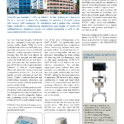
Founded in 1991, Mindray is one of the leading global providers of medical devices, committed to innovation in the fields of patient monitoring & life support, in-vitro diagnostics, and medical imaging. International Hospital’s editor in chief met David Yin, Group Vice President and General Manager of International Sales and Marketing on the Mindray stand and reviewed their latest products on display at CMEF.
Headquartered in Shenzhen, China, Mindray possesses a global marketing and service network with subsidiaries and branch offices in 32 countries in North and Latin America, Europe, Africa and Asia-Pacific, as well as 31 branch offices in China. To date, Mindray has 7,600 employees. Particularly strong is its R&D department which employs 1,700 engineers and accounts for a spend of almost 10% of annual revenue. The company is dedicated to adopting advanced technologies and transforming them into accessible innovation, improving the quality of care, while helping to reduce its cost and make it more accessible to a larger part of humanity. Today, Mindray’s products and services can be found in healthcare facilities in over 190 countries besides China.
Mindray is the perfect example of a company built on growth from the domestic to the international market. Key milestones in its development include the New York Stock Exchange listing in 2006, the Datascope acquisisition in 2008 and the Zonare takeover of 2014.
Among the many products on show at CMEF was the cutting edge design BeneVision patient monitor with its rotatable landscape and portrait layout as well as its innovative clinical decision support tools like HemoSight. On the ultrasound imaging side, the Resona 6 premium system was developed with Zonare and is powered by the innovative ZONE Sonography Technology. At the other end, the M6 hand-carried ultrasound system offers a wide range of tools that maximize diagnostic capabilities at the bedside. Another highlight at CMEF was the WATO EX65 Pro anesthesia workstation which is newly launched in the Chinese market.


Cancer remains the second leading cause of death in Europe after cardiovascular diseases with approximately 3.5 million new cases diagnosed every year and an annual death toll of 1.5 million. However, the good news is that the trend of total cancer mortality levels is downwards for both men and women and also children for which the progress of 5-year leukemia survival has been spectacular.
Breast cancer provides a good example of this trend, being not just the most common female cancer globally but also the number one diagnosed cancer in Europe (13%). Its 5-year survival rate has more than doubled in 40 years, from 40% of patients in 1970 to 90% in 2013. Looking into the future there are also some encouraging signs for certain types of cancer, particularly cervical cancer as the full impact of the HPV vaccination programmes becomes measurable.
In Europe, some of the credit for these positive developments should go to the European Organization for Research and Treatment of Cancer (EORTC), founded in 1962. Over the years, EORTC’s clinical research has helped make significant progress in the treatment and management of cancer, evaluating new molecules, refining existing treatment regimens, identifying biomarkers and assessing patients’ qualify of life. In 2016, the EORTC research network counted more than 4850 physicians from about 870 institutions while patient accrual from 2000 to 2016 totalled over 89,000 patients in clinical studies.
The bad news is that the overall burden of cancer continues to increase not just because of progress in early detection but largely because of the ageing of the population (65% of new cancer cases are diagnosed in patients who are 65 or older). Also, smoking, particularly in women, is linked to a rising incidence of lung cancer.
There are still a number of challenges to be met if the promises of translational research and personalized medicine for cancer therapy are to be fulfilled. Effective coordination in Europe of advances in basic research and quality clinical research programmes is essential. New models of partnerships between academia and the pharma industry are also required as well as public funding for research on rare cancers. Prevention is paramount, though, as no cancer research will have a bigger and quicker impact than smoking cessation. Tobacco kills over one third of its users and studies have shown that smokers lose at least 10 years of life expectancy compared to non-smokers and that quitting smoking before the age of 40 reduces the risk of tobacco-related death by 90%.
Prins Hendrikstraat 1
5611HH Eindhoven
The Netherlands
info@interhospi.com
PanGlobal Media IS not responsible for any error or omission that might occur in the electronic display of product or company data.
