
Prins Hendrikstraat 1
5611HH Eindhoven
The Netherlands
info@interhospi.com
PanGlobal Media IS not responsible for any error or omission that might occur in the electronic display of product or company data.

Breast cancer is the most common female cancer globally, with one woman in eight in the West eventually developing the disease. However improved screening programmes allowing earlier detection and treatment have greatly contributed to the steadily decreasing breast cancer mortality observed from the 1980s on. Although alternative imaging modalities are available, the current gold standard screening tool is mammography. In the majority of EU countries women are invited for screening from the age of 50 until they reach 70. Currently only France extends screening to include women up to 75, though studies are ongoing in several European countries to assess whether a higher cut-off age would lead to a net benefit for older women.
The main limitation of mammography is that the X-rays from different angles can only provide a 2D image of a 3D structure. This leads to false negative results as normal breast tissue can mask tumours. In addition false positive results augment both patient anxiety and hospital workload; the rationale for only screening women from age 50 is that there is a higher rate of false positives in younger women and a much lower incidence of the disease prior to the onset of the menopause. However because randomized, controlled trials have excluded women over 75, there has been a paucity of data concerning the benefits of continuing to screen all older women. One major concern has been that breast cancers that would not pose a threat to women’s health during their lifetime would be diagnosed and aggressively treated. In addition the U.S. Preventive Services Task Force notes that routine exposure to radiation from mammography results in a slightly higher risk of developing breast cancer; this would increase if women were offered screening for possibly an additional 25 years.
Now two recent studies have provided the data that were lacking. The first, a retrospective cohort study carried out in France, compared patient prognosis in women of 75 and older who presented at an oncology center because of a positive mammography result with older women who had been clinically diagnosed. It was found that the former group had improved, disease-free survival rates. The second robust study utilized data from the US National Mammography Database. Over 5.6 million mammograms from 150 facilities in 31 different States over a seven-year period were analysed, with data sorted according to demographics, mammography results and biopsy results. The cancer detection rate and positive predictive values increased with age up to 90, and the recall rate reduced. Surely the jury is no longer out?

In spite of alarm bells that artificial intelligence (AI) would decimate the radiology profession, a host of barriers – both technical and regulatory – make this unlikely to happen for the foreseeable future. Instead, over the coming decade, AI is at best likely to help radiologists do their jobs more quickly and lead to improved patient outcomes.
From CAD to AI
AI in radiology, in some senses, has tended to raise the same level of expectation as computer-aided detection (CAD) did for the profession in the 1990s. Indeed, there is now a distinction between computer aided detection which reduces observational oversight and false negatives in interpreting medical images, and computer aided diagnosis (also called CAD) – by virtue of which software is used to analyse a radiographic finding to estimate the likelihood of a specific disease process (e.g. a benign versus malignant tumour).
As a result, in spite of tens of thousands of machine-learning algorithms, there is little connection to clinical application. Most remain confined to the realms of research.
The Black Box barrier
Radiologists, for example, use visual pattern matching. However, few object recognition algorithms have yet been tested on gray-scale images, such as those widely used in radiology.
Though specific algorithms could in principle be tailored for specific tasks, they use different assumptions and targets, and often are written to function in different modalities. Consolidating a set of algorithms into one package and then using this to underpin image or data analysis is not feasible.
In effect, the key problem with CAD detection is its black box’ nature, which means they cannot explain why an object has been identified as abnormal. Many users remain suspicious about sharing the already-grey zone between detection and diagnosis with a machine, which only provides probabilities.
Sensitivity and specificity
The above kind of issues also hinder AI. Nevertheless, the technology is rapidly evolving and may offer some solutions to new challenges.
Like radiologists, AI faces the twin pulls of sensitivity and specificity, between false positives which overcall disease and false negatives which undercall it. It is clear that it will favour sensitivity over specificity.
Technology creates its own momentum
In recent years, radiologists have been forced to cope with an explosion in the stock of medical images, thanks to modern imaging technologies and PACS storage capacity. In the UK, for example, almost 5 million CT scans are performed per year by the NHS. At the upper end, a single pan scan’ CT of a trauma patient, for example, renders about 4,000 images. Indeed, a busy radiologist can read about 20,000 studies a year.
To deal with this burden – both physical and visual – radiologists clearly need help. AI seems to have become one of the most optimal.
There is, nevertheless, some irony here. Technology, in this case consisting of new imaging modalities, has led to an increase in the workload on radiologists. This is in spite of the fact that the disease burden has remained more or less the same, as has the prevalence on imaging of clinically significant pathology. However, the growth of imaging stock has led to a sharp rise in the presence of detectable and potentially significant pathology. Radiologists therefore face the massive challenge of finding ways to use the latter. This is where yet another technology, AI, steps in.
Industry push combines with radiologist pull
While the need to handle the imaging data explosion will see radiologists pulling’ AI, industry has chosen radiology to push’ for clinical validation. There are two reasons for this: the sheer volume of the imaging data and its continuing growth make it a huge market, while the fact that it is stored in structured and computer-readable DICOM format means it is a ready one.
AI’s own dynamics in change
Meanwhile, AI itself has seen some changes. Although, fuelled by science fiction and Hollywood, the popular imagination associates AI with self-awareness, what we really still have is more accurately machine intelligence. The implications of even such a toned-down definition should, however, not be under-estimated. Neither should some recent developments.
From Deep Blue to AlphaGo
In the late 1990s, IBM’s Deep Blue supercomputer defeated grandmaster Garry Kasparov in a chess game. In March 2016, Google DeepMind’s AlphaGo defeated Lee Sedol, a 9th level Go grandmaster 4-1. For AI experts, the AlphaGo win is far more impressive than Deep Blue because Go is less rules-bound than chess.
Due to these constraints, Deep Blue analysed millions of potential combinations and outcomes, in what IT professionals call brute force’ calculation. No computer can yet achieve this with Go, which according to Business Insider’ (March 10, 2016) has ‘more than 300 times the number of plays as chess. Alongside continuous scenario analysis, top Go players require both experience and intuition’. This is why AlphaGo’s win was seen as a paradigm shift in AI.
Deep learning
Unlike Deep Blue’s brute force, AlphaGo used a programming method called deep learning’, with so-called neural networks, which are far more similar to human thought processes than traditional computing. Rather than seeking to map out every possible move combination, deep learning (DL) is a relatively-unregulated process by which a computer figures out why something is what it is, after being shown several examples. It uses a large but still-finite sample of data, draws conclusions from that sample, and then, along with some human inputs, repeat the process over and over again, to simulate millions of games into a decision-making system.
Technically, AlphaGo’s deep neural networks consisted of a 12-layer network of neuron-like connections with a policy network’ to select the next move and a value network’ to predict the winner of the game.
A new benchmark
Neural network-based deep learning is now the benchmark for AI in radiology, with IBM’s poster child Watson leading the way. At the 2015 RSNA meeting, Watson showed its capacity to find clots in brightly shining pulmonary arteries.
Watson, however, has a DL rival in Australia’s Enlitic, which has developed a lung nodule detector claimed to achieve positive predictive values that are 50percent higher than those of a radiologist. As the detection model analyses images, it learns from those images. It not only finds lung nodules, it also provides a probability score for malignancy. Enlitic is now conducting a trial on a model to detect fractures using X-ray images overlaid with a heat map to highlight their location within a conventional PACS viewer. The clinical application will eventually encompass X-ray, CT, and possibly MRI. At the moment, Enlitic is working to incorporate ACR guidelines into it.
Although both Watson and Enlitic use deep learning, the approach is different. Watson seeks to understand’ a disease, Enlitic simply seeks to find source problem data, solve it, and produce a diagnosis.
Another DL developer is MetaMind, since last year part of CRM (customer relationship management) giant Salesforce.com. MetaMind has an alliance with teleradiology provider vRad to identify key radiology elements associated with critical medical conditions, especially in the latter’s focus area of emergency departments (EDs). The first tool to emerge from the partnership was an algorithm to identify intracranial hemorrhage (ICH), often seen in ED patients and requiring prompt action. vRad, which has put the algorithm into a beta phase that will allow it to collect data to demonstrate outcomes, is adapting it to identify other critical conditions, such as pulmonary embolisms and aortic tears.
Swarm AI
Apart from deep learning, radiology is also seeing the first successful experiments with swarm AI, which helps form a diagnostic consensus by turning groups of human experts into super experts. The technology borrows from nature, which sees species accomplishing more by participating in a flock, school or colony (a swarm’) than they can individually. One study, published in Public Library of Science (PLOS)’, stated that swarm intelligence could improve mammography screening and has the potential to improve other types of medical decision-making, ‘including many areas of diagnostic imaging.’ Another study found that accuracy in distinguishing normal versus abnormal patients was significantly higher with swarm AI than the radiologists’ mean accuracy.
Challenges ahead
Nevertheless, there is much more to be achieved before AI becomes an everyday tool in radiology.
The biggest roadblock will consist of regulators, who are unlikely to sanction the use or marketing of intelligent’ machines. In the US, as first of their kind, they lack the predicate devices needed to be regulated under the FDA’s 510(k) rules, and it would take decades to get approval for each algorithm.
A second issue is the time and cost to get datasets to fine-tune the algorithms. Watson, for example, has a backlog of 30 billion medical images to review.
Thirdly, the algorithms would also raise significant legal and ethical issues, such as knowing when they could be trusted.
Finally, even were such machines to become available, referring physicians are unlikely to accept conclusions or interpretations drawn solely by them.
The scale of such challenges has already been seen by developers of computer-aided detection (CAD) algorithms – and the change of CAD to detection’ rather than diagnosis’, as it was called in the early days.
Need and benefit, reality checks
In short, for now, radiologists need AI just as much as AI needs them.
Radiologists will have to begin to work with AI, both to improve the technology itself and to reduce routine, repetitive tasks such as confirming line placements and looking at scans to find nodules.
On its part, AI is likely to become an increasingly smarter tool, to improve efficiency, for example by prioritizing cases, putting thresholds on data acquisition, improving workflow by escalating cases with critical findings to the worklist of a radiologist and providing automatic alerts to both radiologists and other concerned clinicians.
In the longer term, DL algorithms are likely to be trained to recognize disease patterns, identify, outline and measure nodules and possibly highlight suspicious areas in images. This is likely to be followed by the use of DL-based AI as clinical decision tools, for example to help referring physicians select or narrow choices of scans, based on clinical observations in an EMR. Such steps would not only free up resources for additional testing but also improve patient care, thereby making radiologists even more integral in the care management process.
In the final count, a resonant reality check on AI has been provided by Eliot Siegel, MD, professor of radiology at the University of Maryland. He has offered to wash the car of anyone who develops a program than can segment adrenal glands on a CT scan as reliably as a 7-year-old.
Since the late 1990s, emergency radiology has become one of the fastest developing areas of medicine. It is now commonplace not only in Europe, the US and Japan but also in major urban centres of several developing countries.
The appropriate use of emergency radiology expedites patient care, prevents unnecessary hospital admissions and emergency surgery and therefore reduces costs.
Sub-specialty of radiology
Formally, emergency radiology is a relatively new sub-specialty of radiology. It is defined by the imaging and subsequent management of trauma patients, as well as those who are acutely ill. In effect, it is associated with real-time diagnostic imaging and online interpretation of data, which are conducted and completed in the ED setting itself. Emergency radiologists, on their part, need to be available and provide interpretations of imaging around the clock, including all off-hours shifts.
Professional societies set up very recently
The American Society of Emergency Radiology (ASER) was founded in 1988, with a mission to ‘advance the quality of diagnosis and treatment of acutely ill or injured patients by means of medical imaging and to enhance teaching and research in emergency radiology.’ ASER publishes the journal Emergency Radiology’ and has more than 700 members, both from the US and overseas.
The European Society of Emergency Radiology (ESER) was established in 2011, or over a decade later than its US counterpart. Based in Vienna, ESER seeks to foster education and training in emergency radiology, and collaborate both with the ASER and the British Society of Emergency Radiology (BSER), which was set up in 2014.
Radiography and fluoroscopy: limitations
Traditionally, ED imaging consisted of radiography and fluoroscopy. The procedure began with chest, abdominal and skeletal radiographs, accompanied sometimes by intravenous urograms and barium examinations. Emergency angiography was used in patients with central nervous system or vascular conditions.
Many trauma patients were, however, unable to have completion of imaging examinations in the ED, and several presenting diagnostic uncertainty were admitted to the hospital for fluoroscopic or angiographic procedures.
CT impact dramatic
Emergency medicine practice was revolutionized in the 1990s by the increase in availability of ultrasound, MRI and above all, CT. In spite of some lingering concerns, the speed of CT dramatically altered the equation in emergency radiology. A whole-body trauma CT requires just two minutes, providing information about all major injuries to the head, spine, thorax, abdomen and pelvis and increasing the probability of survival for trauma patients.
These new imaging modalities effectively served to bridge emergency medicine and diagnosis. Dramatic improvements in image quality and acquisition times have since enhanced the role of radiology in diagnosis, and as a bridge to minimally invasive procedures.
Shortening TAT
Such developments, in turn, catalysed an increase in expectations, with emergency physicians demanding quick availability of all imaging modalities, high-quality imaging examinations, real-time 3D post-processing and round-the-clock service – in effect, shortened turn-around times (TAT).
It has for long been a maxim that care provided to a trauma patient in the first few hours can be critical in terms of predicting longer-term recovery and that good trauma care involves getting the patient to the right place at the right time for the right treatment.
Professional societies have anchored such thinking. For example, guidelines from the Royal College of Radiology in Britain recognize that in the overall management of the severely injured patient, ‘diagnostic and therapeutic radiology plays a pivotal role’, although it is but a small part of ‘the whole management process.’
CT poses logistical challenges
Accompanying the increased emphasis on TAT and demands from physicians, emergency radiology facilities began to steadily reduce conventional radiography or replace it with digital X-ray. Instead, CT began to be moved to emergency departments. For example, the Royal College of Radiology guidelines mentioned above specify that CT should be adjacent to, or in, the emergency room (Standard 3) and that digital radiography should be available in the emergency room (Standard 4).
The move to relocate CT has also been driven by a need to reverse some of the major problems associated with scanners in a hospital-based trauma setting – the result of a combination of high technology and poor logistics.
Logistical problems centred upon the need for optimal location of a scanner and the capacity to receive severely injured patients within a very short period of time. This, in turn, required the availability of sufficient radiographers, a seasoned transfer procedure and resuscitation teams to be familiar with a CT environment and ready to accompany the patient during the scan.
Most traditional’ hospitals, dating back to the radiography and fluoroscopy era, were unable to cope with the dramatic changes which CT brought – above all in speed and imaging data sensitivity. This resulted in serious bottlenecks in workflow, which impacted adversely on patient outcomes.
Such shortcomings were enhanced by spikes in the volume of patient visits – e.g. during weekends and over holidays – when accident rates are far higher.
New standards for emergency radiology
To help CT relocate and become more efficient, emergency radiology facilities are being subject to exacting, new standards.
For example, the University of Amsterdam’s Academic Medical Centre (AMC) has been designed to enhance workflow efficiency and prevent dangers in the transfer of critically ill patients, while avoiding or reducing delays for non-emergency patients with scheduled appointments in the radiology department. By enabling proper equipment, transfer and support, AMC has sought to address concerns in emergency departments that, in spite of its benefits, CT might be a dangerous place for the critically ill. This was largely due to perceived limits in ventilation, resuscitation and monitoring during scanning.
One of the most visible innovations at the AMC is a sliding CT gantry on rails which serves two emergency rooms. A radiation-shielding wall closes behind the gantry, allowing the scan to be performed feet-first so IV-lines and monitors do not have to be re-positioned.
In terms of staffing, emergency radiologists at AMC are supported by a dedicated anesthesiologist who initiates ventilation, surgical residents or nurses to insert chest tubes and and radiology residents to help interpret the imaging data. This team interfaces with the trauma surgeon.
Staffing issues
Non-physician staffing is also crucial to an efficient emergency radiology facility. These range from technicians, supervisors and ED managers to receptionists, schedulers as well as ambulance personnel. State-of-the-art facilities strive to make such staff aware of the unique workflow and requirements of emergency imaging. For example, technicians need to have the skills to use different modalities and image multiple body parts. Beyond this, non-physician staff need also to be well versed in other, point-of-care medical equipment and manage a diverse range of patients – from the acutely ill to the pregnant, from children to the elderly.
A key role is also played by IT support staff, who need to be on call round-the-clock. Given the pressures to reduce TAT, they need to be well versed in RIS/PACS solutions and their suite of integrated tools, such as speech-to-text, 3D visualization, and others. More recently, IT professionals have also played a major role in data mining, in order to identify workflow bottlenecks and special situations.
Decision support tools
Another related and fast-emerging sphere consists of decision support tools, which communicate the clinical presentation, physical examination, and laboratory tests. They also confirm imaging appropriateness and selection of the optimal examination protocol.
Decision support is also seen as a means to reduce common causes of superfluous radiation in ED patients, for example, by avoiding repeat CTs (e.g. in referring hospitals). Indeed, one of the most closely-watched debates about emergency radiology concerns CT.
CT versus the rest
CT has undoubtedly been the centrepiece of the emergency radiology revolution. In 2016, a prospective study in Radiology’ showed that CT influenced the leading diagnoses in 25percent-50percent of patients and admission decisions in 20percent-25percent of patients.
Nevertheless, radiography continues to remain the most widely used imaging modality. In the US (for which data is available from a study in the American Journal of Roentgenology’ ), CT was used in 268 of 1,000 ED visits in 2012, compared to 76 for ultrasound, 64 for MRI, and 510 for X-ray.
The study, published in August 2014, also drew some other notable conclusions.
CT use in the ED peaked in 2005, while this happened two years later for MRI. Compared to 1993, CT use grew 457percent by 2005 and then declined by 49percent to 2012. For MRI, growth from 1993 to its 2007 peak was sharper, at 1,750percent, while the fall between 2007 and 2012 was 23percent, half the rate of CT. This was, nevertheless, from a much smaller user base, and as mentioned above, MRI use in the ED is outstripped more than 4-to-1 by CT (64 to 268 per 1,000 visits).
Ultrasound, on the other hand, has shown a steady but less remarkable increase in ED use between 1993 and 2012, by just 35percent. Conversely, although X-ray was used in over half ED visits in 2012, it has fallen steadily since 1993, by 26percent.
REACT-2: reality check for CT
Future trends in emergency radiology are likely to be heavily influenced by a randomized controlled trial trial at four hospitals in the Netherlands and one in Switzerland. Known as REACT-2, the trial sought to determine the effect of total-body CT scanning compared with standard work-up on patients with trauma and compromised vital parameters, clinical suspicion of life-threatening injuries, or severe injury.
The primary endpoint was in-hospital mortality, analysed in the intention-to-treat population and in subgroups of patients with polytrauma and those with traumatic brain injury.
Between April 2011 and Jan 1, 2014, the trial assessed 5,475 eligible patients and randomly assigned 1,403, 702 to immediate total-body CT scanning and 701 to the standard work-up. A total of 541 patients in the immediate total-body CT scanning group and 542 in the standard work-up group were included in the primary analysis. The study found that in-hospital mortality did not differ between groups.
As The Lancet’ reported on August 13, 2016, ‘Diagnosing patients with an immediate total-body CT scan does not reduce in-hospital mortality compared with the standard radiological work-up. Because of the increased radiation dose, future research should focus on the selection of patients who will benefit from immediate total-body CT.’
More MR?
Alongside such selection, it is also likely that there is an increase in demand for MR scanning in the ED, whose decline from its peak has been half the rate of CT (in the American Journal of Roentgenology’ study mentioned previously).
So far, MR is not indicated in an acute trauma care setting. In Britain, for example, Royal College of Radiology trauma radiology guidelines specify that MRI can be available in a different building. However, it states that ‘protocols should be in place for the transfer of critically injured patients if further management is dependent on MRI in the first 12 hours.’
Some of the benefits of MRI versus CT include acute musculoskeletal injuries, and in imaging of acute abdominal conditions in pregnant women and children.
Three-dimensional (3D) printing for medical applications has grown in recent years at a feverish pace. The technology has long made a significant impact in manufacturing and is also revolutionizing healthcare. For some of its proponents, this would be rather like the Gutenberg printing press did with publishing. Indeed, the respected Gartner Group estimates that 30percent of internal medical implants and devices will be 3D printed by 2020.
3D printing was founded in the 1980s as stereo-lithography’ (STL) and the first commercial 3D printer came to market in 1988. Since the 1990s, manufacturers have used the technique principally for rapid prototyping, or the production of models and moulds.
Implants and prosthetics
Medical 3D printing took off in the early 2000s for producing dental implants and custom prosthetics, with a rapid pace of acceptance in areas such as hearing aids and dental braces. Currently, almost all hearing aids fitted into the ear in industrialiZed countries are made with 3D printers and orthodontic braces, too, are almost entirely 3D printed.
CustomiZed 3D-printed prosthetics and implants were made possible by translation of CT and MRI scans into digital STL print files, and imaging continues to play a central role in medical 3D printing.
Orthopedics and neurosurgery applications
The customization offered by 3D printing also quickly made its case for orthopedic patients being fitted with a standardized hip or spinal prostheses, which required the cumbersome process of shaving of pieces of metal and plastic with scalpels and drills afterwards, in order to achieve best fit.
Neurosurgeons too quickly saw the potential of 3D printing to address the drawbacks of variation in skull shape and the difficulties in using standard cranial implants. In head injury victims, for example, it is important to remove bone to provide space for the brain room to swell and the cranial plate must be perfect in fit.
In situ, in the OR
In the operating room, 3D printing has thoroughly transformed the manufacture of patient models to facilitate planning of surgical procedures. In 2016, a 3D printed model was used by Blythedale Children’s Hospital in Westchester in a 27-hour operation to separate twins conjoined at the head. According to many reports, recovery of the infants was accelerated due to the 3D model.
At the University of Michigan, CT images of a patient’s airway were used with a 3D printer to fabricate a precisely modelled, bioresorbable tracheal splint that was surgically implanted in a baby. The baby recovered, and full resorption of the splint is expected to occur within three years.
3D printing has also been used for making surgical tools such as forceps, hemostats, scalpel handles and clamps. They are formed sterile, and some estimates report that they cost only a tenth of a stainless steel equivalent.
In situ printing, by which implants, tissue (and eventually organs) are 3D printed in the human body during operations is anticipated in the future trend. Such a trend is being reinforced by rapid developments in miniaturized robotic bioprinters and robot-assisted surgery.
Personalized pills
3D printing technologies are also used for personalized medicine, with precision in dose (matched to patient profile and response). Some firms are experimenting with complex drug-release profiles, such as poly-pills with multiple active ingredients in a multilayered form.
This is seen as promising new standards of care for patients with several chronic diseases. Extended to one poly-pill per day for everyday medications, such a step would reduce a bane of medical practitioners – namely patient non-compliance.
In 2016, Spritam levetiracetam, a new drug to control seizures brought on by epilepsy, was approved by the US Food and Drug Administration (FDA). The pill, the world’s first to be 3D printed, is based on a trademarked ZipDose technology developed by Ohio-based Aprecia, and provides more porosity than alternative dosage forms.
Industry experts foresee drugs manufacturing being done eventually at the point-of-care, with physicians emailing medication formulations to pharmacies for on-demand drug printing.
Post-industrial production
The logic of 3D printing is in some ways truly revolutionary. What it brings is an end to the idea that there is commercial sense in only large runs of standardized products, a cornerstone of 19th/20th century manufacturing tradition as well as the Industrial Revolution. The first 3D manufactured product, in other words, costs approximately the same as the next one.
3D printing also reduces cost in certain cases. For example, a 5-mg pharmaceutical tablet can be custom-fabricated on demand as a smaller and less expensive 2.5-mg tablet rather than being broken up and left unused.
Speed
Speed too is a major asset of medical 3D manufacturing, and a spin-off from the fact that large production runs are not required. Customized products like prosthetics and implants, in particular, can be made within hours.
As with pharmacy pills, some expect on-site 3D printing at, or adjacent to, a hospital, to eventually emerge, for making patient-specific products.
Basic technology
The basic technique of 3D printing, which is also known as additive manufacturing, involves the successive deposit of layers of materials, typically plastic and ceramics or metal and powders, to make the final product.
One of the most exciting innovations, however, consists of using live cells as the printing material.
Types of 3D printer
The type of 3D printer chosen for an application often depends on the material used and the method for bonding the layers in the final product. Key technologies for medical applications include selective laser sintering (SLS) and thermal inkjet (TIJ) printing. Another widely-used 3D printing technology is fused deposition modelling (FDM).
Though relatively basic and inexpensive, FDM was one of the earliest examples of successful medical 3D printing in the late 1990s/early 2000s when it was used to construct cranial implants. FDM remains widely used for rapid modelling and prototyping in orthopedics and dentistry.
FDM printers use a print-head similar to an inkjet printer. Rather than ink, however, beads of thermoplastic (similar to those used in injection moulding) are released to form a thin layer. The process is repeated continuously. Since the plastic is heated, it fuses to the layers below, and then hardens as it cools to create the final product.
More complex medical uses of 3D printing are based on SLS and TIJ.
SLS uses metal, plastic or ceramics as material. A laser draws out the shape of the object and this is then fused to a powdered metal substrate. The process is repeated until the product is formed. The degree of detail in SLS is directly linked to the precision of the laser and the powder’s fineness.
On its part, TIJ uses thermal (as well as electromagnetic or piezoelectric) technology to deposit tiny droplets of ink or even cells (bio-ink) on a substrate. Unlike office inkjet printers, 3D TIJ heats a print-head to create collapsing air bubbles, which in turn create pressure pulses to eject the droplets from nozzles. The size of the droplets can be adjusted by temperature, pulse frequency or material viscosity and volumes can be as little as 10-20 picolitres. Multiple-head TIJ is especially promising for producing tissue and simple organs in the process of bioprinting’ (discussed below). Other applications under study include drug delivery and gene transfection.
Bioprinting – the final frontier
While implants and prosthetics have convincingly demonstrated the real-world relevance of 3D printing, the maximum excitement is currently focused on its use in tissue and organ fabrication.
Ageing, accidents, disease and birth problems often cause tissue and organ failure. Treatment is largely based on donor transplants. However, there is a chronic shortage of supply, not least of suitable donors (e.g. with matching tissue). In addition, surgery and follow-up is complex and expensive.
One recent approach to finding a solution consists of tissue engineering and regenerative medicine, based on mixing growth factors into isolated stem cells, multiplying them in a lab and then seeding the cells on scaffolds which transform direct cell proliferation and differentiation into functioning tissues.
Beyond regenerative medicine
Bioprinting takes traditional regenerative technologies further than scaffold support alone by using 3D printing technology to produce layers of cells, biomaterials, and cell-laden biomaterials. This is then precisely placed by the printer in tissue-like structures. As mentioned previously, inkjet-based bioprinting is the most commonly used technique for bioprinting.
Tissues and organs
German researchers have been developing skin cell bioprinting since 2010. In January 2017, a team from Spain’s Universidad Carlos III de Madrid (UC3M) reported in the journal Biofabrication’ they had developed 3D-printed human skin adequate for transplant into patients, and for testing drugs and cosmetics. Their product is currently undergoing European approval. Meanwhile, in the US, Organovo too has developed 3D-printed skin. Demonstrating the potential of such markets, French cosmetics giant L’Oreal has begun collaborating with Organovo.
Researchers have so far also successfully printed a knee meniscus, heart valves, bone and an artificial liver. In 2016, scientists at Cambridge University’s Centre for Brain Repair reported the 3D printing of a retina using a piezoelectric TIJ printer.
One application area is to use 3D printing to create tissues and organs for medical research, and rapidly screen candidate drugs, cutting research costs and time. Organovo is developing strips of printed kidney and liver tissue for exactly such a purpose, while Russia’s 3D Bioprinting Solution has 3D printed a functional thyroid in a mouse and claims to be ready to do the same in humans.
20 years to a 3D-printed heart?
Nevertheless, most bio-printed organs have so far been relatively small and simple, with no vascularity or nerve system and nourishment provided wholly by diffusion from the host vasculature. Such diffusion seems to suffice for thicknesses of 150-200 micrometers. Beyond it, there is none. In future, the bioprinting of 3D organs such as an entire kidney or heart will require precise multicellular structures with full vascular network integration.
Such a process may not be that far away. Collaborators from a network of academic institutions, including the Harvard University, Stanford University, the Massachusetts Institute of Technology and the University of Sydney recently announced they had bioprinted a perfusable network of capillaries, marking a significant stride toward overcoming the limits to diffusion.
According to some projections, we may be less than 20 years from a fully functioning printable heart.
Challenges ahead
As with many other frontiers of medicine, an immediate challenge for medical 3D printing consists of regulatory acceptance. Though a hundred-odd 3D-printed products had been approved in the US and Europe by the end of 2016, these consist almost entirely of prosthetics, surgical tools and artificial bone replacement.
Fulfilling regulatory requirements for more complex products is likely to be much more demanding. Included here are the need for large randomized controlled trials, which require funding and time – for instance to determine the biocompatibility of several of the new materials being used.
Breast tomosynthesis exams, Hologic calls the exams 3D MAMMOGRAPHYTM, have shown to be an advance over digital mammography, with higher cancer detection rates and fewer patient recalls for additional testing.
The new Affirm prone system, which was installed for the first time in Europe earlier last year, is widely considered one of the most significant advance in biopsy technology since the first prone biopsy system was introduced more than 20 years ago. It uses the same proven detector technology as the Hologic Selenia Dimensions breast tomosynthesis system, a top selling breast cancer screening and diagnostic system in the U.S. and in many other countries around the world. With a significantly larger field of view than the MultiCare Platinum system, along with its translucent paddles, the new prone system is designed to deliver exceptional 2D and 3DTM images and better target lesions found during 3D MAMMOGRAPHYTM exams, as well as other screening modalities. In addition, the Affirm prone system allows full 360degree Celsius access to the breast to accommodate most lesion locations. Users can go from a standard to lateral needle approach in seconds to accelerate procedures and ensure reaching targeted lesions.
Doctors in Spain report handling complex biopsies that they were only able to see with breast tomosynthesis imaging with the new system
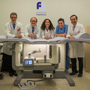
As Doctor Tejerina, a radiologist with the Centro de Patologia de la Mama, Tejerina Foundation, in Madrid, Spain, reports, feedback from the first wave of patients is very positive. ‘We have been suffering to handle complex biopsies of subtle lesions like faint calcifications or distortions that we were only able to see on 3D images,’ Dr. Tejerina says. ‘Older breast biopsy systems are restricted to 2D imaging with a narrow window for targeting the lesions. Often they require multiple X-ray exposures to find and position the suspect tumour for the biopsy needle. With tomosynthesis imaging on the new Affirm prone system, there is a much wider field of view. So the biopsy device can be positioned anywhere in a 360-degree circle, and areas of suspicion seen only with 3D imaging can be easily biopsied.’
Dr. Tejerina also notes that with the previous Hologic biopsy table, the tube head of the biopsy device had to be positioned manually. ‘The new system does this for us automatically, which saves time,’ he says. ‘The software really streamlines our workflow, so the procedure goes faster.’ And he adds, the Affirm Prone table, with its translucent paddles and wider detector, ‘helps us see lesions in the first scout and significantly reduce the number of images needed to get to the lesion.’
The Centro de Patologia de la Mama, Tejerina Foundation has been leading the way in women’s breast health for over 40 years. In 1997, the Centre was first centerein Spain to install a stereotactic guided prone biopsy table. In 2010 the Centre installed a Hologic Selenia Dimensions breast tomosynthesis system, the first site in Spain to use the innovative technology. In 2010 the Centre was also the first site in Spain to combine the Hologic AffirmTM upright biopsy system with the Hologic tomosynthesis system. The Centre was also one of the first sites in the world to offer prone biopsies on the new Affirm system from Hologic.
Doctors in the Netherlands say Affirm system is fast and comfortable for patients
Dr. Henebiens, a radiologist at Spaarne Gasthuis Hospital in Hoofddorp-the first Affirm prone user in the Netherlands-commented on how fast doctors can do a procedure on the Affirm system and how comfortable the new system is for patients.
‘We make fewer exposures on the new Affirm prone system, compared to the older MultiCare Platinum table,’ she notes. ‘And because the table uses 3DTM technology, we use fewer steps getting to the target and getting biopsies.’
Dr. Henebiens also likes how easy it was to get up to speed on the table. ‘The learning curve for the new table was very fast. Training was scheduled for two days, but in one day, the staff knew how to use it.’
The Spaarne Gasthuis Hospital staff had completed over 60 procedures on the table in their first 7 months of use.
Doctors in Italy report faster and lower patient dose biopsies with the new system
Doctor Gianfranco Scaperrotta, Chief of the Breast Imaging and Interventional Radiology at Fondazione IRCCS Istituto Nazionale dei Tumori (INT) in Milano, Italy was an early adopter of the Affirm prone system.
‘The Affirm prone system is a quick, effective and easy to use system,’ he says. ‘The image quality is high, comparable to the Hologic Selenia Dimensions digital mammography system. Workflow is quick thanks to a dedicated workstation and the system’s fully integrated C-arm and automated tube-head. Procedures are faster and safer with the new system thanks to the programmed needle parameters and automated calculations such as the display of safety margins and relative distance in real time.’
After 73 procedures on the new system, INT has seen a 20percent reduction in the time needed for performing a biopsy (patient time under compression) and approximately a 50percent drop in the mean glandular patient dose when they compare the new system to the older Hologic system.
The Fondazione IRCCS Istituto Nazionale dei Tumori is the largest oncology site in Lombardia, the most populous region in Italy. The research and cancer treatment site draws patients from throughout Italy.
In sum, doctors at the first three European Affirm prone install sites reported that the new system offers significant benefits to the patient, the doctor and the technologist.
So what’s next from Hologic in 2D and 3DTM Breast Biopsy after The AffirmTM Prone system? Hologic will show at ECR an all-integrated breast biopsy system that combines tissue acquisition, real-time imagining, and tissue handling. The new system is designed to work in synergy with imaging guidance systems like the AffirmTM Prone table and provide actionable real-time information in the procedure room and improve biopsy workflow.
For specific information on what products are available for sale in a particular country, please contact your local Hologic representative or write to iims@hologic.com
While mammography remains the gold standard for detecting breast cancer, research has shown it is not equally effective in all women. In the 40percent of with dense breast tissue, mammography can miss up to one third of breast cancers. This may lead to a delay in diagnosis and a worse prognosis for women with dense breast tissue. Mammography has been shown to miss 30percent of cancer in dense breasts. Using screening ultrasound for women with dense breasts is helping address this challenge. However, the limitations of traditional hand-held ultrasound (HHUS), which include operator dependency, variability and long acquisition times, make it inefficient for broad-scale breast cancer screening. With the introduction of ABUS (automated breast ultrasound), clinicians are able to address these variables and shorten both exam and read times, while increasing sensitivity with a multi-modality approach.
New findings from a Swedish study show a 57percent relative increase in breast cancer detection in women with dense breast tissue when ABUS was used together with mammography.
The system is found to have significantly improved cancer detection in women with dense breast tissue when used together with mammography.
The European Asymptomatic Screening Study (EASY) aimed to evaluate the impact of ABUS in conjunction with full field digital screening mammography (FFDSM) in 1,668 women aged 40-74 with dense breasts. The study showed a 57 percent relative increase in breast cancer detection in dense breast tissue, compared with mammography alone.
‘If ABUS would be a part of national screening programmes in dense breasts, more cancers could be detected at an earlier stage. Many countries are working to try to optimize screening so that each woman can get examinations according to her assessed risk,’ said Dr Brigitte Wilczek, lead researcher on the EASY study.
Dense breast tissue is linked with an increase in the risk of developing cancer. It also makes detecting cancer more difficult. This is because both masses and breast tissue appear white in the mammogram, which makes the search for masses like a search for a snowball in a snowstorm. By contrast, masses appear dark against white tissue with ultrasound technology.
Dense breasts are particularly common in younger women and seems to reduce with age, as on average 74percent of women in their 40s, 57percent of women in their 50s, 44percent of women in their 60s and 36percent of women in their 70s have dense breast tissue.
In the study, published in the European Journal of Radiology, FFDSM was first used in the examination followed by a 3D ABUS exam which took 15 minutes to complete per patient. The inclusion criteria for the women in the study was that they be 40 years or older, asymptomatic, and have heterogeneously dense parenchyma or extremely dense breast on assessment by the radiographer in the screening.
‘The study shows that it is feasible to implement 3D ABUS into a high volume mammography center and increase the cancer detection rate while maintaining an acceptable low recall rate,’ said Dr Wilczek.
The recall rate for ABUS and FFDSM combined was only +0.9percent compared to FFDSM alone. This is an acceptable low recall rate well within the recommendations of the European guidelines for quality assurance in breast cancer screening.
www.gehealthcare.com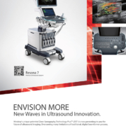

Prins Hendrikstraat 1
5611HH Eindhoven
The Netherlands
info@interhospi.com
PanGlobal Media IS not responsible for any error or omission that might occur in the electronic display of product or company data.