The world of medical technology innovation
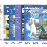
This year, International Hospital celebrates its 40th anniversary

This year, International Hospital celebrates its 40th anniversary

Eighty years ago around one woman in 200 giving birth in the West died, mainly from post-partum sepsis as well as from hemorrhage and hypertensive disease. With the advent of sulphonamides and antibiotics, the maternal death rate plummeted. From the 1940s the introduction of organized pre-natal care, methods to predict which patients were moderate or high risk, procedures for appropriate interventions, and effective reduction of post-partum bleeding resulted in a steady decrease in maternal mortality. Maternal deaths in the West are currently so rare that standards of care are assessed by reference to the perinatal mortality rate.
However the incidence of one potentially life-threatening obstetric condition is seriously increasing, namely Placenta accreta, when either part or all of the placenta invades the uterus wall and becomes inseparable from it, even involving adjacent organs in some cases. The placenta cannot be delivered normally, causing massive hemorrhage even when a hysterectomy is performed to control bleeding. In the 1950s Placenta accreta occurred in one in 30,000 Western pregnancies

The recently published article in
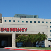
Emergency medicine departments are often the first point of call or
Technological breakthroughs in diagnostic imaging and interventional radiology have revolutionized patient care. However, these benefits seem to be accompanied by risks. In 2009, a report in the
Hybrid imaging is the fusion of medical images, most commonly from CT (computed tomography), PET (positron emission tomography) and MRI (magnetic resonance imaging), in order to enhance visualisation. It addresses both anatomical detail and functional processes, thereby providing superior accuracy for diagnosis and the monitoring of interventional procedures. In several cases, it is also accompanied by lower radiation exposure for patients.
Hybrid imaging is now being used to combine structural and molecular imaging – revealing molecular processes in vivo while depicting their anatomic location. Some proponents believe the technique marks the dawn of the era of personal medicine.
Medical Invention of the Year
The age of hybrid imaging could be considered to have begun in the year 2000, after PET/CT was heralded as the
Medical imaging is confronted with heterogeneities in many areas, including education and training, research, and quality of equipment and practice, which calls for a pan-European approach.
by Prof. Guy FRIJA, Past-President of the European Society of Radiology (ESR)
In 2014, the European Commissions Expert Panel advising the European Commission on effective ways of investing in health recently adopted an opinion1 recommending specific actions to improve quality of care and patient safety.
The actions proposed in the expert panel
The ALARA principle (As Low As Reasonably Achievable) will remain the key method used to determine the proper exposure technique for a given examination. However, the technology and the methods used to achieve the lowest reasonably achievable dose will continue to evolve.
New, more efficient technology can have a significant impact on required dose levels. To confirm this, Agfa HealthCare conducted both a technical assessment and an image quality evaluation with radiologists. The goal of this evaluation was to determine by how much patient exposure (and dose) could be reduced while providing the same or similar image quality, comparing conventional BaFBr plate CR systems to CsBr needle plate CR systems and CsI needle scintillator DR detectors using Agfa HealthCare
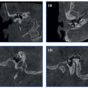
The Cone Beam CT (CBCT) technique is normally used in head and neck area for dento-maxillofacial, sinonasal and cervical spine imaging. Until recently, temporal bone imaging has typically been done with multidetector CT (MDCT). An issue related to CBCT in this anatomical area is the thick bony mass that surrounds the small details of the middle and inner ear. When the narrow cone shaped beam is localized to the small field-of-view (FOV) , this bony mass, which is outside of the target area, significantly attenuates the X-rays before and after they pass the region of interest and therefore pose challenges to the image formation. The MDCT technique covers axially the whole head and hence does not have this problem, but at the expense of a considerably higher radiation dose. The usefulness and diagnostic capability of the CBCT technique in the temporal bone area is demonstrated in the following three case reports.
Case reports
The CBCT unit used in this study is SCANORA
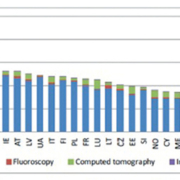
Each year approximately 3.6 billion x-ray examinations are performed worldwide, leading to earlier and more accurate diagnosis of medical diseases. However, concern has been raised regarding the stochastic and even deterministic impact on both patients and medical staff. Authorized bodies have emphasized the importance of ensuring the proper performance of X-ray equipment and of keeping the dose to medical staff and patients as low as reasonably achievable (ALARA). Unfors RaySafe has developed comprehensive solutions to monitor and reduce radiation in the X-ray room.
Personal dosimetry
The most significant personnel doses in the hospital environment often occur in the interventional suite where fluoroscopy is performed. Recent studies have shown hospital workers who are routinely exposed to ionizing radiation are at an increased risk to cancer, cataracts and other health problems. These findings have forced radiation safety professionals to look at more effective methods to reduce personnel exposures.
In the past, health and medical physicists relied primarily on training and the use of shielding to reduce personnel exposures. They would then review passive dosimeter results months later in hopes of seeing a dose reduction. . However, as interventional techniques grow longer and more complicated, this delayed method has become less effective. Some hospitals have tried to use electronic dosimeters to monitor staff exposures during interventional procedures to obtain more accurate dose measurements. However, this method is only minimally accurate. Some electronic dosimeters are not capable of accurately monitoring cumulative exposure in a pulsed radiation field as found in the fluoroscopy suite. Additionally, some are too heavy and are uncomfortable to wear, and all electronic dosimeters require the user to retrieve and view the LED display, which can occasionally be difficult to read. As a result, efforts to achieve ALARA for staff in the interventional suite have been challenging and largely unsuccessful.
Unfors RaySafe, a Fluke Biomedical company, recognized the need for a more accurate dosimeter, and developed the RaySafe i2 system to overcome the limitations of passive and conventional electronic dosimeters. In 2012, RaySafe introduced the i2, an active dosimetry system that gives real-time insight about personal radiation exposure, as well as access to time stamped dose date. The i2 wirelessly transmits exposure rate data to a large display monitor at the centre of every interventional suite. Displaying exposure rate and cumulative data allows each individual to make real-time adjustments such as shielding to immediately see the results of their efforts to achieve ALARA during interventional procedures. A study by the University of Rochester showed a 50 percent reduction in staff dose over a nine month period as the result of using these real-time personal dosimeters.
A primary challenge of real-time dose monitoring is in pricing to compete with passive dosimetry. However, as any radiation safety officer knows, the more significant cost of a dosimetry programme lies not in the cost of the dosimeters, but in the time and resources needed to collect and distribute dosimeters on a monthly and quarterly basis, and in the unpopular task of chasing after unreturned badges. Anything which can reduce or remove this administrative burden will be welcomed. The move from passive dosimeters being used only
Prins Hendrikstraat 1
5611HH Eindhoven
The Netherlands
info@interhospi.com
PanGlobal Media IS not responsible for any error or omission that might occur in the electronic display of product or company data.
