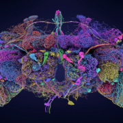
Unveiling the neural labyrinth: Fruit fly brain map sets new benchmark in neuroscience
Researchers unveil the most comprehensive neural map of an adult animal brain to date, offering unprecedented insights into brain circuitry and function.

Researchers unveil the most comprehensive neural map of an adult animal brain to date, offering unprecedented insights into brain circuitry and function.

Researchers at Karolinska Institutet have uncovered key neural processes behind morphine’s analgesic effects, potentially paving the way for safer pain management strategies.
A groundbreaking neuroimaging study has identified distinct patterns of brain activation associated with various forms of love, offering new insights into the neural underpinnings of this complex emotion.
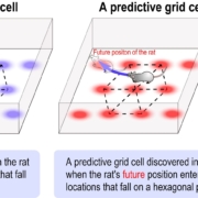
Researchers at the RIKEN Center for Brain Science in Japan have identified a new type of grid cell in the brain that predicts an animal’s future position, shedding light on how we navigate a spatial environment.

A groundbreaking study from Finnish researchers uncovers alarming trends in early-onset dementia among working-age adults, with Alzheimer’s disease cases nearly doubling over the past decade.
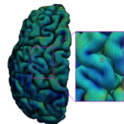
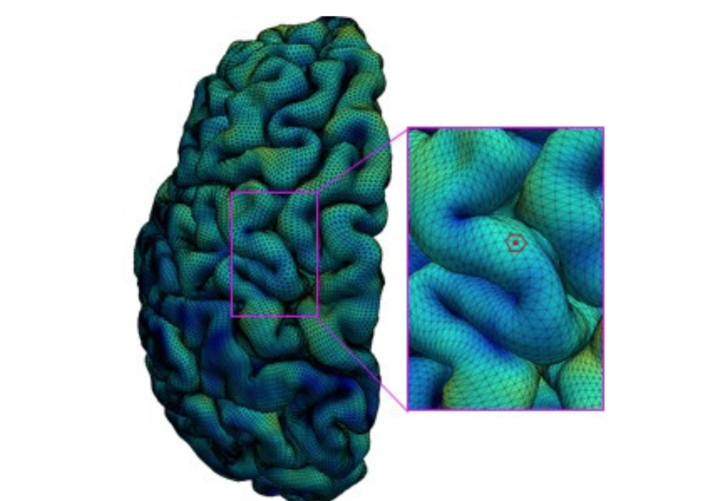
Brain cortical surfaces are represented by triangular meshes. The surface area of each vertex (e.g., the red point) is one third of the total surface area of its one-ring neighboring triangles (the red hexagon). Credit: Gang Li Lab, UNC School of Medicine
Scientists at the University of North Carolina (UNC) School of Medicine have mapped the surface of the cortex of the young human brain with unprecedented resolution, revealing the development of key functional regions from two months before birth to two years after.
The new cortical development mapping, reported online in the Proceedings of the National Academy of Sciences, represents a valuable resource for further research on brain development and offers a powerful new approach to the study of brain-development conditions such as autism and schizophrenia.
“These results provide an important reference for exploring and understanding the dynamics of early brain development,” said study senior author Gang Li, PhD, associate professor of radiology at the UNC School of Medicine.
The study’s first author was Ying Huang, a PhD candidate in Li’s laboratory.
The cortex is a sheet of brain cells that wraps around much of the rest of the brain. The most evolutionarily advanced brain region, it is proportionately larger in humans than in other mammals, and is responsible for higher, distinctively human functions including language abilities and abstract reasoning.
The third trimester of pregnancy through the first two years of life is the most dynamic period in cortical development. The cortex thickens markedly during this interval, and grows at an even faster pace in terms of surface area, by forming complicated cortical folds.
Disruptions to cortical thickening and expansion in this phase have been linked to autism and schizophrenia. However, neuroscientists haven’t had as detailed an understanding of this developmental phase as they would like. In particular, they’ve had a need for more comprehensive, high-resolution mapping, across the foetal-to-toddler age range, that divides or “parcellates” the developing cortex into distinct regions with their own growth rates – especially surface area growth rates.
In the study, Li and colleagues performed just such a mapping. They first gathered a set of 1,037 high-quality magnetic resonance imaging (MRI) scans of infants in the third-trimester-to-two-year age interval. The scans came from two other research projects, the UNC/UMN Baby Connectome Project (BCP) and the Developing Human Connectome Project. The team analyzed the scan data using state-of-the-art, computer-based image-processing methods, essentially dividing the cortical surface into a virtual mesh containing thousands of tiny circular areas, and calculating the surface expansion rate for each of these areas.
The analysis didn’t start with assumptions about the locations of brain structures or functional regions, but this regionalization of the brain became evident anyway from the resulting maps, based solely on the different rates at which areas of the surface expanded. In all, the researchers defined 18 distinct regions, which they found correlated well with what is already known about the developing cortex’s functional regions.
“All these regions show dramatic expansion in surface area during this developmental window, with each region having a distinct trajectory,” Li said.
The maps revealed that each region tended to have the same developmental path as its counterpart in the cortex’s opposite hemisphere. Sex differences were apparent too. Even when controlling for sex differences in overall surface area – male brains having greater area – there remained differences in multiple regions. For example, the medial prefrontal region in the left hemisphere, which is believed to host important functions such as attention and working memory, became proportionately larger in males early in the second year of postnatal life.
The analysis also showed that the patterns of cortical surface area expansion in this early period of life were very different from the patterns of cortical thickness development, suggesting that these two measures of brain development involve distinct mechanisms.
All in all, Li said, the mapping provides fundamental new insights into brain development.
He and his team now plan to extend this approach with MRI scan datasets that start at earlier ages and end at older ones. They also hope eventually to study scan datasets covering children who have autism-spectrum or other neurodevelopmental conditions. Such analyses might offer not only clues to the origins of these conditions, but also the identification of early signs or biomarkers, which in the future could be used to administer early and more effective treatments.
Ying Huang, Zhengwang Wu, Fan Wang, et. al. “Mapping developmental regionalization and patterns of cortical surface area from 29 post-menstrual weeks to 2 years of age.” PNAS, August 8, 2022. doi: https://doi.org/10.1073/pnas.2121748119

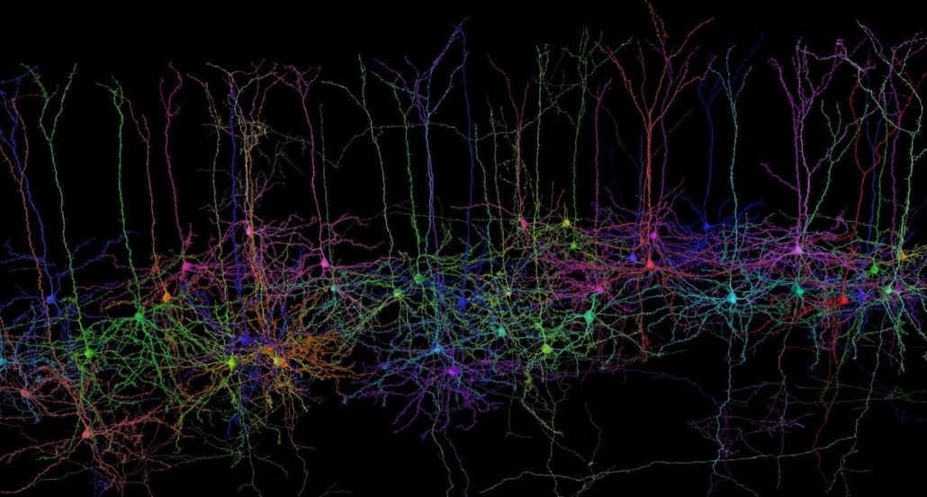
Prof. Ecker and his team want to develop machine learning techniques to describe the shape and function of neurons in the cerebral cortex and find out how the shape relates to the function. © MICrONS Consortium
The European Research Council (ERC) has awarded €1.5 million to computational neuroscientist Professor Alexander Ecker from the University of Göttingen and the Max Planck Institute for Dynamics and Self-Organization (MPIDS) to conduct research on how the shape of a neuron in the cerebral cortex relates to its function.
The funding, a Starting Grant to be provided over five years, is for Prof Ecker and his team’s project: “Deep Neuron Embeddings: Data-driven multi-modal discovery of cell types in the neocortex”.
The “form follows function” principle was proclaimed by the early 20th century architect Louis Sullivan to be a universal law of nature and later formed the basis of the Bauhaus style.

The computational neuroscientist Professor Alexander Ecker from the University of Göttingen and the Max Planck Institute for Dynamics and Self-Organization (MPIDS) has received a Starting Grant from the European Research Council.
“Whether this also applies to the brain, however, is currently unknown,” says Prof. Ecker, describing the background to his work. “For example, it is not yet clear exactly how the shape and function of the neurons in the cerebral cortex are related.”
Building upon recent scientific advances, Ecker and his team can now address this question. For a long time, it was only possible to measure either the morphology (shape) of a neuron or its functional activity, but not both at the same time.
Cortical neurons exhibit a wide diversity of shapes with complex branching patterns and other morphological features. Similarly, they exhibit a great degree of diversity in how they process stimuli during visual perception. To find out how features of a neuron’s shape relate to its role in sensory information processing, complex mathematical descriptions are needed. The research team will draw upon a large dataset from an earlier collaboration funded by the US Brain Initiative, which investigated the anatomy and activity of about 100,000 neurons of the visual cortex of a mouse.
Prof. Ecker and his team of scientists – with the help of the ERC funding – want to develop machine-learning methods to describe both the neurons’ shape and their function mathematically, recognise patterns in the data, and relate their form and function to each other.
This research is also made possible through the collaboration of a number of research institutions across the Göttingen Campus.
Prof. Ecker has been Professor of Data Science at Göttingen University since 2019 and is also a Max Planck Fellow at the MPI for Dynamics and Self-Organization. At the University, he holds his professorship at the Institute of Computer Science and also serves as a board member of the Campus Institute Data Science (CIDAS), the interface for collaboration in the field of Data Science at the Göttingen Campus.
Prins Hendrikstraat 1
5611HH Eindhoven
The Netherlands
info@interhospi.com
PanGlobal Media IS not responsible for any error or omission that might occur in the electronic display of product or company data.
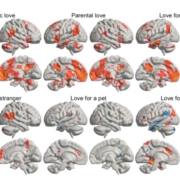 Pärttyli Rinne et al 2024, Aalto University