Researchers use AI to identify new high-risk endometrial cancer subtype – and show how to diagnose it
Researchers at the University of British Columbia have made a significant breakthrough in endometrial cancer diagnosis, utilising artificial intelligence (AI) to identify a previously unrecognised high-risk subtype. This discovery promises to enhance patient care for those with the most common gynaecological malignancy.

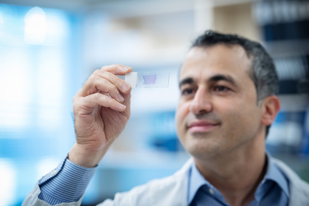
Dr Ali Bashashati, assistant professor of biomedical engineering and pathology and laboratory medicine at UBC, observes an endometrial cancer sample on a microscope slide.
Advancing molecular subtyping
The study, published in Nature Communications [1], builds upon previous work by the B.C. Gynecologic Cancer Initiative. In 2013, this multi-institutional collaboration demonstrated that endometrial cancer could be classified into four molecular subtypes, each associated with different risk levels.
Subsequently, the team developed ProMiSE, an innovative molecular diagnostic tool now used internationally to guide treatment decisions. However, challenges persisted, particularly with the most prevalent molecular subtype, which accounts for approximately 50% of all cases and lacks discernible molecular features.
Dr Jessica McAlpine, professor and Dr Chew Wei Chair in Gynaecologic Oncology at UBC, and surgeon-scientist at BC Cancer and Vancouver General Hospital, explained the problem: “There are patients in this very large category who have extremely good outcomes, and others whose cancer outcomes are highly unfavourable. But until now, we have lacked the tools to identify those at-risk so that we can offer them appropriate treatment.”
Harnessing AI for precision medicine
To address this challenge, Dr McAlpine collaborated with Dr Ali Bashashati, an assistant professor of biomedical engineering and pathology and laboratory medicine at UBC. Dr Bashashati’s team developed a deep learning AI model capable of analysing images of tissue samples collected from patients.
The AI was trained to differentiate between different subtypes and, after analysing over 2,300 cancer tissue images, successfully identified a new subgroup exhibiting markedly inferior survival rates.
Dr Bashashati highlighted the unique capabilities of AI in this context: “The power of AI is that it can objectively look at large sets of images and identify patterns that elude human pathologists. It’s finding the needle in the haystack. It tells us this group of cancers with these characteristics are the worst offenders and represent a higher risk for patients.”
Clinical integration and implications
The research team is now exploring how to integrate this AI tool into clinical practice alongside traditional molecular and pathology diagnostics, supported by a grant from the Terry Fox Research Institute.
Dr McAlpine emphasised the complementary nature of the new approach: “The two work hand-in-hand, with AI providing an additional layer on top of the testing we’re already doing.”
One significant advantage of the AI-based approach is its cost-efficiency and ease of deployment across diverse geographical locations. The AI analyses images routinely gathered by pathologists and healthcare providers, even at smaller hospital sites in rural and remote communities.
Enhancing equity in cancer care
The combined use of molecular and AI-based analysis could allow many patients to remain in their home communities for less intensive surgery while ensuring those who need treatment at a larger cancer centre can access it.
Dr Bashashati highlighted the potential for improved healthcare equity: “What is really compelling to us is the opportunity for greater equity and access. The AI doesn’t care if you’re in a large urban centre or rural community, it would just be available, so our hope is that this could really transform how we diagnose and treat endometrial cancer for patients everywhere.”
This innovative approach to endometrial cancer diagnosis and risk stratification represents a significant step forward in personalised medicine. By leveraging AI to identify high-risk patients who might otherwise go unrecognised, clinicians can offer more targeted and potentially life-saving interventions.
Reference:
- McAlpine, J., Bashashati, A., et al. (2024). AI-based histopathology image analysis reveals a novel subset of endometrial cancers with distinct genomic alterations and unfavourable outcome. Nature Communications, 15.
https://doi.org/10.1038/s41467-024-49017-2

