
PACS and imaging: seeking the fourth dimension
Given their pervasiveness in hospitals, one could be forgiven to take picture archiving and communication systems (PACS) for granted. However, in reality, they have been around for less than two decades. The first PACS prototype was set up in 1990 at the University of California, and soon after at Hammersmith Hospital in London. Commercial products began to enter the market 3-4 years later but serious growth in PACS use was seen only in the early 2000s.
PACS: boost to productivity and imaging demand
PACS revolutionized workflow between radiology and other hospital departments, resulting in a gigantic leap in productivity. PACS also catalysed the growth of imaging, especially computed tomography (CT) and magnetic resonance imaging (MRI) scanners, whose huge masses of data it was able to process, store and forward to different users on demand.
Along with older X-Rays and ultrasound (whose data are also managed by PACS), CT and MRI devices have become integral to nearly every single surgical procedure, as well as the medical management of chronic diseases ranging from arthritis to cancer. In acute care settings such as A&E, their availability has directly contributed to a fall in patient waiting times and increased the efficiency of triaging.
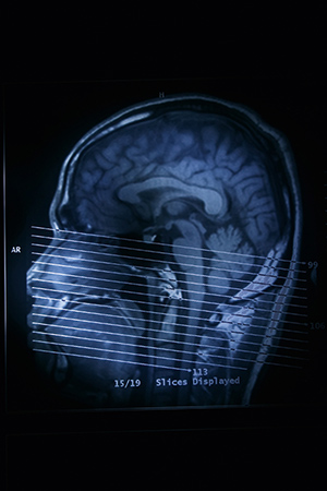
2007: 3D video of beating heart
Until recently, 2D PACS was the only realistic choice. In spite of an explosion in core processing power and storage capacity, PACS technologies had been consistently outpaced by developments in CT, which permitted imaging of increasingly thinner slices, outputting exponentially larger volumes of data.
In 2007, Toshiba launched Aquilion One, with a capability of acquiring five 320 slices per second and attaining a highly-symbolic milestone
