Novel advanced brain imaging technique reveals complex blood flow patterns in microscopic detail
Scientists have developed a revolutionary optical imaging technique that provides unprecedented views of blood vessels in the living brain, offering new ways to study neurological conditions like stroke and Alzheimer’s disease.
Mapping the brain’s blood supply
Researchers from the University of Zurich and ETH Zurich have created an advanced microscopy system that can capture detailed three-dimensional images of blood vessels in the brain while simultaneously measuring blood flow velocities across thousands of vessels. The breakthrough technology, called extended-focus optical coherence microscopy, provides a comprehensive view of the brain’s vascular network from its largest vessels down to its tiniest capillaries.

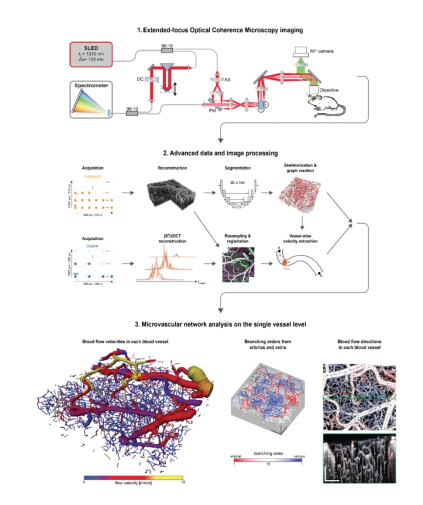
Research overview. 1. The extended-focus optical coherence microscopy system. 2. The data acquisition and post-processing pipeline. 3. Exemplary results, showing a volumetric render of the imaged microvascular network and its flow velocity, arterial and venous vessel trees and the blood flow direction within multiple blood vessels. © Light: Science & Applications
Technical innovation enables deeper insights
The new system uses a specially designed “Bessel beam” to extend the focus of the microscope, allowing it to image much larger sections of brain tissue than conventional methods. Unlike traditional approaches that either look at tiny volumes or lose detail over larger areas, this technique maintains high resolution across a substantial viewing area of 1000 × 1000 × 360 micrometres.
“By integrating high-sensitivity Doppler optical coherence tomography with advanced data processing algorithms, we can precisely measure blood flow velocities and directions across thousands of brain vessels,” the researchers explain in their paper published in Light: Science & Applications.
Artificial intelligence enhances analysis
The team incorporated artificial intelligence into their system to help process and analyse the vast amount of data generated by the imaging. A deep learning algorithm helps accurately identify and trace blood vessels throughout the captured volume, while sophisticated processing techniques determine blood flow direction and velocity in each vessel.
Understanding blood flow patterns
The research revealed that blood flow velocities vary significantly across different types of vessels. The team found that pial arteries, which sit on the brain’s surface, exhibit the highest flow velocities of up to 30 mm/s while accounting for 21% of the total blood volume. In comparison, pial veins showed reduced blood flow velocities but represented a larger portion (33%) of the blood volume.
Applications for neurological research
This new imaging capability has important implications for studying various neurological conditions. In Alzheimer’s disease, for instance, changes in brain blood flow can occur long before other symptoms appear. The technology could help researchers better understand how these early changes develop and potentially lead to new therapeutic strategies.
Similarly, in stroke research, the technique could provide valuable insights into how blood flow changes in affected brain regions and how potential treatments might influence recovery. The ability to visualise these changes in an intact living brain marks a significant advancement in neurological research.
Future directions
The researchers plan to extend their studies to examine a range of neurovascular conditions, particularly focusing on stroke and its treatments. They believe their technology could also be applied to studying blood flow in other organs, potentially advancing vascular research across multiple medical fields.
The research represents a significant step forward in our ability to study the brain’s vascular system in unprecedented detail. By providing both structural and functional information about blood vessels at multiple scales, it opens new possibilities for understanding and treating various neurological conditions.
Reference:
Glandorf, L., Wittmann, B., Droux, J., et. al. (2024). Bessel beam optical coherence microscopy enables multiscale assessment of cerebrovascular network morphology and function. Light: Science & Applications, 13, 307. https://doi.org/10.1038/s41377-024-01649-1

