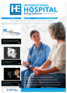
Optimal management of the ICU: some problems and potential solutions
The costly critical care provided by highly qualified personnel using state-of-the-art equipment in today

The costly critical care provided by highly qualified personnel using state-of-the-art equipment in today

Currently both North America and more affluent areas of Europe are facing an increasing shortage of physicians in primary healthcare and in various hospital specialities, a shortage which in many of these countries is being alleviated by the excessively aggressive recruitment of foreign doctors. For example in the UK national health service, poor planning and under-investment in medical education has lead to a third of physicians now being recruited from overseas. Frequently these doctors are citizens of poorer EU nations or commonwealth countries in Africa and the Indian sub-continent that can ill afford to lose such expensively educated personnel. Yet sadly these poorer nations are now effectively subsidising the healthcare of the wealthy West. The UK is of course not alone in its reliance on foreign physicians- the same strategy has been employed in the US and Canada, as well as affluent EU countries such as Germany and Sweden. While the rights of individual workers to migrate should be protected, clearly health services in developed countries should be moving towards more ethical ways of solving their physician shortage.
The long-term solution is to increase the supply of domestically educated medical doctors, but policy makers could also consider ways of reducing the current demand on physicians by developing systems and work flows that allow other healthcare professionals and hospital workers to perform those tasks where the input of a doctor is not essential. Currently there are artificial barriers developed decades ago preventing highly educated healthcare workers from providing services that they are competent to offer. For instance specialist nurses and medical assistants could triage patients, order lab tests and either treat patients directly or refer them to doctors as necessary, and the bulk of a physician

In Europe prostate cancer (PCa) is currently the most commonly diagnosed cancer in men- around one in six in the West will eventually be diagnosed with the disease- but the majority of patients will die of unrelated causes. There are two major problems related to PCa diagnosis: firstly, because of the lack of a highly sensitive and specific biomarker, many elderly men without cancer, or with clinically insignificant tumours confined to the prostate gland, are still undergoing unnecessary biopsies. Secondly because of its random nature, the standard method of biopsy, the transrectal ultrasound biopsy (TRUS), frequently detects low risk cancers that do not need aggressive therapy but fails to detect many clinically significant tumours in the less accessible areas of the gland. Recent studies suggest that multiparametric MRI (mp-MRI) pre-biopsy, to identify suspicious areas, followed by targetted biopsy using MR-ultrasound fusion, which combines detailed MRI scans with real-time ultrasound images of the prostate, is the better approach.
One preliminary prospective cohort study reported last year was carried out at the US National Cancer Institute. During a seven year period more than 1000 men underwent mp-MRI followed by MR-ultrasound fusion and concurrent TRUS. Whole-gland pathology of the prostate was also carried out after any prostatectomies. It was found that MR-ultrasound fusion diagnosed 30% more high risk tumours and 17% fewer low-risk tumours than TRUS. However data are still needed on disease recurrence and PCa mortality, and the authors consider that random clinical trials should be carried out to determine eventual clinical outcomes. A retrospective analysis involving more than 600 Brazilian patients with suspected PCa was also reported recently with 286 patients undergoing MR-ultrasound fusion biopsies and 331 patients undergoing random ultrasound-guided biopsies. Again the former technique detected significantly more patients with high-risk cancer requiring surgery and significantly fewer low-risk tumours where only surveillance was needed. There are of course financial impacts of purchasing and using such technology, but improved patient risk stratification may well result in a negligible net cost increase.
Hopefully MR-ultrasound fusion can replace techniques such as TRUS for suspected PCa diagnosis, but a question still remains. Is it really always necessary to treat clinically significant PCa by radical prostatectomy or radiotherapy of the entire organ when erectile dysfunction, urinary incontinence and intestinal problems are such common side effects? Would it not be possible to utilize appropriate imaging technologies and confine treatment to the affected area of the gland?

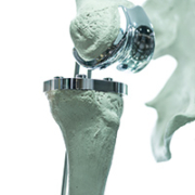
Three-dimensional (3D) printing for medical applications has grown in recent years at a feverish pace. The technology has long made a significant impact in manufacturing and is also revolutionizing healthcare. For some of its proponents, this would be rather like the Gutenberg printing press did with publishing. Indeed, the respected Gartner Group estimates that 30percent of internal medical implants and devices will be 3D printed by 2020.
3D printing was founded in the 1980s as stereo-lithography’ (STL) and the first commercial 3D printer came to market in 1988. Since the 1990s, manufacturers have used the technique principally for rapid prototyping, or the production of models and moulds.
Implants and prosthetics
Medical 3D printing took off in the early 2000s for producing dental implants and custom prosthetics, with a rapid pace of acceptance in areas such as hearing aids and dental braces. Currently, almost all hearing aids fitted into the ear in industrialiZed countries are made with 3D printers and orthodontic braces, too, are almost entirely 3D printed.
CustomiZed 3D-printed prosthetics and implants were made possible by translation of CT and MRI scans into digital STL print files, and imaging continues to play a central role in medical 3D printing.
Orthopedics and neurosurgery applications
The customization offered by 3D printing also quickly made its case for orthopedic patients being fitted with a standardized hip or spinal prostheses, which required the cumbersome process of shaving of pieces of metal and plastic with scalpels and drills afterwards, in order to achieve best fit.
Neurosurgeons too quickly saw the potential of 3D printing to address the drawbacks of variation in skull shape and the difficulties in using standard cranial implants. In head injury victims, for example, it is important to remove bone to provide space for the brain room to swell and the cranial plate must be perfect in fit.
In situ, in the OR
In the operating room, 3D printing has thoroughly transformed the manufacture of patient models to facilitate planning of surgical procedures. In 2016, a 3D printed model was used by Blythedale Children’s Hospital in Westchester in a 27-hour operation to separate twins conjoined at the head. According to many reports, recovery of the infants was accelerated due to the 3D model.
At the University of Michigan, CT images of a patient’s airway were used with a 3D printer to fabricate a precisely modelled, bioresorbable tracheal splint that was surgically implanted in a baby. The baby recovered, and full resorption of the splint is expected to occur within three years.
3D printing has also been used for making surgical tools such as forceps, hemostats, scalpel handles and clamps. They are formed sterile, and some estimates report that they cost only a tenth of a stainless steel equivalent.
In situ printing, by which implants, tissue (and eventually organs) are 3D printed in the human body during operations is anticipated in the future trend. Such a trend is being reinforced by rapid developments in miniaturized robotic bioprinters and robot-assisted surgery.
Personalized pills
3D printing technologies are also used for personalized medicine, with precision in dose (matched to patient profile and response). Some firms are experimenting with complex drug-release profiles, such as poly-pills with multiple active ingredients in a multilayered form.
This is seen as promising new standards of care for patients with several chronic diseases. Extended to one poly-pill per day for everyday medications, such a step would reduce a bane of medical practitioners – namely patient non-compliance.
In 2016, Spritam levetiracetam, a new drug to control seizures brought on by epilepsy, was approved by the US Food and Drug Administration (FDA). The pill, the world’s first to be 3D printed, is based on a trademarked ZipDose technology developed by Ohio-based Aprecia, and provides more porosity than alternative dosage forms.
Industry experts foresee drugs manufacturing being done eventually at the point-of-care, with physicians emailing medication formulations to pharmacies for on-demand drug printing.
Post-industrial production
The logic of 3D printing is in some ways truly revolutionary. What it brings is an end to the idea that there is commercial sense in only large runs of standardized products, a cornerstone of 19th/20th century manufacturing tradition as well as the Industrial Revolution. The first 3D manufactured product, in other words, costs approximately the same as the next one.
3D printing also reduces cost in certain cases. For example, a 5-mg pharmaceutical tablet can be custom-fabricated on demand as a smaller and less expensive 2.5-mg tablet rather than being broken up and left unused.
Speed
Speed too is a major asset of medical 3D manufacturing, and a spin-off from the fact that large production runs are not required. Customized products like prosthetics and implants, in particular, can be made within hours.
As with pharmacy pills, some expect on-site 3D printing at, or adjacent to, a hospital, to eventually emerge, for making patient-specific products.
Basic technology
The basic technique of 3D printing, which is also known as additive manufacturing, involves the successive deposit of layers of materials, typically plastic and ceramics or metal and powders, to make the final product.
One of the most exciting innovations, however, consists of using live cells as the printing material.
Types of 3D printer
The type of 3D printer chosen for an application often depends on the material used and the method for bonding the layers in the final product. Key technologies for medical applications include selective laser sintering (SLS) and thermal inkjet (TIJ) printing. Another widely-used 3D printing technology is fused deposition modelling (FDM).
Though relatively basic and inexpensive, FDM was one of the earliest examples of successful medical 3D printing in the late 1990s/early 2000s when it was used to construct cranial implants. FDM remains widely used for rapid modelling and prototyping in orthopedics and dentistry.
FDM printers use a print-head similar to an inkjet printer. Rather than ink, however, beads of thermoplastic (similar to those used in injection moulding) are released to form a thin layer. The process is repeated continuously. Since the plastic is heated, it fuses to the layers below, and then hardens as it cools to create the final product.
More complex medical uses of 3D printing are based on SLS and TIJ.
SLS uses metal, plastic or ceramics as material. A laser draws out the shape of the object and this is then fused to a powdered metal substrate. The process is repeated until the product is formed. The degree of detail in SLS is directly linked to the precision of the laser and the powder’s fineness.
On its part, TIJ uses thermal (as well as electromagnetic or piezoelectric) technology to deposit tiny droplets of ink or even cells (bio-ink) on a substrate. Unlike office inkjet printers, 3D TIJ heats a print-head to create collapsing air bubbles, which in turn create pressure pulses to eject the droplets from nozzles. The size of the droplets can be adjusted by temperature, pulse frequency or material viscosity and volumes can be as little as 10-20 picolitres. Multiple-head TIJ is especially promising for producing tissue and simple organs in the process of bioprinting’ (discussed below). Other applications under study include drug delivery and gene transfection.
Bioprinting – the final frontier
While implants and prosthetics have convincingly demonstrated the real-world relevance of 3D printing, the maximum excitement is currently focused on its use in tissue and organ fabrication.
Ageing, accidents, disease and birth problems often cause tissue and organ failure. Treatment is largely based on donor transplants. However, there is a chronic shortage of supply, not least of suitable donors (e.g. with matching tissue). In addition, surgery and follow-up is complex and expensive.
One recent approach to finding a solution consists of tissue engineering and regenerative medicine, based on mixing growth factors into isolated stem cells, multiplying them in a lab and then seeding the cells on scaffolds which transform direct cell proliferation and differentiation into functioning tissues.
Beyond regenerative medicine
Bioprinting takes traditional regenerative technologies further than scaffold support alone by using 3D printing technology to produce layers of cells, biomaterials, and cell-laden biomaterials. This is then precisely placed by the printer in tissue-like structures. As mentioned previously, inkjet-based bioprinting is the most commonly used technique for bioprinting.
Tissues and organs
German researchers have been developing skin cell bioprinting since 2010. In January 2017, a team from Spain’s Universidad Carlos III de Madrid (UC3M) reported in the journal Biofabrication’ they had developed 3D-printed human skin adequate for transplant into patients, and for testing drugs and cosmetics. Their product is currently undergoing European approval. Meanwhile, in the US, Organovo too has developed 3D-printed skin. Demonstrating the potential of such markets, French cosmetics giant L’Oreal has begun collaborating with Organovo.
Researchers have so far also successfully printed a knee meniscus, heart valves, bone and an artificial liver. In 2016, scientists at Cambridge University’s Centre for Brain Repair reported the 3D printing of a retina using a piezoelectric TIJ printer.
One application area is to use 3D printing to create tissues and organs for medical research, and rapidly screen candidate drugs, cutting research costs and time. Organovo is developing strips of printed kidney and liver tissue for exactly such a purpose, while Russia’s 3D Bioprinting Solution has 3D printed a functional thyroid in a mouse and claims to be ready to do the same in humans.
20 years to a 3D-printed heart?
Nevertheless, most bio-printed organs have so far been relatively small and simple, with no vascularity or nerve system and nourishment provided wholly by diffusion from the host vasculature. Such diffusion seems to suffice for thicknesses of 150-200 micrometers. Beyond it, there is none. In future, the bioprinting of 3D organs such as an entire kidney or heart will require precise multicellular structures with full vascular network integration.
Such a process may not be that far away. Collaborators from a network of academic institutions, including the Harvard University, Stanford University, the Massachusetts Institute of Technology and the University of Sydney recently announced they had bioprinted a perfusable network of capillaries, marking a significant stride toward overcoming the limits to diffusion.
According to some projections, we may be less than 20 years from a fully functioning printable heart.
Challenges ahead
As with many other frontiers of medicine, an immediate challenge for medical 3D printing consists of regulatory acceptance. Though a hundred-odd 3D-printed products had been approved in the US and Europe by the end of 2016, these consist almost entirely of prosthetics, surgical tools and artificial bone replacement.
Fulfilling regulatory requirements for more complex products is likely to be much more demanding. Included here are the need for large randomized controlled trials, which require funding and time – for instance to determine the biocompatibility of several of the new materials being used.
The safe use of health technology-from basic infusion pumps to large, complex imaging systems-requires identifying possible sources of danger or difficulty with those technologies and taking steps to minimize the likelihood that adverse events will occur. This list will help healthcare facilities do that.
Produced each year by ECRI Institute’s Health Devices Group, the Top 10 Health Technology Hazards list identifies the potential sources of danger that it believes warrant the greatest attention for the coming year. The list does not enumerate the most frequently reported problems or the ones associated with the most severe consequences-although such information is certainly considered in the analysis. Rather, the list reflects the Health Devices Group’s judgment about which risks should receive priority now.
All the items on the list represent problems that can be avoided or risks that can be minimized through the careful management of technologies. Additional content provided with the full article, which is available separately to members of certain ECRI Institute programmes, provides guidance to help manage the risks. In this way, the list serves as a tool that healthcare facilities can use to prioritize their patient safety efforts.
International Hospital presents here the abridged version of ECRI Institute’s 2017 Top 10 list of health technology hazards which is available as a free public service to inform healthcare facilities about important safety issues involving the use of medical devices and systems.
1. Infusion errors can be deadly if simple safety steps are overlooked
Most large-volume infusion pumps incorporate safety mechanisms for reducing the risks of potentially deadly intravenous (IV) infusion errors. These mechanisms have greatly improved infusion safety, but can’t eliminate all potential errors. And the mechanisms themselves have been known to fail.
ECRI Institute continues to learn about and investigate incidents of infusion errors involving pump or administration set failures, staff unknowingly defeating a safety mechanism, or incorrect infusion programming. Such errors- particularly those that result in the uncontrolled flow of medication to the patient, known as ‘IV free flow’-can lead to patient harm and even death.
In many of these incidents, harm could have been averted if staff had:
Once commonplace, these simple practices are now often overlooked-perhaps because staff implicitly trust the pump’s advanced safety features.
2. Inadequate cleaning of complex reusable instruments can lead to infections
The use of contaminated medical instruments can lead to disabling or deadly patient infections or instrument malfunctions.
Outbreaks associated with the use of contaminated duodenoscopes-such as those that caused headlines in recent years-illustrate the severity of this issue. But duodenoscopes are not the only devices that warrant attention. ECRI Institute has received reports involving a variety of contaminated medical instruments that have been used, or almost used, on patients.
Complex, reusable instruments-such as endoscopes, cannulated drills, and arthroscopic shavers-are of particular concern. They can be difficult to clean and then disinfect or sterilize (i.e., reprocess) between uses, and the presence of any lingering contamination on, or in, the instrument can be difficult to detect.
Often, we find that inattention to the cleaning steps within the reprocessing protocol is a contributing factor. Healthcare facilities should verify that comprehensive reprocessing instructions are available to staff and that all steps are consistently followed, including precleaning of the device at the point of use.
3. Missed ventilator alarms can lead to patient harm
Ventilator alarm management challenges complicate efforts to prevent patient harm resulting from missed alarms. Ventilators deliver life-sustaining therapy, and a missed alarm could be deadly. Concerns include:
These concerns, and the ways to manage them, are similar to those that exist with physiologic monitoring systems, which we have addressed in previous Top 10 Health Technology Hazards lists. Ventilators, however, pose some unique challenges. For example: Collecting and analysing ventilator alarm data can be difficult, making it harder for hospitals to identify where their vulnerabilities lie. And the options for supplementing a ventilator’s alarms-so that the alarm can be noticed outside the patient’s room, for example-are limited.
As a result, ventilators will require different methods for studying the problem and different strategies for addressing it.
4. Undetected opioid-induced respiratory depression
Patients receiving opioids-such as morphine, hydromorphone, or fentanyl-are at risk for drug-induced respiratory depression. If not detected, this condition can quickly lead to anoxic brain injury or death. Thus, spot checks every few hours of a patient’s oxygenation and ventilation are inadequate.
Drug-induced respiratory depression is of particular concern for patients receiving parenteral and neuraxial opioids in medical-surgical and general care areas. However, it is also of concern for hospital or ambulatory surgery/endoscopy facility patients receiving opioids during procedural sedation and while in the postanesthesia care unit (PACU).
Even if they are otherwise healthy, such patients can be at risk if, for example:
ECRI Institute recommends that healthcare facilities implement measures to continuously monitor the adequacy of ventilation of these patients and has recently tested and rated monitoring devices for this application.
5. Infection risks with heater-cooler devices used in cardiothoracic surgery
Heater-cooler systems have been identified as a potential source of nontuberculous mycobacteria (NTM) infections in heart surgery. The likelihood of infection during surgery is not fully understood. However, these infections can be life-threatening and have resulted in patient deaths.
Heater-cooler systems are used in cardiothoracic surgeries to warm or cool the patient by extracorporeal heat exchange with the patient’s blood during heart-lung bypass procedures. These devices circulate warm or cold water through a closed circuit. Water in the circuit is not intended to come into direct contact with the patient or the patient’s circulating blood. However, aerosolized water carried by air from the exhaust vents of contaminated heater-coolers has been suggested as a cause of NTM infections.
Initial reports focused on one specific model of heater-cooler, but models from other suppliers could likewise become contaminated under certain circumstances and if appropriate precautions are not taken.
The U.S. Food and Drug Administration has issued recommendations for all heater-cooler devices; they are intended to help prevent and manage device contamination risks and to minimize patient exposure to heater-cooler exhaust air, which may contain aerosolized contaminated water.
6. Software management gaps put patients, and patient data, at risk
Inadequate medical device software management can delay a facility’s responses to safety alerts, allow cybersecurity vulnerabilities to be exploited, and impact patient safety.
Maintaining a central repository of up-to-date and easily retrievable information about the software versions used in a healthcare facility’s medical devices is challenging. But failure to do so leaves the facility ill-prepared to effectively manage software updates and alerts.
Mismanagement of software updates and alerts can adversely affect patient care or impact patient/staff safety- for example, by:
To address the hazard, a healthcare facility should verify that its computerized maintenance management system (CMMS) provides the capabilities needed to effectively track software versions for its medical devices and systems. In addition, the facility should establish practices for keeping the software version information in the CMMS current and complete.
7. Occupational radiation hazards in hybrid ORs
Clinicians working in hybrid ORs-operating suites that include built-in x-ray imaging systems-are at risk of unnecessary occupational exposures to ionizing radiation if appropriate precautions are not consistently followed.
Particular concern exists in this environment because hybrid OR staff may be less knowledgeable than radiology and interventional radiology staff about the risks of radiation exposure, and they may be less experienced at taking appropriate precautions.
In addition, with the increasing reliance on X-ray imaging systems during complex OR procedures, an increasing number of specialists and staff members who previously would have had little exposure to ionizing radiation during surgeries are now participating in these procedures.
Because long-term exposure to radiation increases the risk of cancer, it is imperative that hybrid OR staff obtain OR-specific radiation protection training, that they put this training into action, and that available tools and methods be used to minimize radiation exposures.
8. Automated dispensing cabinet setup and use errors may cause medication mishaps
Poor choices made when setting up automated dispensing cabinets (ADCs), as well as mistakes made during use, can lead to harmful medication errors.
Medication errors and near misses associated with ADCs have been traced to insufficient planning when setting up medication drawers, as well as errors made when stocking them. Incidents reported to ECRI Institute include: the presence of the wrong drug or dose in an ADC pocket, the availability of high-alert drugs in unsecured areas of the cabinet, and the unavailability of needed drugs.
Problems such as these have resulted in delays in patient care and the administration of incorrect drugs or drug concentrations, leading in some cases to severe patient injury.
Careful planning is required to determine:
9. Surgical stapler misuse and malfunctions
Problems associated with the use and functioning of surgical staplers can lead to intraoperative hemorrhaging, tissue damage, unexpected postoperative bleeding, failed anastomoses, and other forms of patient harm.
Surgical staplers require meticulous technique to operate, and problems during use are not uncommon. The U.S. Food and Drug Administration receives thousands of adverse event reports related to surgical staplers each year, and ECRI Institute likewise consistently receives reports of surgical stapler problems. Although severe injuries are infrequent, they do occur: We have investigated fatalities and other cases of serious patient harm.
Commonly reported problems include: misfiring or difficulty in firing, misapplied staples, unusual sounds during firing (which can indicate a damaged or malfunctioning mechanism), and tissue becoming ‘jammed’ in the mechanism.
To prevent patient harm, users must be familiar with device operation, they must carefully select the appropriate staple size for the patient and tissue type, and they must be alert to the signs that the stapler may not be functioning as intended.
10. Device failures caused by cleaning products and practices
The use of cleaning agents or cleaning practices that are incompatible with the materials used in a medical device’s construction, or that are otherwise inappropriate for the device’s design, can cause the device to malfunction or to fail prematurely, possibly affecting patient care. Specifically:
Because there is no single cleaner or cleaning process that will work with all devices, hospitals must stock and use multiple cleaning products and familiarize staff with device-specific cleaning methods-tasks that pose a significant burden. Nevertheless, failure to do so can lead to ineffective cleaning (a potentially deadly circumstance), as well as excessive component breakage and premature equipment failures (which can affect patient care and be a significant financial burden).
www.ecri.org.uk www.ecri.org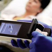
Point-of-care testing (POCT) refers to diagnostic tests which are performed physically close to a patient, with the results obtained on site. They are conducted at primary care centres and at hospital bed sides (increasingly, in emergency departments and intensive care units, too).
POCTs are also used in the field in settings such as natural or man-made disasters, and accompanied by telemedicine, in patients’ homes.
Saving time and space
While traditional diagnostic tests involve taking patient specimens, transporting them to a laboratory for analysis and then returning the results to a physician, POCTs cut out both the transport and laboratory. As a result, they provide quicker turnaround time (TAT), sometimes near-instantaneously.
In the past, the traditional laboratory-centric process was unavoidable due to the sheer size of equipment required for diagnostic tests. In recent years, technology developments – especially in terms of miniaturization – have made it possible to perform a growing number of tests outside of the laboratory. One recent book on biomedical engineering (D. Issadore and R.M. Westervelt (eds.), Point-of-Care Diagnostics on a Chip, Biological and Medical Physics, Biomedical Engineering’, Springer-Verlag, Berlin 2013) notes the array of sophisticated, low-power and small ‘microfilters, microchannels, microarrays, micropumps, microvalves and microelectronics …. integrated onto chips to analyse and control biological objects at the microscale’, that have made decentralized diagnostics possible.
Impact on efficiency, outcomes – and costs
Such time savings can have a dramatic impact on downstream clinical efficiency and patient outcomes. In many cases (although not universally or under all circumstances), they also save costs.
For example, POCT can reduce revenue losses due to workflow delays of test-dependent medical procedures – such as disruptions in magnetic resonance imaging (MRI) or computer tomography (CT) queue. This is not a rare occurrence, and delays in radiology testing have been shown to extend total length of stay in the emergency department (ED).
From lab downscaling to targeted solutions
Early POCTs were based on the simple transfer of traditional methods from a central laboratory, accompanied by their downscaling to smaller platforms. At a later stage, unique, innovative assays were designed specifically for POCT (such as the rapid streptococcal antigen test). This was accompanied by the development of wide arrays of POCT-specific analytic methods, ranging from the simple (such as pH paper for assessing amniotic fluid) to the ultra-sophisticated (for example, thromboelastogram for intra-operative coagulation assessment).
Today, the typical POCT test arsenal includes cardiac biomarkers, hemoglobin concentrations, differential complete blood count (CBC), blood glucose concentrations, coagulation testing, platelet function, pregnancy testing as well as tests for streptococcus, HIV, malaria etc.
Beside and near-bedside POCT
POCT devices are used in a wide range of healthcare settings. They can be divided into two broad groups, depending on size and portability – bedside and near-bedside.
Bedside POCT devices are smaller, usually hand-held, and offer the greatest mobility. Due to their compact nature they are often more specialized and limited in overall functionally. Many are enclosed in test cassettes (such as easy-to-use membrane-based strips) and based on portable, sometimes handheld, instruments. This family of POCT requires only a single drop of whole blood, urine or saliva, and the tests can be performed and interpreted by a general physician in minutes. Nevertheless, some of them can be quite sophisticated.
New POCTs for early detection of rheumatoid arthritis, for example, require only a single drop of whole blood, urine or saliva, and can be performed and interpreted by any general physician within minutes. Two of the earliest efforts in this area were made in Europe. The first, from Sweden’s Euro-Diagnostica detects antibodies to CCP, while Rheuma-Chec from Orgentec in Germany combines two biomarkers – rheumatoid factor and antibodies to MCV. These tests are targeted at primary care.
Near-bedside (or neighbourhood) devices are larger and typically located in a designated testing area. They provide higher calibration sensitivity and quality control and are used for more complex diagnostic tests than their smaller bedside counterparts.
They are themselves also far more complex, with high degrees of automation in comparison to their bedside POCT counterparts. This automation contributes to the increased speed and ease-of-use of the devices. However, it also leads to challenges in training users.
The imperatives of turnaround time
As mentioned, the principal interest in POCT is to reduce turnaround time (TAT) – the duration between a test and the obtaining of results which aid in making clinical decisions. The impact of this has been profound in the emergency department.
Already in 1998, a randomized, controlled trial in the A&E department of a British teaching hospital assessed the impact of POCT on health management decisions. The results, published in British Medical Journal’ in 1998, found that physicians using POCT reached patient management decisions an average of 1 hour and 14 minutes faster than patients evaluated through traditional means.
Use in emergency departments
Though the bulk of POCT is conducted by primary care physicians, one of its fastest growing users has been hospital EDs, which the British Medical Journal’ study hinted at almost 20 years ago.
POCT’s relevance for emergency departments is multi-faceted.
In the ED, prolonged wait times and overcrowding directly correlate to reduced patient satisfaction and adverse clinical outcomes. Several European countries have regulations on length-of-stay time targets in EDs, requiring that patients must transit through four to 8 hours. Though there are several factors at play here, no one would argue that reducing the delay between sample collection and test results can enable healthcare professionals to arrive at quicker decisions and increase patient throughput. POCTs make this possible.
One study in Switzerland evaluated adding POCT to B-type natriuretic peptide levels for ED patients presenting acute dyspnea as their primary symptom. POCT was not only associated with significant decreases in time to treatment initiation, but was also associated with a shorter length of stay and a 26percent reduction in total treatment costs.
Another study on D-dimer POCT in the ED found a 79percent reduction in TAT compared to central laboratory testing and resulted in shorter ED lengths of stay and reduced hospital admissions, while a randomized study in coagulopathic cardiac surgery patients found that POCT-guided hemostatic therapy led to reduction in transfusion and complication rates, and improved survival.
From ACS to pregnancy tests, and overcrowding
Favourable perspectives on POCT in the ED have strengthened over time. One recent study in Critical Care’ found POCT increased the number of patients discharged in a timely manner, expedited triage of urgent but non-emergency patients, and decreased delays to treatment initiation. The study quantitatively assessed several conditions such as acute coronary syndrome (ACS), venous thromboembolic disease, severe sepsis and stroke, and concluded that POCT, when used effectively, ‘may alleviate the negative impacts of overcrowding on the safety, effectiveness, and person-centeredness of care in the ED.’
A great deal of attention has been given to the use of POCT in emergency settings for screening patients who presented with symptoms of acute coronary syndromes (ACS). The rapid identification and treatment of ACS patients is critical.
Due to the time-sensitive nature of ACS, reduced TATs can offer a clear advantage. POCT has been shown to increase the speed at which positive cases of ACS are accurately identified, allowing physicians the ability to admit and initiate treatment at a faster rate than previously possible. Decreased TATs also can result in the earlier identification of negative cases of ACS, thereby increasing the number of successful discharges, and allowing for more efficient use of hospital resources .
The ICU and POCT
Unlike the ED, the use of POCT in intensive care units is still in its infancy. In 2013, researchers at Germany’s Klinikum rechts der Isar in Munich sought to retrospectively investigate whether POCT predicted hospital mortality in over 1,500 ICU admissions. The results were mixed. Lactate and glucose seemed to independently predict mortality. So did some forms of metabolic acidosis, especially lactic acidosis. However, anion gap (AG)-acidosis failed to show any use as a biomarker.
One of the most important areas for POCT focus in the ICU consists of sepsis – which is directly correlated to poor outcomes. ICU patients often have other ongoing disease processes whose biomarkers are shared with sepsis, such as raised white blood cell count and fever. More crucially, many ICU patients are already on antibiotics at admission, making microbiological cultures redundant.
POCT as part of health management strategy
Overall, POCTs have an impact and make most sense when utilized as part of an overall health management strategy which enhances the efficiency if clinical decision-making. Indeed, the rapid TAT provided by POCT allows for accelerated identification and classification of patients into high-risk and low-risk groups, improving quality of care and increasing clinical throughput.
POCT results are often available in minutes. However, decreased TATs on their own mean nothing, until they provide clinical pathways means to impact on workflow. The latter varies widely across healthcare settings.
Differences in practice
Such a scenario is by no means straightforward. In Europe, for example, POCT use is highly irregular and differs greatly between institutions and countries. Though differences in operating procedures are natural by-products of institutional cultures, there are some oversight and quality control issues which healthcare leaders must address to take maximum advantage of POCT.
Answers to the above are not a question of if’ but when’.
Regulation – the future ?
The future of POCT may well be shaped by regulators, and their response to the kind of pressures mentioned above.
In Europe, POCT devices are regulated under the 1998 European Directive 98/79/EC on in vitro diagnostic medical devices, which became operational in 2001. POCT devices are not specifically mentioned or referred to in this directive, and at the European level, coverage of POCT is referred by international standard ISO 22870:2006, used in conjunction with ISO 15189 which covers competence and quality in medical laboratories.
In the US, CLIA88 (Clinical Laboratory Improvement Amendments of 1988) provided a major impetus for growth in POCT. The rules, published in 1992, expanded the definition of laboratory’ to include any site where a clinical laboratory test occurred (including a patient’s bedside or clinic) and specified quality standards for personnel, patient test management and quality.
One of CLIA88’s biggest contributions to POCT growth was to define tests by complexity (waived, moderate complexity and high complexity control), with minimal quality assurance for the waived category.
CLIA88 has been followed by US federal and state regulations, along with accreditation standards developed by the College of American Pathologists and The Joint Commission. These have established POCT performance guidelines and provided strong incentives to ensure the quality of testing.

Prins Hendrikstraat 1
5611HH Eindhoven
The Netherlands
info@interhospi.com
PanGlobal Media IS not responsible for any error or omission that might occur in the electronic display of product or company data.