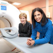
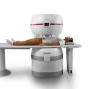
The results of Olympus’ ongoing joint research programme to create an AI-based pathology diagnostic tool with the potential to streamline pathologists’ workloads were announced at the Japan Society of Digital Pathology Study annual meeting. The diagnostic tool achieved 100% sensitivity and 50% or more specificity for all gastric biopsy pathology specimens analysed.
https://interhospi.com/wp-content/uploads/sites/3/2022/02/shutterstock_598193240-scaled.jpg17072560panglobalhttps://interhospi.com/wp-content/uploads/sites/3/2020/06/Component-6-–-1.pngpanglobal2022-02-15 09:26:542022-02-15 09:26:54Olympus announces first results of its AI-based pathology diagnostic tool for gastric cancer
Philips has signed an agreement to acquire Vesper Medical, a US-based medical technology company that develops minimally-invasive peripheral vascular devices. Vesper Medical will further expand Philips’ portfolio of diagnostic and therapeutic devices with an advanced venous stent portfolio for the treatment of deep venous disease. The transaction, which is subject to customary closing conditions, is […]
42 Technology (42T), FIND, the global alliance for diagnostics, and Rutgers University have worked together to develop an innovative stool sample processing kit that has played a central role in informing the World Health Organization’s (WHO) recent policy update to improve the diagnosis of tuberculosis (TB) in children.
Cline Scientific AB has received clearance from the Swedish Ethical Review Authority for the collection and testing of human cartilage tissue as part of the StemCART project.
https://interhospi.com/wp-content/uploads/sites/3/2022/02/dreamstime_m_191827942.jpg14732036panglobalhttps://interhospi.com/wp-content/uploads/sites/3/2020/06/Component-6-–-1.pngpanglobal2022-02-15 09:17:552022-02-15 09:18:50Cline receives clearance for ex-vivo testing of StemCART product on human cartilage tissue
Owen Mumford, a global leader in the design and manufacture of medical devices, has announced its emissions reduction targets through the Science Based Targets initiative (SBTi). The company has set a target to achieve net zero by 2045 (to mirror the UK NHS target) in alignment with the recommendations of the 2015 Paris Agreement.
https://interhospi.com/wp-content/uploads/sites/3/2022/02/Schermafbeelding-2022-02-15-om-10.14.47.png7731130panglobalhttps://interhospi.com/wp-content/uploads/sites/3/2020/06/Component-6-–-1.pngpanglobal2022-02-15 09:15:482022-02-15 09:15:48Owen Mumford sets verified targets to achieve net zero greenhouse gas emissions
New estimates reveal that at least 1.27 million deaths per year are directly attributable to antimicrobial resistance (AMR), requiring urgent action from policymakers and health communities to avoid further preventable deaths.
Based on estimates across 204 countries and territories the ‘Global burden of bacterial antimicrobial resistance in 204 countries and territories in 2019’ paper provides the most comprehensive estimates of the global impact of antibiotic resistance to date and reveals that AMR has now become a leading cause of death globally. Published in The Lancet following rigorous external peer review by independent experts, the paper highlights specific areas of concern and equips governments and health communities with the information they need to act quickly and develop a proportionate response.
Estimates included in the paper show that AMR is a leading cause of death globally, higher than HIV/AIDs or Malaria.
Antimicrobial Resistance (AMR) occurs when bacteria, viruses, fungi and parasites change over time and no longer respond to medicines, making infections harder to treat and increasing the risk of disease spread, severe illness and death.
Common infections from AMR
Common infections such as lower respiratory tract infections, bloodstream infections, and intra-abdominal infections are now killing hundreds of thousands of people every year because bacteria have become resistant to treatment. This includes historically treatable illnesses, such as pneumonia, hospital-acquired infections, and foodborne ailments. An estimated 4.95 million people who died in 2019 suffered from at least one drug-resistant infection and AMR directly caused 1.27 million of those deaths.
Everyone is at risk from AMR, but the data shows that young children are particularly affected. In 2019, one in five deaths attributable to AMR occurred in children under the age of five – often from previously treatable infections.
Sub-Saharan Africa faces the highest burden, with 255,000 deaths due to AMR in just one year and a particularly high number from vaccine preventable pneumococcal bacterial disease. However, high income countries also face alarmingly high levels, most notably for Escherichia coli, which commonly causes kidney infections, and Staphylococcus aureus, which is often acquired in hospitals and can cause bloodstream infections.
More ambitious plans needed to tackle AMR
Past projections estimated that as many as 10 million annual deaths from AMR could occur by 2050. In 2022, as we now have access to these robust estimates, we know that we are already far closer to this figure than expected. AMR is already threatening the ability of hospitals to keep patients safe from infections and undermining the ability of doctors to carry out essential medical practice safely, including surgery, childbirth and cancer treatment since infection is a risk following these procedures.
We are not innovating fast enough to develop effective vaccines, medicines and treatments:
Between 1980 and 2000, 63 new antibiotics were approved for clinical use. Between 2000 and 2018, just 15 additional antibiotics were approved.
Out of the seven deadliest drug-resistant bacteria, vaccines are only available for two (Streptococcus pneumoniae and Mycobacterium tuberculosis).
Whilst all seven of the leading bacteria have been identified as ‘priority pathogens’ by the World Health Organization (WHO) only two have been a focus of major global health intervention programmes – S. pneumoniae (primarily through pneumococcal vaccination) and M. tuberculosis.
And with high levels of hospitalisations from COVID-19, there is a risk that the burden of AMR has already accelerated due to increased use of antibiotics.However, more evidence is needed to ascertain the true impact.
“This paper is a critical step that allows us to see the full scale of the challenge. We now need to leverage these estimates to course-correct action and drive innovation if we want to stay ahead in the race against AMR.” — Professor Chris Murray, Director of the Institute for Health Metrics and Evaluation
The GRAM leadership team at the University of Oxford, Prof Christiane Dolecek, Dr Catrin Moore, and Prof Benn Sartorius, also commented: “Being able to measure AMR, and compare it with other major health threats, is essential to addressing its serious consequences. This work incorporates the best available data and provides reliable evidence describing the substantial mortality and morbidity caused by AMR globally. We anticipate that policymakers will use these results as we intended them, to drive action.”
New estimates from the paper were pre-released to Health Ministers at the G7 in June 2021, who agreed that AMR must be prioritised as part of plans to strengthen global health systems.
Talking about the need for political action on AMR, UK Special Envoy on Antimicrobial Resistance, Dame Sally Davies said: “AMR is already one of the greatest challenges facing humanity. Behind these new numbers are families and communities who are tragically bearing the brunt of the silent AMR pandemic. We must use this data as a warning signal to spur on action at every level.”
Immediate actions that can be taken
There are immediate actions that can help countries around the world protect their health systems against the threat of AMR:
We need to take greater action to monitor and control infections, globally, nationally and within individual hospitals.
We must accelerate our support for infection prevention and control, as well as expand access to vaccines and clean water and sanitation.
We must optimise our use of antibiotics unrelated to treating human disease, such as in food and animal production, taking a One Health* approach and recognising the interconnection between human and animal health.
It’s time to be more thoughtful about our use of antimicrobial treatments – expanding access to lifesaving antibiotics where needed, minimising use where they are not necessary to improve human health, and acting according to WHO Global Action Plan and AWaRE guidelines.
We must increase funding at every stage of the development pipeline for new antimicrobials targeting priority pathogens – from research for high priority bacteria, such as K. pneumoniae and E. coli, to securing access through innovative market solutions.
Authors of the paper are hopeful that, with this new data, the world is better equipped to correct the trajectory. COVID-19 has demonstrated the importance of global commitments to infection and control measures, such as hand washing and surveillance, and rapid investments in treatments.
Talking about the lessons learnt from COVID-19, Head of Intervention, Infectious Disease at Wellcome Trust, Tim Jinks said: “The COVID-19 pandemic has highlighted the importance of global collaboration: political leaders, the healthcare community, the private sector and the public working together to tackle a global health threat. Like COVID-19, we know what needs to be done to address AMR, but we must now come together with a sense of urgency and global solidarity if we are to be successful.”
The paper was authored by the Global Research on Antimicrobial Resistance (GRAM) Project, a partnership between the Institute for Health Metrics and Evaluation (IHME) and the University of Oxford. GRAM was launched with support from the Fleming Fund, the Wellcome Trust, and the Bill & Melinda Gates Foundation.
https://interhospi.com/wp-content/uploads/sites/3/2022/01/antibiotic.jpg13391417panglobalhttps://interhospi.com/wp-content/uploads/sites/3/2020/06/Component-6-–-1.pngpanglobal2022-01-28 07:55:192022-01-28 07:55:19Antimicrobial resistance becoming a leading cause of death globally
Hani Najm, M.D., who led the heart surgery team, inserts an IV line in the foetus’s right arm to deliver fluids and medications as needed. — courtesy Cleveland Clinic
A multidisciplinary team of Cleveland Clinic and Cleveland Clinic Children’s doctors and nurses performed a rare and complex lifesaving foetal surgery to remove a tumour attached to the heart of a 26-week-old foetus.
Few studies about the rare condition – an intrapericardial teratoma with foetal hydrops (i.e., fluid accumulation) evolving to foetal heart failure – have been reported in medical journals. “Only one previous incidence of continued pregnancy and delivery after foetal intrapericardial teratoma resection is documented in the world’s medical literature,” said Darrell Cass, M.D., director of Cleveland Clinic’s Fetal Surgery and Fetal Care Center. “As far as we know, Cleveland Clinic is the second academic medical centre in the world to have performed this foetal surgery successfully with continued pregnancy and delivery.”
During the procedure in May, surgeons successfully removed the malignant tumour, which had been compressing the left side of the foetus’ heart and impairing circulation, leading to fluid accumulation around the heart and other organs of the foetus.
The surgery included paediatric and congenital heart surgeons, a paediatric cardiologist, obstetric and paediatric anaesthesiologists, and a maternal-foetal medicine specialist. — courtesy Cleveland Clinic
Recovery with no complications
Following the operation, the mother and foetus recovered well with no complications and no sign of tumour recurrence during prenatal check-ups. On July 13, at 36 weeks and two days, a baby boy was delivered near full term by Caesarean section. Both mother and baby are doing well.
“I am very proud of our talented congenital heart surgery and foetal surgery teams that integrated seamlessly to successfully perform a complex lifesaving foetal surgery,” said Dr. Cass. “This tumour was growing rapidly in the exact wrong spot. It was compressing the heart of the foetus, causing fluid accumulation, and we started seeing signs that the cardiac function was deteriorating. We needed to act quickly and decisively to rescue the foetus.”
To perform this foetal surgery, Dr. Cass led a team that included paediatric and congenital heart surgeons Hani Najm, M.D., and Alistair Phillips, M.D.; paediatric cardiologist Francine Erenberg, M.D.; obstetric and paediatric anaesthesiologists McCallum Hoyt, M.D., Tara Hata, M.D., Yael Dahan, M.D.; and maternal-foetal medicine specialist, Amanda Kalan, M.D., who provided the mother’s care, including delivery of the newborn baby boy 10 weeks after the foetal surgery.
Once the mother was anesthetized with an approach to provide the safest environment for the foetal heart surgery to be successful, a Caesarean section-like incision was made to expose the mother’s uterus. Ultrasound was used to carefully locate the placenta and foetus, and best location for entry. The uterus was then opened about 12 cm, and the arms of the foetus were brought out to expose the chest.
Dr. Najm, who led the heart surgery team, inserted an IV line in a blood vessel of the foetus’s right arm to deliver fluids and medications as needed. Then, he carefully opened the chest and pericardium, and removed the tumour from the beating foetal heart. “As soon as the tumour was removed, the compression of the left atrium disappeared, and there was a nice blood flow that was almost back to normal,” said Dr. Najm.
Following the completion of the foetal heart surgery, the chest of the foetus was closed and the foetus was positioned back in the uterus. The uterus was then closed, followed by closure of the mother’s abdomen. The surgery lasted 3 ½ hours.
Rylan Harrison Drinnon was born on July 13 to Sam and Dave Drinnon.
Both mother and foetus recovered well following the surgery. The foetus’s cardiac function immediately improved, and the foetus stayed in the womb for the remainder of the pregnancy. Ten weeks later, the baby was born.
“Such an innovative foetal surgery provides hope to other families who may receive a similar devastating diagnosis,” said Dr. Najm, chair of Pediatric and Congenital Heart Surgery at Cleveland Clinic Children’s. “Clinical teams from Cleveland Clinic and Cleveland Clinic Children’s are consistently collaborating and remain dedicated to innovation and teamwork to ensure our patients of all ages can feel safe when entrusting their care to us.”
Moving forward, the infant’s healthcare team will monitor his heart health and check that there are no signs of tumour recurrence. In the future, the child will likely need surgery to reposition together his sternum that did not properly heal in utero.
“In this case, time was of the essence,” said Dr. Cass. “Shortly after the patient arrived at Cleveland Clinic, imaging tests showed that the tumour kept growing and the foetus’s heart function was deteriorating. It is important to acknowledge the whole care team. This family’s maternal foetal medicine specialist accurately diagnosed the condition and reached out to us because of our expertise in foetal care and treatment.”
https://interhospi.com/wp-content/uploads/sites/3/2022/01/heart_surgery_3.jpg17072560panglobalhttps://interhospi.com/wp-content/uploads/sites/3/2020/06/Component-6-–-1.pngpanglobal2022-01-20 11:59:032023-09-06 08:29:49Cleveland Clinic team perform complex foetal surgery to remove rare heart tumour
Being overweight may cause more hospital admissions and higher incidences of disease and mortality than previous studies report, according to new University of Bristol-led research. The study, published in Economics and Human Biology, used a genetic technique to identify the sole impact of body composition on hospital admissions from over 300,000 people.
The study, led by researchers from Bristol Medical School’s Population Health Sciences, aimed to find out the impact of excess body fat on the yearly hospital admission rate in the UK by analysing body mass index (BMI) data — a marker of overall body fat — and waist-hip ratio (WHR) data — a marker of regional body fat — from 310,471 individuals within the UK Biobank cohort. Linked with this data was information on 550,000 UK inpatient hospital admissions, with participants followed up for an average of six years.
Using this data, researchers compared estimates from conventional epidemiological analyses with a method called Mendelian randomisation, a genetic technique which allows scientists to quantify how being overweight may be causally related to disease and mortality. The method uses genetic changes in the genome linked to body composition to estimate the causal effect of being overweight on a health outcome and remove the effects of other factors that may jointly influence body composition and rates of hospital admission.
Waist-hip-ratio Their results found evidence for a direct causal effect of higher BMI and WHR on higher yearly hospital admission rates, with estimates that were larger than those obtained from existing research. One of the team’s most striking discoveries showed the relationship was largely driven by an adverse fat distribution in a certain area (measured by waist-hip ratio) rather than overall BMI.
They found that people were between 16 per cent to 26 per cent more likely to be admitted to hospital with each 0.09-unit higher waist-hip ratio compared to 8 per cent to 16 per cent with each 4.74 kg/m2 higher BMI. For example, for a woman of average height (163 cm) and weight (65 kg) in this study, and average measurements of waist (86 cm) and hip (103 cm) in this study, this would be the equivalent of gaining 9.3 cm (or 3.7 inches) in waist circumference, and just under 13 kg in weight, respectively. For a man of average height (177 cm) and weight (79 kg) in this study, and average measurements of waist (94 cm) and hip (102 cm) in this study, this would correspond to a 9.2 cm (or 3.6 inches) increase in waist circumference and a 15 kg increase in weight.
Dea Hazewinkel, the study’s lead researcher from the University’s Bristol Medical School and Population Health Sciences Institute, said: “We live in increasingly obesogenic environments with the World Health Organisation identifying 39 per cent of men and 40 per cent of women as being overweight, and 11 per cent of men and 15 per cent of women as obese worldwide. Finding causal effect estimates between fatty tissue and hospital admissions larger than those previously reported in existing studies emphasises the necessity of exploring policies aimed at reducing obesity in the population.
“The results also suggest that a preference should be given to waist-hip ratio as a measure of body fat over BMI as this may be more important for predicting hospital admissions,” said Hazewinkel.
https://interhospi.com/wp-content/uploads/sites/3/2022/01/doctors-obesity_web.jpg15772362panglobalhttps://interhospi.com/wp-content/uploads/sites/3/2020/06/Component-6-–-1.pngpanglobal2022-01-17 09:40:452022-01-17 09:40:45New study shows overweight may cause more UK hospital admissions than previous studies indicate
The European Research Council (ERC) has awarded €1.5 million to computational neuroscientist Professor Alexander Ecker from the University of Göttingen and the Max Planck Institute for Dynamics and Self-Organization (MPIDS) to conduct research on how the shape of a neuron in the cerebral cortex relates to its function.
The funding, a Starting Grant to be provided over five years, is for Prof Ecker and his team’s project: “Deep Neuron Embeddings: Data-driven multi-modal discovery of cell types in the neocortex”.
The “form follows function” principle was proclaimed by the early 20th century architect Louis Sullivan to be a universal law of nature and later formed the basis of the Bauhaus style.
The computational neuroscientist Professor Alexander Ecker from the University of Göttingen and the Max Planck Institute for Dynamics and Self-Organization (MPIDS) has received a Starting Grant from the European Research Council.
“Whether this also applies to the brain, however, is currently unknown,” says Prof. Ecker, describing the background to his work. “For example, it is not yet clear exactly how the shape and function of the neurons in the cerebral cortex are related.”
Building upon recent scientific advances, Ecker and his team can now address this question. For a long time, it was only possible to measure either the morphology (shape) of a neuron or its functional activity, but not both at the same time.
Diversity of morphological features
Cortical neurons exhibit a wide diversity of shapes with complex branching patterns and other morphological features. Similarly, they exhibit a great degree of diversity in how they process stimuli during visual perception. To find out how features of a neuron’s shape relate to its role in sensory information processing, complex mathematical descriptions are needed. The research team will draw upon a large dataset from an earlier collaboration funded by the US Brain Initiative, which investigated the anatomy and activity of about 100,000 neurons of the visual cortex of a mouse.
Prof. Ecker and his team of scientists – with the help of the ERC funding – want to develop machine-learning methods to describe both the neurons’ shape and their function mathematically, recognise patterns in the data, and relate their form and function to each other.
This research is also made possible through the collaboration of a number of research institutions across the Göttingen Campus.
Prof. Ecker has been Professor of Data Science at Göttingen University since 2019 and is also a Max Planck Fellow at the MPI for Dynamics and Self-Organization. At the University, he holds his professorship at the Institute of Computer Science and also serves as a board member of the Campus Institute Data Science (CIDAS), the interface for collaboration in the field of Data Science at the Göttingen Campus.
https://interhospi.com/wp-content/uploads/sites/3/2022/01/prof_ecker.jpg15001002panglobalhttps://interhospi.com/wp-content/uploads/sites/3/2020/06/Component-6-–-1.pngpanglobal2022-01-17 09:17:102022-01-17 09:17:10How does the shape of a cortical neuron relate to its function? European research team gets funding to find out
Bartley P. Griffith, MD and the patient, David Bennett — University of Maryland School of Medicine
In a major medical breakthrough, a man in the United States has received a heart from a genetically modified pig in a successful transplant.
The 57-year-old man, David Bennett, who was not eligible for a routine heart transplant, opted for the experimental surgical procedure, saying: “It was either die or do this transplant. I want to live. I know it’s a shot in the dark, but it’s my last choice.”
The eight-hour surgery took place on Friday 7 January and at the time of this publication, the patient was reportedly doing well.
The historic surgery was conducted by a team lead by Bartley P. Griffith, MD, and Muhammad M. Mohiuddin, MD, at the University of Maryland Medicine.
This experimental transplant procedure shows that a genetically-modified animal heart can function like a human heart without immediate rejection by the body and has major implications for the future of transplant surgery.
The U.S. Food and Drug Administration granted emergency authorization for the surgery on New Year’s Eve through its expanded access (compassionate use) provision. It is used when an experimental medical product, in this case the genetically-modified pig’s heart, is the only option available for a patient faced with a serious or life-threatening medical condition.
Dr Griffith, who is the Thomas E. and Alice Marie Hales Distinguished Professor in Transplant Surgery at University of Maryland School of Medicine (UMSOM), commented: “This was a breakthrough surgery and brings us one step closer to solving the organ shortage crisis. There are simply not enough donor human hearts available to meet the long list of potential recipients. We are proceeding cautiously, but we are also optimistic that this first-in-the-world surgery will provide an important new option for patients in the future.”
Prior to transplant, the heart from the genetically modified pig is removed from a machine perfusion device to keep it preserved until surgery — University of Maryland School of Medicine
Dr Mohiuddin, Professor of Surgery at UMSOM is considered one of the world’s foremost experts on transplanting animal organs, known as xenotransplantation. He established the Xenotransplantation Program five years ago with Dr. Griffith at UMSOM.
“This is the culmination of years of highly complicated research to hone this technique in animals with survival times that have reached beyond nine months. The FDA used our data and data on the experimental pig to authorize the transplant in an end-stage heart disease patient who had no other treatment options,” Dr Mohiuddin said. “The successful procedure provided valuable information to help the medical community improve this potentially life-saving method in future patients.”
Genetically modified pig
The genetically modified pig was provided by Revivicor, a regenerative medicine company based in Blacksburg, Virginia, US.
Three genes – responsible for rapid antibody-mediated rejection of pig organs by humans – were knocked out in the donor pig. Six human genes responsible for immune acceptance of the pig heart were inserted into the genome. Lastly, one additional gene in the pig was knocked out to prevent excessive growth of the pig heart tissue.
The transplant surgical team was lead by Bartley P. Griffith, MD, and Muhammad M. Mohiuddin, MD. — University of Maryland School of Medicine
Dr. Mohiuddin, Dr. Griffith, and their research team have spent the past five years perfecting the surgical technique for transplantation of pig hearts into non-human primates.
“As a cardiothoracic surgeon who does lung transplants, this is an amazing moment in the history of our field. Decades of research here at Maryland and elsewhere have gone into this achievement. This has the potential to revolutionize the field of transplantation by eventually eliminating the organ shortage crisis,” said Christine Lau, MD, MBA the Dr. Robert W. Buxton Professor and Chair of the Department of Surgery at UMSOM and Surgeon-in-Chief at UMMC. “This is a continuation of steps to making xenotransplantation a life-saving reality for patients in need.”
We can’t give you a human heart
The New York Times quoted Dr. Griffith as saying he first broached the experimental treatment in mid-December. It was a “memorable” and “pretty strange” conversation.
“I said, ‘We can’t give you a human heart; you don’t qualify. But maybe we can use one from an animal, a pig,” Dr. Griffith recalled. “It’s never been done before, but we think we can do it.’”
“I wasn’t sure he was understanding me,” Dr. Griffith added. “Then he said, ‘Well, will I oink?’”
Organs from genetically modified pigs have been the focus of much of the research in xenotransplantation, in part because of physiologic similarities between pigs, human, and nonhuman primates.
UMSOM received $15.7 million sponsored research grant to evaluate Revivicor genetically-modified pig ‘UHearts’ in baboon studies.
Bartley Griffith, MD, performs the historic transplant — University of Maryland School of Medicine
A new experimental drug made by Kiniksa Pharmaceuticals, along with conventional anti-rejection drugs, which are designed to suppress the immune system and prevent the body from rejecting the foreign organ, were used in the procedure.
Bruce Jarrell, MD, President of the University of Maryland, Baltimore, who himself is a transplant surgeon, recalled: “Dr. Griffith and I began as organ transplant surgeons when it was in its infancy. Back then, it was the dream of every transplant surgeon, myself included, to achieve xenotransplantation and it is now personally gratifying to me to see this long-sought goal clearly in view. It is a spectacular achievement.”
https://interhospi.com/wp-content/uploads/sites/3/2022/01/Bartley-Griffith-MD-web.jpg1444964panglobalhttps://interhospi.com/wp-content/uploads/sites/3/2020/06/Component-6-–-1.pngpanglobal2022-01-11 06:57:592023-09-06 08:30:05Patient receives pig heart in world-first transplant
We may ask you to place cookies on your device. We use cookies to let us know when you visit our websites, how you interact with us, to enrich your user experience and to customise your relationship with our website.
Click on the different sections for more information. You can also change some of your preferences. Please note that blocking some types of cookies may affect your experience on our websites and the services we can provide.
Essential Website Cookies
These cookies are strictly necessary to provide you with services available through our website and to use some of its features.
Because these cookies are strictly necessary to provide the website, refusing them will affect the functioning of our site. You can always block or delete cookies by changing your browser settings and block all cookies on this website forcibly. But this will always ask you to accept/refuse cookies when you visit our site again.
We fully respect if you want to refuse cookies, but to avoid asking you each time again to kindly allow us to store a cookie for that purpose. You are always free to unsubscribe or other cookies to get a better experience. If you refuse cookies, we will delete all cookies set in our domain.
We provide you with a list of cookies stored on your computer in our domain, so that you can check what we have stored. For security reasons, we cannot display or modify cookies from other domains. You can check these in your browser's security settings.
.
Google Analytics Cookies
These cookies collect information that is used in aggregate form to help us understand how our website is used or how effective our marketing campaigns are, or to help us customise our website and application for you to improve your experience.
If you do not want us to track your visit to our site, you can disable this in your browser here:
.
Other external services
We also use various external services such as Google Webfonts, Google Maps and external video providers. Since these providers may collect personal data such as your IP address, you can block them here. Please note that this may significantly reduce the functionality and appearance of our site. Changes will only be effective once you reload the page
Google Webfont Settings:
Google Maps Settings:
Google reCaptcha settings:
Vimeo and Youtube videos embedding:
.
Privacy Beleid
U kunt meer lezen over onze cookies en privacy-instellingen op onze Privacybeleid-pagina.