The German Federal Government asked domestic vehicle manufacturers to produce medical equipment, such as masks and ventilators, to help fight Covid-19.
In a statement, the Volkswagen Group announced 20 March it will be providing about 200,000 category FFP-2 and FFP-3 protective face masks for public health protection in the near future. The donation is being made in close cooperation with Federal Minister of Health Jens Spahn.
A spokesperson said the company has more than 125 industrial 3-D printers which could be repurposed to make respirators or other necessary devices, once they receive the required info.
Kathrin Schnurr, spokesperson for Daimler AG Human Resources and External Affairs Communications told International Hospital: “We have inquiries from the medical technology sector. We are currently examining how we can contribute, for example by providing 3D printers or our production expertise.
“In addition, we are in constant communication with the authorities about how and where we can help, for example to sustain the supply infrastructure.”
This follows a trend across Europe as companies unrelated to the medical-device industry offer to retool factories to help make equipment to combat the shortage of devices such as respirators and face masks.
https://interhospi.com/wp-content/uploads/sites/3/2020/06/logo-footer.png442003wmediahttps://interhospi.com/wp-content/uploads/sites/3/2020/06/Component-6-–-1.png3wmedia2020-08-26 14:35:292020-08-26 14:35:38Vehicle manufacturers retool to help medical device industry cope with demand
Thirona, a Dutch start-up company specialising in AI to analyse medical images, is offering one of their products for free to medical imaging specialists in an effort to combat the COVID-19 pandemic. International Hospital speaks to Dr Eva van Rikxoort, the Managing Director and founder of Thirona, about the company and its products. International Hospital: Thirona is a Dutch company established in 2014. Can you give our readers a bit of background about the company? Eva van Rikxoort: Thirona started in 2014 as a spin-off from the Radboud University in Nijmegen. We started with two full-time employees and we have built our company to 25 full-time employees and 20 part-time medical analysists. Together, we developed artificial intelligence software to analyse chest CT scans, chest X-ray images and retina images. Healthcare specialists around the world use our AI software for diseases like asthma, COPD, tuberculosis and diabetic retinopathy. IH: What led you to set it up? EVR: I was doing research on chest CT analysis at the Radboud University with my co-founder Prof. Bram van Ginneken. We saw that there was a gap between research that was being done on potential clinical solutions and putting those into clinical practice; Thirona was founded with the vision to bridge that gap. IH: What products did you have at that time? EVR: We started with two launching customers for one solution – our AI solution for chest CT analysis. IH: Where does the name Thirona come from? EVR: The name Thirona comes from the Celtic goddess named Thirona, worshipped for healing. Although more commonly spelled as ‘Sirona’ in the Latin alphabet, the spelling Thirona was chosen to reflect our roots in thoracic image analysis. IH: Delft Imaging appears to be part of Thirona. Can you tell us a bit more about Delft Imaging and the relationship between Thirona and Delft Imaging? EVR: Technically, Thirona and Delft Imaging are separate companies, although we collaborate extensively and practically work together as sister companies. Where Thirona specialises in artificial intelligence software for medical imaging, Delft Imaging specialises in diagnostic innovations that can be used in developing countries. For example, our AI solution for chest X-ray analysis (CAD4TB) is developed by Thirona and distributed by Delft Imaging. IH: Can you tell us briefly about your key products and where they are being used? EVR: We essentially offer three categories of products. AI software for chest CT analysis focusing on COPD and asthma, called LungQ; AI software for X-ray analysis – focusing on tuberculosis, called CAD4TB; and AI software for the analysis of retinal images – focusing on DR, AMD and Glaucoma, called RetCAD. LungQ allows for the quantification of chest CTs and is used for patient monitoring, treatment planning and clinical trial analysis.It is mostly used across the United States and European Union. CAD4TB is used in 40 countries around the world and has screened more than 6 million people for tuberculosis. RetCAD is being rolled out across Europe and Asia. IH: If we look at how the company has grown over the past 5-6 years — can you explain what has been the driving force behind the growth? EVR: As a spin-off of the Radboud University, our company (and our solutions) is rooted in science. Every software product we develop is thoroughly validated (through 150+ publications to date) and that level of validation drives our growth, I believe; our customers know the type of quality we aim to offer. IH: What guided your research and development? In other words, why did you take the product development route you have taken? EVR: We are a demand-driven organisation. Meaning, we develop and work on where our customers and partners have needs to be supported. That has led us on the route we have been on for the past sevearl years. IH: What are the main challenges have you faced and how have you overcome them? EVR: One of the main challenges was keeping the same culture in the company during the growth from a small team of a few people with similar backgrounds to a larger more diverse team. We did this by installing a management team structure. By making it a focus for each team allowed us to overcome any difficulties. IH: Most recently, you are offering a free AI-powered COVID-19 tool – the CAD4COVID-Xray. I understand it has been developed on the back of your successful TB-screening AI tool, CAD4TB. Can you explain how the CAD4TB tool works and how you adapted it for COVID-19 screening. EVR: Yes, we developed two AI-powered COVID-19 tools actually: one for chest X-ray analysis and one for chest CT. Both have been built on the technical foundation of our existing and proven CAD4TB and LungQ solutions. Because there were underlying algorithms already in place, we were able to rapidly pivot these for the detection of COVID-19. IH: How will this tool help healthcare facilities and COVID-19 patients? EVR: CAD4COVID-XRay and CAD4COVID-CT automatically detect COVID-19 related abnormalities and thereby help with triage before any follow-up testing, like RT-PCR. This helps to reduce the workload of healthcare personnel and alleviate the burden on RT-PCR tests. Furthermore, both solutions show the percentage of affected lung tissue, thereby helping to track disease progress and recovery. IH: How has the AI tool been validated? Has it been approved for use in Europe? In which other countries / regions has it been approved? EVR: We have done several studies for both solutions (a publication on CAD4COVID-XRay was recently published in Radiology), through which we were able to prove that the software performs on par with expert human readers. For both solutions we have applied for class IIa CE certification, which we expect to receive soon. IH: Are the CAD4COVID tools specific to certain platforms? EVR: Both solutions are system agnostic. They process DICOM images which can be from any type of system. IH: Are you receiving many requests for the CAD4COVID-Xray AI tool? EVR: The response has been tremendous, which is probably also because we made the software available free-of-charge. CAD4COVIDXRay was launched first (March 31st) and has since been made available to 30+ healthcare facilities across more than 20 countries. We are especially focusing on resource-constrained settings (mostly in developing countries) because in those settings CT often has limited availability, making X-ray all the more important. CAD4COVID-CT was launched a month later and is available at 15+ facilities across 10 countries. We are also integrating it in several platforms through collaborations with partner companies like Smart Reporting. IH: Why are you offering it for free? EVR: We knew that for CAD4COVID to have the biggest impact during the pandemic and provide the most support to healthcare specialists globally, we needed to roll it out rapidly. In order to do that, we wanted to avoid as many hurdles as possible that could cause a delay in facilities being able to use the software. We believe cost is a big factor in that. That’s why we, supported by several organisations, have made it available to use free-of-charge. IH: Can it be shared easily with healthcare facilities that want to use it? EVR: Yes, people can fill in a form on www.delft.care/cad4covid (for CAD4COVID-XRay) and www.thirona.eu/cad4covid (for CAD4COVID-CT) and our team will reach out to them to help them set it up for their facility. IH: Is any training required to use the CAD4COVID tools? EVR: The tool is designed to be very intuitive, but we have developed an onboarding tool to guide new users on how to use the software effectively. IH: Lastly, what’s in the pipeline for Thirona and how do you envisage development of the field of AI in medical imaging? EVR: We are quickly expanding into other areas like cystic fibrosis on chest CT analysis, silicosis on chest X-ray analysis and cataract on retinal images. I believe that AI still has to prove itself in many settings, for many different use cases, but that it will become more and more accepted over time, and we already see this happening at incredible speed. In time, AI will leverage the efforts of our healthcare specialists, helping them to diagnose and determine treatment planning quicker, more effectively and reduce their workload in the meantime.
https://interhospi.com/wp-content/uploads/sites/3/2020/06/logo-footer.png442003wmediahttps://interhospi.com/wp-content/uploads/sites/3/2020/06/Component-6-–-1.png3wmedia2020-08-26 14:35:292020-08-26 14:35:31Thirona, taking AI from spin-off to 40 countries in 6 years
https://interhospi.com/wp-content/uploads/sites/3/2020/06/logo-footer.png442003wmediahttps://interhospi.com/wp-content/uploads/sites/3/2020/06/Component-6-–-1.png3wmedia2020-08-26 14:35:292020-08-26 14:35:36COVID-19: Mid-field lessons from the pandemic
The Nellcor Bedside SpO2 System technology continuously monitors oxygen saturation (SpO2) and pulse rate for adult, paediatric and neonatal patients. The monitoring system gives clinicians instant access to comprehensive trending respiratory information, enabling them to detect subtle yet critical variations and respiratory complications earlier. The system features enhanced digital signal processing for precise SpO2 readings during low perfusion or other challenging conditions. It also incorporates an alarm management technology to differentiate between serious and minor events and reduce clinically insignificant oxygen desaturation alarms. The monitor further offers an intuitive, multicolour screen that is easy to read in any light and from many angles. Hospital technicians can set institutional defaults, replace the battery, perform diagnostics and generally maintain the monitor within the hospital, saving time and resources. Read more
https://interhospi.com/wp-content/uploads/sites/3/2020/08/IH196_Covidien.jpg661503wmediahttps://interhospi.com/wp-content/uploads/sites/3/2020/06/Component-6-–-1.png3wmedia2020-08-26 14:34:092021-01-08 12:12:22Bedside patient monitoring system
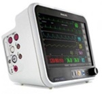
The IntelliVue patient monitoring platform is now available with Masimo rainbow SET technology. Philips also offers its customers the option to add rainbow SET to existing IntelliVue monitors, and the company is working to integrate the technology into its Philips SureSigns and Philips Efficia patient monitoring platforms. In conjunction with the appropriate patient monitoring platform, Masimo rainbow SET technology analyses multiple wavelengths of light to accurately measure total hemoglobin (SpHb), oxygen content (SpOC), carboxyhemoglobin (SpCO), methemoglobin (SpMet) and Pleth Variability Index (PVI) noninvasively and continuously. Continuous monitoring of rainbow SpHb on a Philips monitor at the point-of-care provides clinicians with real-time visibility to changes in hemoglobin in between invasive blood sampling. To ensure that customers have choice of SpO2 pulse oximetry measurement technology, the company continues to offer Philips FAST SpO2 pulse oximetry and Covidien’s OxiMax SpO2 pulse oximetry, depending on the patient monitoring platform. Read more
https://interhospi.com/wp-content/uploads/sites/3/2020/08/IH168_Philips.jpg1381503wmediahttps://interhospi.com/wp-content/uploads/sites/3/2020/06/Component-6-–-1.png3wmedia2020-08-26 14:32:122021-01-08 12:10:54Patient monitoring platform with additional measurement technologies
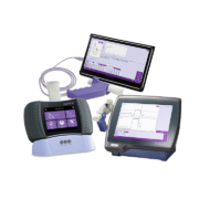
ndd Medical Technologies (ndd), a leading provider of diagnostic technology enabling healthcare professionals in the early detection of COPD and other chronic lung diseases, has introduced new single patient-use, inline filters for its lung function testing devices, in response to the global COVID-19 pandemic. Ensuring an added level of safety, the new filters can be used with ndd’s portable EasyOne spirometry range, including the EasyOne Air, EasyOne Pro, and EasyOne Pro LAB. EasyOne spirometers are popular among pulmonologists and medical researchers as they provide sensitive, reliable, real-time lung function results and diagnosis of chronic lung diseases, even at the point of care.
The EasyOne product line was originally designed with a strong focus on infection control with easy-to-wipe surfaces, single-use parts for anything that comes into contact with a patient’s breath, and ndd’s unique spirette and flow tube design which protects the flow sensor from cross-contamination. Now, when performing spirometry and testing of the diffusing capacity of the lungs for carbon monoxide (DLCO tests), the new EasyOne inline, single patient-use filters provide “double protection” for technicians and patients by keeping the ambient environment clean, without significantly affecting the EasyOne’s sensitivity.
ndd’s rigorous sensitivity tests have shown that the new inline filters do not significantly affect the device’s sensitivity and show comparable results in 24 waveform testing, (as required by ATS/ERS) and pass testing requirements. World-renowned medical research organizations – such as Johns Hopkins University, University Hospital Zurich and University of Cape Town – are partnering with ndd to assess the long-term lung damage caused by SARS-CoV-2.
Dr Trishul Siddharthan, Assistant Professor of Pulmonary and Critical Care Medicine at Johns Hopkins University, commented: “There is a lot of research underway to investigate the long-term effects of COVID-19. Our research group has been partnering with ndd thanks to the reliability of these spirometers in global settings and the ease of cleaning these devices during the pandemic to ensure safety.”
Prof. Friedrich Thienemann, General Medicine & Global Health research group, University of Cape Town and University Hospital Zurich, also noted: “In order to measure lung functions in resource-constrained settings like South African townships, one needs a device that is portable, point-of-care, easy-to-use, low-maintenance, and reliable without complicated calibration. EasyOne Pro LAB is that device.”
Incorporating ndd’s patented TrueFlow ultrasound technology, the EasyOne translates airflow into ultrasound signals which are measured by the sensors, so the patient’s breath does not come into contact with the sensors. This helps to prevent contamination and minimizes the cleaning required for safe use, while enabling a lifetime of calibration-free accuracy for flow and volume measurements.
To learn more about ndd, visit: www.nddmed.com Read more
https://interhospi.com/wp-content/uploads/sites/3/2020/08/IH_PN_ndd.jpg7207203wmediahttps://interhospi.com/wp-content/uploads/sites/3/2020/06/Component-6-–-1.png3wmedia2020-08-26 14:31:322021-01-08 12:10:16ndd’s new inline filters offer additional protection against COVID-19 during lung function tests
Xenios AG, a company of the Fresenius Medical Care Group, reports that it has seen a significant growth in demand for its extracorporeal membrane oxygenation (ECMO) devices, which can be used for the treatment of patients who develop severe pneumonia and acute respiratory distress syndrome (ARDS) due to COVID-19 infection.
Jürgen Böhm, CMO of Xenios, explained that for critically ill COVID-19 patients with acute lung failure and refractory hypoxemia – despite use of standard therapy – “our treatment often remains the last therapeutic option and has been a lifesaver for many patients”.
Xenios’s ECMO therapy bypasses the function of the lungs. The patient’s blood is freed from carbon dioxide outside the body and enriched with oxygen. The lungs are thus given time to heal. Because of the increase of critically ill COVID-19 patients, more physicians are opting for ECMO therapy, and thus the increase in demand for Xenios’s ECMO devices.
To meet the demand, the company has increased production of its ECMO devices. “We have put many measures in place to maximize the utilization of our capacity to manufacture ECMO devices as well as patient kits. Our biggest challenge right now is the availability of specific components for our products,” said Andreas Terpin, CEO of Xenios.
For more information, visit: www.xenios-ag.com Read more
https://interhospi.com/wp-content/uploads/sites/3/2020/08/PRODUCT_XENIOS.jpg74910003wmediahttps://interhospi.com/wp-content/uploads/sites/3/2020/06/Component-6-–-1.png3wmedia2020-08-26 14:31:322021-01-08 12:10:17Xenios sees growing demand for ECMO devices
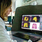
Pisa University Hospital has received support from Fujifilm Italia to improve and speed up the analysis of lung CT images which are important for the diagnosis of COVID-19. This has been possible thanks to an update by Fujifilm of the configuration of the Synapse 3D software used by emergency room radiologists.
The hospital says this new technological development has been useful to improving management of patients affected by COVID-19, during the emergency.
Synapse3D is a 3D medical image analysis system that uses Fujifilm’s image recognition technology to construct and analyze highprecision 3D images, compiled from tomographic images from CT and MRI. It delivers 3D visualization of medical images and has applications in image-based diagnosis and surgery simulation.
Synapse 3D delivers clinical value through fast, accurate, efficient and robust image processing for radiology, cardiology and surgical preoperation simulation.
It was developed in Japan (where it is named Synapse VINCENT) and it consists of more than 50 different processing modules.
The Synapse3D Lung Analysis/Airway module enables the analysis of density ranges in the lung in a quick, easy, objective and reproducible way.
At Pisa University Hospital the configuration of Synapse 3D was completed quickly, with the study of density ranges and a feasibility analysis carried out in a few days, meaning that this updated integrated workflow has been possible to use in the hospital since April 1.
The Pisa University Hospital had already set up three levels of assessing Coronavirus patients, and with the support of Synapse 3D software, doctors have had the ability to quickly and easily identify the stage of pneumonia, and therefore hospitalise patients accordingly:
normal hospitalisation for patients with mild pneumonia not requiring respiratory support
assessment by a pulmonologist or intensive care doctor for patients with moderate pneumonia to plan suitable respiratory support
intensive care assessment for patients with severe pneumonia with a view to transferring them to the intensive care unit
The density analysis provided by Synapse 3D has made it is possible to analyse the lung according to the different pixel densities of the CT images. Three groups were therefore defined based on different density ranges that allow the radiologist to evaluate the percentage of lung with lower density (emphysema), higher density (interstitial effort) and normal lung.
Dr. Chiara Romei, MD, PhD, Radiologist at the Pisa University Hospital, explained: “In a time of emergency such as this, it is crucial for us radiologists to detect accurate data as quickly as possible. Synapse 3D has enabled a quantitative analysis that is much faster and more objective than the visual analysis of the radiologist; in a couple of minutes it is possible to obtain data relating to the percentage of lung with greater and lesser density and to have a precise and objective, reproducible and shareable value.”
It is important to note that the data obtained by Synapse 3D does not replace the molecular diagnoses made through the nasopharyngeal swab (RT-PCR) and does not replace the analysis and diagnostic work by the radiologist, but instead it supports the reporting of daily exams to monitor and study the evolution of the disease, thus optimising workflow.
For medical professionals – download the take-away: https://synapse.fujifilm.eu/fujifilm-takeaway/ Read more
https://interhospi.com/wp-content/uploads/sites/3/2020/08/PRODUCT_FUJIFILM_1.jpg42010003wmediahttps://interhospi.com/wp-content/uploads/sites/3/2020/06/Component-6-–-1.png3wmedia2020-08-26 14:31:322021-01-08 12:10:17Fujifilm’s Synapse 3D provides clinical decision support, speeds up workflow during COVID-19 emergency at Pisa University Hospital
Siemens is bringing a new and flexible telemedicine solution to the market with its teamplay myCare Companion software. The software enables remote care for patients with chronic diseases. The teamplay myCare Companion allows healthcare providers to design their own telemedicine programs and flexibly adapt the underlying technology to their needs.
The solution has initially been used at the German Heart and Diabetes Center in Bad Oeynhausen for the care of patients with chronic heart failure.
During the COVID-19 pandemic, the occupational health and safety medical center at Mannheim University Hospital has been using teamplay myCare Companions’ documentation and communication components as a “coronavirus diary” for clinical. The staff records their health status every day and medical officers can respond quickly to any changes.
“Previously we used to receive piles of diaries in paper form, but now everything is nicely arranged for software to analyse”, said Dr. Margit Eisenbarth. medical officer. “Employees can enter their data conveniently via an app or website. That saves them valuable time, and we get an immediate overview.”
Wido Menhardt, Executive Vice President Digital Health at Siemens Healthineers, said: “Thanks to state-of-the-art telemedicine solutions like teamplay myCare Companion, a large number of patients can be cared for with a relatively low use of employee resources, and the number of unscheduled hospital visits can be reduced. That can substantially lower costs and is also conducive to better quality of life for chronically ill patients. Additional uses for the program that make life a little easier for medical officers and clinical employees in the current situation show just how flexible this software is.”
Teamplay myCare Companion basically consists of two components: an intuitive smartphone app for patients and a special workstation for medical care providers. Patients use the app to report their health status, wellness, the medications they’re taking, and their vital parameters to the nursing staff. Among other things, the program helps employees quickly identify patients whose state of health may have worsened. The attending doctors can then decidewhether a scheduled hospital visit should be moved forward or if more intensive remote medical care would be beneficial.
Siemens Healthineers supplies the software and supports healthcare providers with the design, installation, and operation of their own telemedicine centre. In addition, the company can assist with procurement of the measuring devices needed to monitor patients’ vital parameters at home (such as scales and blood pressure devices).
This telemedicine solution was developed in a partnership between Siemens Healthineers, the Austrian Institute of Technology (AIT), and TELBIOMED Medizintechnik und IT Service in Austria. The partners agreed to set up a global sales and development partnership at the beginning of 2020. Read more
Azienda Socio Sanitaria Territoriale (ASST) Vimercate Hospital in Italy has implemented Fujifilm’s REiLI artificial intelligence system to assist healthcare practitioners on the frontline in the fight against the COVID-19 pandemic.
Due to the pandemic, operations and workflows in Italian hospitals were disrupted, which significantly increased workloads. As a consolidated partner of Vimercate, Fujifilm wanted to ensure its cutting-edge tools were available to reduce the impact caused by the rapid spread of COVID-19 by assisting in speeding up workflows.
REiLI’s processing of CT scans and chest x-rays provides important support for radiologists, offering them an extremely rapid, quantitative and objective assessment of the various zones of the lungs. The chest x-ray, in particular, is the best example of applying the new AI technology, in that it produces a quick and simple examination which provides a large amount of useful information for evaluating the presence of the pulmonary parenchymal consolidation caused by the virus.
The data obtained from artificial intelligence does not replace the molecular diagnoses performed using the nasopharyngeal swab (RT-PCR), nor does it replace the analyses and diagnoses performed by the radiologist. The data, however, does provide support in reporting on daily examinations conducted to monitor and study the development of the disease, and constitute a second opinion for the operators.
Commenting on the REiLI artificial intelligence system, Marcello Intotero, Head of Radiology Structure and Diagnostic Services Department at ASST Vimercate Hospital, said: “REiLI’s support has allowed our radiologists to report examinations with greater speed and efficiency. This new technology has given great support to the workflow; all images that the artificial intelligence system identified as suspected COVID-19 were subjected to processing by the radiologist in a faster and more urgent manner.”
REiLI is integrated with the Lunit Insight CXR module for the analysis and detection of the main types of pulmonary disease. The Lunit Insight CXR3 module was updated to provide support in specifically identifying the pulmonary parenchymal consolidation caused by the virus.
Giovanni Delgrossi, Head of the IT Department at ASST Vimercate noted that during the health emergency, more than 80 chest x-rays of patients were analysed each day at the hospital. “If no priority is set for the examinations to be reported, it may take hours to identify a particular x-ray which may require prompt, immediate action from doctors. In an emergency like the current one, we need to react and act even more quickly,” he said.
Discussing Fujifilm’s REiLI AI system, Nicola Bilibio, Clinical Specialist Medical Informatics (CSE) Fujifilm Europe GmbH explained that AI is a new operating concept projected towards collaborative intelligence between machine and man. “This is a fundamental tool for optimizing workflow and for screening patients for COVID-19. During the emergency, we promptly made our most advanced technologies available to make a concrete contribution to the crisis.”
For more information, visit: http://reili.fujifilm.com
About REiLI on YouTube: https://youtu.be/M6ISuRmvQbY
We may ask you to place cookies on your device. We use cookies to let us know when you visit our websites, how you interact with us, to enrich your user experience and to customise your relationship with our website.
Click on the different sections for more information. You can also change some of your preferences. Please note that blocking some types of cookies may affect your experience on our websites and the services we can provide.
Essential Website Cookies
These cookies are strictly necessary to provide you with services available through our website and to use some of its features.
Because these cookies are strictly necessary to provide the website, refusing them will affect the functioning of our site. You can always block or delete cookies by changing your browser settings and block all cookies on this website forcibly. But this will always ask you to accept/refuse cookies when you visit our site again.
We fully respect if you want to refuse cookies, but to avoid asking you each time again to kindly allow us to store a cookie for that purpose. You are always free to unsubscribe or other cookies to get a better experience. If you refuse cookies, we will delete all cookies set in our domain.
We provide you with a list of cookies stored on your computer in our domain, so that you can check what we have stored. For security reasons, we cannot display or modify cookies from other domains. You can check these in your browser's security settings.
.
Google Analytics Cookies
These cookies collect information that is used in aggregate form to help us understand how our website is used or how effective our marketing campaigns are, or to help us customise our website and application for you to improve your experience.
If you do not want us to track your visit to our site, you can disable this in your browser here:
.
Other external services
We also use various external services such as Google Webfonts, Google Maps and external video providers. Since these providers may collect personal data such as your IP address, you can block them here. Please note that this may significantly reduce the functionality and appearance of our site. Changes will only be effective once you reload the page
Google Webfont Settings:
Google Maps Settings:
Google reCaptcha settings:
Vimeo and Youtube videos embedding:
.
Privacy Beleid
U kunt meer lezen over onze cookies en privacy-instellingen op onze Privacybeleid-pagina.