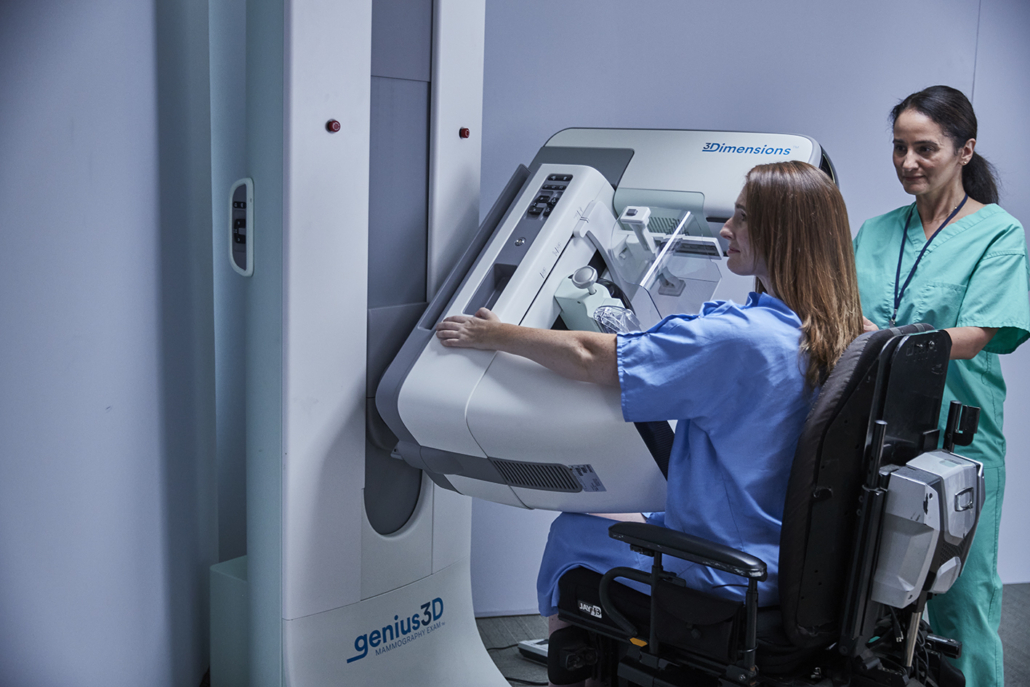
Bridging the mammography gap for a better patient experience
It is proven and widely accepted that mammography is an essential tool in breast cancer screening. Over the years, the development of the technology used to image breast tissue has enabled breast cancer to be detected earlier and more often.
Detecting cancer earlier and more often, increases a woman’s options for earlier treatment and decreases breast cancer mortality rates. Imaging technology companies in the breast health space, have continued to innovate mammography solutions that deliver ever higher quality images, cancer detection software and technologies, workflow efficiencies for healthcare facilities and automation that could help clinicians identify risk factors, detect more cancers even earlier, support constrained healthcare environments and improve the overall patient experience.
Digital breast tomosynthesis
One of these advancements is digital breast tomosynthesis (DBT), also known as 3D mammography or TOMO. For all women, DBT is more accurate for finding breast cancer. The technology has been shown to detect up to 65% more invasive cancers than a 2D mammogram alone[i], and to reduce false positive rates compared to full field digital mammography[ii]. Early detection can save lives, and DBT can help achieve this outcome for women by producing a more accurate, higher quality breast tissue images.
Traditional 2D mammography systems capture a single image of the breast tissue, which can sometimes mask small lesions – particularly for patients with dense breasts. Breast density is defined by the amount of fibrous tissue in the breast. Fibrous tissue appears white on a mammogram and can mask or hide small lesions which also appear white on a mammogram – leading to a delay in detecting the abnormalities and potentially increasing an individual’s risk of breast cancer. When DBT is utilised in place of 2D, the system captures progressive image “slices” of the breast tissue that help radiologists and clinicians see the content of breast tissue layer by layer. These new sliced images can show lesions that might be hidden beneath dense tissue.
Until now, DBT has not been used in population-based screening programmes for breast cancer. However, as screening technology has advanced, many healthcare organisations have updated their guidelines for when, how and how often a woman should be screened for breast cancer. In fact, in 2021, the European Commission Initiative on Breast Cancer (ECIBC) updated its guidelines, recommending for the first time that women with dense breasts could benefit from screening with DBT. This is an important update as it validates the studies that have shown DBT is beneficial for screening women with dense breast tissue.[iii]
Within Europe, though, there remains a lack of awareness amongst doctors and patients around dense breast tissue and the benefit of using DBT.
Measuring breast density
There are different categories of breast density, and a woman’s tissue density can change based on some life events like pregnancy and breast feeding. As part of the ECIBC guidelines update, the Guidelines Development Group recommended that women who have ongoing high breast density should utilize DBT for their screenings. However, there are three issues that arise with this recommendation.
First, many clinicians and radiologists do not currently record density when a woman has a mammogram, so there is no established protocol to track how a patient’s breast density changes throughout her life. By recording this information in her medical files, doctors can continue to evaluate any changes in a patient’s risk of breast cancer and make more informed decisions regarding whether additional screenings are needed. Additionally, this record provides opportunities for discussion about risk factors to improve patient awareness and offers the potential to personalise breast screening, so women can take responsibility for their health and know their risks.
Second, there is currently no universal standard of measurement for breast density. In the U.S., radiologists utilize the Breast Imaging Reporting & Data System (BI-RADS) to categorize lesions for mammography, ultrasound, and MRI, so there is consistent communication and understanding across the medical team. Despite this being the most common system for recording breast density, it is not the standard of practice in Europe.
Finally, although equally as important, is a lack of patient knowledge of breast density and accessibility of DBT. Currently, breast density may not be communicated at all to patients at their breast screening or in follow-up appointments. It is only if a patient asks about their risk factors that they may be told, but disclosure is not standard.
While the ECIBC guidelines recommend utilising DBT for patients with dense breast tissue, many women have no choice on the type of technology available for their breast screening nor are they aware of all the risk factors, such as dense breasts, that could affect their personal risk. Education around the risk factors has the potential to improve attendance at screening.
Expanding awareness about breast cancer screenings
When most women think of risk factors for breast cancer, the first that often comes to mind is family history. Although it does play a role in a patient’s risk of breast cancer, it is only one of seven factors that can impact a woman’s chances of being diagnosed with breast cancer.
Doctors play a vital role in helping patients understand their risk factors so they can manage their healthcare appropriately. However, in a recent U.S. study, half of participants could not identify more than four risk factors for breast cancer, and none of them could identify all seven.[iv] Additionally, this study also concluded that most women found out about mammography from their mothers, only then followed by their general practitioner or a clinician.4 This only increases the chance that a woman will not have the most accurate information about the importance of mammography and the recent innovations in technology, such as concerns about discomfort or pinching during a screening.
Pain is the number one complaint with mammography[v],[vi] and can prevent women from making regular appointments a priority. Not only can this have a negative impact on patient load for radiologists and clinicians, but it can put women at risk of a cancer being missed or diagnosed at a more advanced stage. However, many DBT machines utilise innovative breast stabilization systems that can deliver a more comfortable compression compared to standard compression5, which could help alleviate the unnecessary anxiety about discomfort that many women experience ahead of their screening.
Doctors who focus on women’s health have an obligation to provide the best care possible to their patients, and this includes making them aware of their risk factors for breast cancer and building awareness around the importance of accurate screenings.
The future of mammography
Despite these obstacles, there is progress – Europe is increasingly adopting more mammography technology that places the patient at the centre of care. The recognition from ECIBC that women with dense breast tissue could benefit from DBT is the first step, and the additional support that has followed by standardising organizations and policies is helping to spread awareness. The European Reference Organisation for Quality Assured Breast Screening and Diagnostic Services (EUREF) Type Test certification of its first DBT gantry only helps emphasize that these types of systems deliver results.
The mammographic type test certification helps assure hospitals and imaging centres that a system passed a rigorous series of mechanical, physics and clinical tests demonstrating it meets the image quality, radiation exposure and stability standards set by EUREF for screening and diagnostic mammography equipment.
The mission of EUREF council is to raise standards by bringing together the best examples of quality control in mammography screening from regional and national breast cancer screening programmes across Europe, so that hospitals and breast health centres can make informed decisions about the systems they use. With these instrumental reference bodies behind the benefits of DBT, the health systems in place across the European Union are awakening to its advantages as well. Hospitals and centres should take this time to have a closer look at their gantries and determine if these systems are not only meeting the new guidelines, but also serving their purpose of delivering the best care to women so they can live healthier lives. Healthier women mean more effective and thriving families, communities, societies and economies.
Mammography is a vital tool in the fight against breast cancer and DBT enables medical professionals to detect lesions earlier, deliver treatment earlier and drive better outcomes. With the new ECIBC guidelines in place, breast health screening programmes are poised to offer women the best opportunity at a healthier life as more doctors, clinicians and patients understand the risks around breast density and place DBT at the centre to drive earlier breast cancer detection.
References:
[i] Results from Friedewald, SM, et al. “Breast cancer screening using tomosynthesis in combination with digital mammography.” JAMA 311.24 (2014): 2499-2507; a multi-site (13), non-randomized, historical control study of 454,000 screening mammograms investigating the initial impact the introduction of the Hologic Selenia® Dimensions® on screening outcomes. Individual results may vary. The study found an average 41% increase and that 1.2 (95% CI: 0.8-1.6) additional invasive breast cancers per 1,000 screening exams were found in women receiving combined 2D FFDM and 3D™ mammograms acquired with the Hologic 3D Mammography™ System versus women receiving 2D FFDM mammograms only.
[ii] Destounis, S. V., Morgan, R., & Arieno, A. (2015). Screening for dense breasts: Digital Breast Tomosynthesis. American Journal of Roentgenology, 204(2), 261–264. https://doi.org/10.2214/ajr.14.13554
[iii] Friedewald SM, Rafferty EA, Rose SL, et al. Breast cancer screening using tomosynthesis in combination with digital mammography. JAMA. 2014 Jun 25;311(24):2499-507.
[iv] MISC-08197 MQD Consulting for Hologic, Mammography Patient Journey: Doorway Study, November 2021, U.S.
[v] Data on file and from public sources, 2017
[vi] Smith, A. Improving Patient Comfort in Mammography. Hologic WP-00119 Rev 003 (2017).
By Tanja Brycker,
Vice President, Strategic Development, Breast & Skeletal Health and Gynaecological Surgical Solutions, Hologic
About the author
Tanja Brycker joined Hologic in 2018 and serves as Vice President of International Strategic Development for Breast and Skeletal Health, and for Gynaecological Surgical Solutions.
In the preceding two decades, she held senior leadership roles in a number of global healthcare companies where she pursued a passion for advancing patient outcomes through high quality medical innovation and excellence in the commercial partnership with healthcare providers. Her desire to improve patient experience is born from over ten years as a midwife and critical care nurse where she witnessed, first hand, the benefit of prevention over cure and how medical technology can definitively enhance health outcomes. Tanja is passionate about driving and improving earlier detection, diagnosis and treatment of women’s health issues.


