
Brain’s waste clearance system definitively imaged in humans for the first time
Researchers at Oregon Health & Science University have provided the first definitive imaging evidence of the glymphatic system in human brains, confirming a long-hypothesised network of waste clearance pathways.
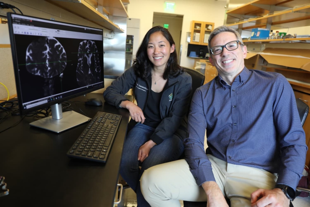
Erin Yamamoto, M.D., and Juan Piantino, M.D., are among the co-authors of a new study from Oregon Health & Science University that used imaging in neurosurgery patients to definitively reveal the existence of waste-clearance pathways in the human brain known as the glymphatic system.
© OHSU/Christine Torres Hicks
In a groundbreaking study published in the Proceedings of the National Academy of Sciences on 7 October 2024, researchers at Oregon Health & Science University (OHSU) have provided the first conclusive imaging evidence of the glymphatic system in human brains. This network of perivascular spaces, long theorised to play a crucial role in clearing metabolic waste from the brain, has been visualised in living human subjects for the first time.
Visualising the invisible
The study, led by Dr Juan Piantino, associate professor of paediatrics (neurology) at OHSU School of Medicine, utilised a novel approach to image the brain’s waste clearance pathways. Five patients undergoing neurosurgery for tumour removal consented to have a gadolinium-based contrast agent injected through a lumbar drain as part of their surgical procedure.
Following the surgery, the patients underwent magnetic resonance imaging (MRI) at 12, 24, and 48 hours post-operation. The researchers employed a specific MRI technique known as fluid attenuated inversion recovery (FLAIR) to trace the movement of cerebrospinal fluid through the brain.
“Nobody has shown it before now,” said Dr Piantino. “I was always sceptical about it myself, and there are still a lot of sceptics out there who still don’t believe it. That’s what makes this finding so remarkable.”
Channelled flow, not random diffusion
The imaging results revealed that cerebrospinal fluid does not diffuse uniformly through brain tissue, as previously thought. Instead, it moves along distinct pathways through perivascular spaces, forming a network of channels.
Dr Erin Yamamoto, a resident in neurological surgery at OHSU and co-lead author of the study, described the phenomenon: “You can actually see dark perivascular spaces in the brain turn bright. It was quite similar to the imaging the Rochester group showed in mice.”
This finding supports the concept of the glymphatic system, first proposed over a decade ago by researchers at the University of Rochester. The term “glymphatic” was coined due to the system’s dependence on glial cells in the brain and its similarity to the body’s lymphatic system.
Implications for neurodegenerative diseases
The confirmation of the glymphatic system’s existence in humans has significant implications for our understanding of brain health and neurodegenerative diseases. Scientists believe this network efficiently flushes the brain of metabolic wastes, including proteins such as amyloid and tau, which are associated with Alzheimer’s disease.
Dr Piantino emphasised the importance of this discovery: “This shows that cerebrospinal fluid doesn’t just get into the brain randomly, as if you put a sponge in a bucket of water. It goes through these channels.”
Potential for therapeutic interventions
While the study does not directly address therapeutic interventions, it provides a foundation for future research into maintaining and enhancing the brain’s waste clearance system. Emerging research suggests that certain medications may be useful in this regard.
However, much of the current focus revolves around lifestyle-based measures to improve sleep quality. These include maintaining a regular sleep schedule, establishing a relaxing bedtime routine, and avoiding screens before bed. Researchers believe that during deep sleep, a well-functioning glymphatic system efficiently carries waste proteins towards veins exiting the brain.
Future directions
This study opens up new avenues for research into brain health and neurodegenerative diseases. By providing a method to visualise the glymphatic system in humans, it may enable future studies to assess the effectiveness of interventions aimed at enhancing waste clearance in the brain.
The research team acknowledged the late Dr Justin Cetas, who initiated the study as an OHSU neurosurgeon before his untimely death in 2022. The study was supported by grants from the Medical Research Foundation of Oregon, the North American Skull Base Society, and the National Heart, Lung and Blood Institute of the National Institutes of Health.
Reference:
Bagley, J. H., Yamamoto, E., Geltzeiler, M., Piantino, J., et. al. (2024). The perivascular space is a conduit for cerebrospinal fluid flow in humans: a proof-of-principle report. Proceedings of the National Academy of Sciences, 121(41). https://doi.org/10.1073/pnas.2407246121
