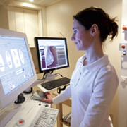
Digital tomosynthesis – the promise of 3D mammograms
Digital tomosynthesis is a new, three-dimensional (3D) technology which promises to address key shortcomings of conventional mammography. It also offers a way to tackle some of the challenges faced by computed tomography (CT) and is under investigation for a variety of new medical and surgical applications.
The concept of digital tomosynthesis is clean and elegant. A 3D slice is made by superimposing (and retrospectively reconstructing) consecutive high-resolution images, taken from different angles, across an arc. The accompanying 2D images are also used for interpretation by radiologists.
Flat-panel technology, CT pave the way
Tomosynthesis was already known in the 1930s, as part of the family of geometric tomography techniques. However, the use of plain film meant that it was procedurally painstaking, since only one image could be acquired at a time. Even more problematic was the high dosage of radiation required to produce more imaging sections.
The emergence of computed tomography (CT) in the 1970s generated a new wave of excitement about tomosynthesis. However, progress remained dormant until the mid-1990s, when the advent of flat-panel digital detectors promised a means for tomosynthesis to acquire both technical traction and momentum.
One of the most important characteristics of flat-panel technology is the lack of distortion, since its geometry (rows and columns) is known. As a result, it is possible to interpolate reconstructions on the exact point in a tomographic layer, from which data has been recorded.
First-generation flat-panel tomosynthesis systems were, nevertheless, handicapped by speed. Experimental devices, even in the late 1990s, could only achieve four to five frames per second (FPS). However, flat panel technology has evolved since then, fuelled by increasingly sophisticated optoelectronics and back-end algorithms to interpret the data. As a result, it is now possible to acquire images at 20-30 FPS with a radiation exposure similar to a chest X-Ray.
Different from CT
In spite of parallels, digital tomosynthesis and CT are two different techniques. Unlike the former, which typically consists of 15 images across a 15 degree arc, CT makes a full 360-degree rotation around a patient to acquire data for image reconstruction.
In digital tomosynthesis, the fewer data sets entail limited depth of field, and an inability to attain the very narrow slice widths of CT. However, given the digital processing of an image, one data set can provide for reconstruction of slices with both different depths and thicknesses; this not only saves time but radiation exposure requirements too.
Addressing limitations in slice width, but cost remains concern
Considerable efforts are underway to address the limits to slice width in digital tomosynthesis, especially in the form of more sophisticated detectors which allow higher in-plane resolution. The algorithms used to reconstruct tomosynthesis data are also more complex than CT. Together, both add to cost.
The Year of 3D Mammography
The application where digital tomosynthesis has drawn maximum attention is mammography. Indeed, digital tomosynthesis is now widely labelled as


