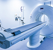
Emergency radiology – has CT growth peaked ?
Since the late 1990s, emergency radiology has become one of the fastest developing areas of medicine. It is now commonplace not only in Europe, the US and Japan but also in major urban centres of several developing countries.
The appropriate use of emergency radiology expedites patient care, prevents unnecessary hospital admissions and emergency surgery and therefore reduces costs.
Sub-specialty of radiology
Formally, emergency radiology is a relatively new sub-specialty of radiology. It is defined by the imaging and subsequent management of trauma patients, as well as those who are acutely ill. In effect, it is associated with real-time diagnostic imaging and online interpretation of data, which are conducted and completed in the ED setting itself. Emergency radiologists, on their part, need to be available and provide interpretations of imaging around the clock, including all off-hours shifts.
Professional societies set up very recently
The American Society of Emergency Radiology (ASER) was founded in 1988, with a mission to ‘advance the quality of diagnosis and treatment of acutely ill or injured patients by means of medical imaging and to enhance teaching and research in emergency radiology.’ ASER publishes the journal Emergency Radiology’ and has more than 700 members, both from the US and overseas.
The European Society of Emergency Radiology (ESER) was established in 2011, or over a decade later than its US counterpart. Based in Vienna, ESER seeks to foster education and training in emergency radiology, and collaborate both with the ASER and the British Society of Emergency Radiology (BSER), which was set up in 2014.
Radiography and fluoroscopy: limitations
Traditionally, ED imaging consisted of radiography and fluoroscopy. The procedure began with chest, abdominal and skeletal radiographs, accompanied sometimes by intravenous urograms and barium examinations. Emergency angiography was used in patients with central nervous system or vascular conditions.
Many trauma patients were, however, unable to have completion of imaging examinations in the ED, and several presenting diagnostic uncertainty were admitted to the hospital for fluoroscopic or angiographic procedures.
CT impact dramatic
Emergency medicine practice was revolutionized in the 1990s by the increase in availability of ultrasound, MRI and above all, CT. In spite of some lingering concerns, the speed of CT dramatically altered the equation in emergency radiology. A whole-body trauma CT requires just two minutes, providing information about all major injuries to the head, spine, thorax, abdomen and pelvis and increasing the probability of survival for trauma patients.
These new imaging modalities effectively served to bridge emergency medicine and diagnosis. Dramatic improvements in image quality and acquisition times have since enhanced the role of radiology in diagnosis, and as a bridge to minimally invasive procedures.
Shortening TAT
Such developments, in turn, catalysed an increase in expectations, with emergency physicians demanding quick availability of all imaging modalities, high-quality imaging examinations, real-time 3D post-processing and round-the-clock service – in effect, shortened turn-around times (TAT).
It has for long been a maxim that care provided to a trauma patient in the first few hours can be critical in terms of predicting longer-term recovery and that good trauma care involves getting the patient to the right place at the right time for the right treatment.
Professional societies have anchored such thinking. For example, guidelines from the Royal College of Radiology in Britain recognize that in the overall management of the severely injured patient, ‘diagnostic and therapeutic radiology plays a pivotal role’, although it is but a small part of ‘the whole management process.’
CT poses logistical challenges
Accompanying the increased emphasis on TAT and demands from physicians, emergency radiology facilities began to steadily reduce conventional radiography or replace it with digital X-ray. Instead, CT began to be moved to emergency departments. For example, the Royal College of Radiology guidelines mentioned above specify that CT should be adjacent to, or in, the emergency room (Standard 3) and that digital radiography should be available in the emergency room (Standard 4).
The move to relocate CT has also been driven by a need to reverse some of the major problems associated with scanners in a hospital-based trauma setting – the result of a combination of high technology and poor logistics.
Logistical problems centred upon the need for optimal location of a scanner and the capacity to receive severely injured patients within a very short period of time. This, in turn, required the availability of sufficient radiographers, a seasoned transfer procedure and resuscitation teams to be familiar with a CT environment and ready to accompany the patient during the scan.
Most traditional’ hospitals, dating back to the radiography and fluoroscopy era, were unable to cope with the dramatic changes which CT brought – above all in speed and imaging data sensitivity. This resulted in serious bottlenecks in workflow, which impacted adversely on patient outcomes.
Such shortcomings were enhanced by spikes in the volume of patient visits – e.g. during weekends and over holidays – when accident rates are far higher.
New standards for emergency radiology
To help CT relocate and become more efficient, emergency radiology facilities are being subject to exacting, new standards.
For example, the University of Amsterdam’s Academic Medical Centre (AMC) has been designed to enhance workflow efficiency and prevent dangers in the transfer of critically ill patients, while avoiding or reducing delays for non-emergency patients with scheduled appointments in the radiology department. By enabling proper equipment, transfer and support, AMC has sought to address concerns in emergency departments that, in spite of its benefits, CT might be a dangerous place for the critically ill. This was largely due to perceived limits in ventilation, resuscitation and monitoring during scanning.
One of the most visible innovations at the AMC is a sliding CT gantry on rails which serves two emergency rooms. A radiation-shielding wall closes behind the gantry, allowing the scan to be performed feet-first so IV-lines and monitors do not have to be re-positioned.
In terms of staffing, emergency radiologists at AMC are supported by a dedicated anesthesiologist who initiates ventilation, surgical residents or nurses to insert chest tubes and and radiology residents to help interpret the imaging data. This team interfaces with the trauma surgeon.
Staffing issues
Non-physician staffing is also crucial to an efficient emergency radiology facility. These range from technicians, supervisors and ED managers to receptionists, schedulers as well as ambulance personnel. State-of-the-art facilities strive to make such staff aware of the unique workflow and requirements of emergency imaging. For example, technicians need to have the skills to use different modalities and image multiple body parts. Beyond this, non-physician staff need also to be well versed in other, point-of-care medical equipment and manage a diverse range of patients – from the acutely ill to the pregnant, from children to the elderly.
A key role is also played by IT support staff, who need to be on call round-the-clock. Given the pressures to reduce TAT, they need to be well versed in RIS/PACS solutions and their suite of integrated tools, such as speech-to-text, 3D visualization, and others. More recently, IT professionals have also played a major role in data mining, in order to identify workflow bottlenecks and special situations.
Decision support tools
Another related and fast-emerging sphere consists of decision support tools, which communicate the clinical presentation, physical examination, and laboratory tests. They also confirm imaging appropriateness and selection of the optimal examination protocol.
Decision support is also seen as a means to reduce common causes of superfluous radiation in ED patients, for example, by avoiding repeat CTs (e.g. in referring hospitals). Indeed, one of the most closely-watched debates about emergency radiology concerns CT.
CT versus the rest
CT has undoubtedly been the centrepiece of the emergency radiology revolution. In 2016, a prospective study in Radiology’ showed that CT influenced the leading diagnoses in 25percent-50percent of patients and admission decisions in 20percent-25percent of patients.
Nevertheless, radiography continues to remain the most widely used imaging modality. In the US (for which data is available from a study in the American Journal of Roentgenology’ ), CT was used in 268 of 1,000 ED visits in 2012, compared to 76 for ultrasound, 64 for MRI, and 510 for X-ray.
The study, published in August 2014, also drew some other notable conclusions.
CT use in the ED peaked in 2005, while this happened two years later for MRI. Compared to 1993, CT use grew 457percent by 2005 and then declined by 49percent to 2012. For MRI, growth from 1993 to its 2007 peak was sharper, at 1,750percent, while the fall between 2007 and 2012 was 23percent, half the rate of CT. This was, nevertheless, from a much smaller user base, and as mentioned above, MRI use in the ED is outstripped more than 4-to-1 by CT (64 to 268 per 1,000 visits).
Ultrasound, on the other hand, has shown a steady but less remarkable increase in ED use between 1993 and 2012, by just 35percent. Conversely, although X-ray was used in over half ED visits in 2012, it has fallen steadily since 1993, by 26percent.
REACT-2: reality check for CT
Future trends in emergency radiology are likely to be heavily influenced by a randomized controlled trial trial at four hospitals in the Netherlands and one in Switzerland. Known as REACT-2, the trial sought to determine the effect of total-body CT scanning compared with standard work-up on patients with trauma and compromised vital parameters, clinical suspicion of life-threatening injuries, or severe injury.
The primary endpoint was in-hospital mortality, analysed in the intention-to-treat population and in subgroups of patients with polytrauma and those with traumatic brain injury.
Between April 2011 and Jan 1, 2014, the trial assessed 5,475 eligible patients and randomly assigned 1,403, 702 to immediate total-body CT scanning and 701 to the standard work-up. A total of 541 patients in the immediate total-body CT scanning group and 542 in the standard work-up group were included in the primary analysis. The study found that in-hospital mortality did not differ between groups.
As The Lancet’ reported on August 13, 2016, ‘Diagnosing patients with an immediate total-body CT scan does not reduce in-hospital mortality compared with the standard radiological work-up. Because of the increased radiation dose, future research should focus on the selection of patients who will benefit from immediate total-body CT.’
More MR?
Alongside such selection, it is also likely that there is an increase in demand for MR scanning in the ED, whose decline from its peak has been half the rate of CT (in the American Journal of Roentgenology’ study mentioned previously).
So far, MR is not indicated in an acute trauma care setting. In Britain, for example, Royal College of Radiology trauma radiology guidelines specify that MRI can be available in a different building. However, it states that ‘protocols should be in place for the transfer of critically injured patients if further management is dependent on MRI in the first 12 hours.’
Some of the benefits of MRI versus CT include acute musculoskeletal injuries, and in imaging of acute abdominal conditions in pregnant women and children.


